
Introduction
The problem of particles flowing through channels is pertinent to a wide range of applications, such as microfluidics, drug delivery, cell sorting and hemostasis.[Reference Qi and Shaqfeh1] In these applications, the particles are typically carriers of active agents, which are key to the critical functions of a device or an organism. The fluid flow has the potential to impact the structural stability of the particles, thereby impacting the ability to transport active molecules. Hence, a fundamental understanding of the key parameters impacting the flow and characteristics of single and multiple particles in channels becomes imperative. Of particular interest is the flow of phospholipid vesicles through channels as these particles are used for the delivery of therapeutics,[Reference Moretti, Zhang, Lee, Dutt and Uhrich2] and can be designed to mimic biological cells.[Reference Chu, Yu, Greenstein, Aydin, Uppaladadium and Dutt3, Reference Aydin, Uppaladadium and Dutt4]
Fluid flow can affect the shape, stability, and relative organization of the phospholipid vesicles with respect to the channel walls and each other (this is in the case of multiple particles flowing in the channel). The shear stresses[Reference Chu, Yu, Greenstein, Aydin, Uppaladadium and Dutt3] can impact the molecular organization and dynamics in the bilayer of the vesicle, thereby translating into changes in the shape and structural stability of the vesicles. External conditions such as temperature,[Reference Leonenko, Finot, Ma, Dahms and Cramb5, Reference Kucerka, Nieh and Katsaras6] salt concentration, and pH directly control the characteristics of the molecules within the bilayer, and therefore impact the bilayer properties. Hence, external conditions can also determine the flow of single and multiple vesicles in a channel.
Various experimental investigations have probed the impact of molecular composition, channel characteristics, and external conditions on the flow of phospholipid vesicles. Experimental studies[Reference Vitkova, Mader and Podgorski7, Reference Pommella, Brooks, Seddon and Garbin8] have investigated the effects of the size and the stretching elasticity on the structural stability of phospholipid vesicles, as a function of the molecular composition. Molecular composition has been determined to play an important role on the deformation of vesicles.[Reference Pommella, Brooks, Seddon and Garbin8] Other investigations[Reference McWhirter, Noguchi and Gompper9, Reference Fahraeus and Lindqvist10] on channel characteristics have demonstrated their impact on the viscosity of the flow and the hydrodynamic decoupling between particles that are flowing sequentially in a channel. Furthermore, flow conditions[Reference Vitkova, Mader and Podgorski7, Reference McWhirter, Noguchi and Gompper11] and temperature[Reference Foo, Liu and Chan12, Reference Foo, Chan and Liu13] have been demonstrated to affect the shape, structural stability, and the orientation of the vesicles. However, experimental studies are constrained by the resolution of dynamical processes occurring at multiple length and time scales. These processes can be resolved using suitable computational tools.
Computational studies have employed particle dynamics-based and continuum-based approaches to study the impact of various key factors on the characteristics of single and multiple vesicles flowing through channels. Particle dynamics-based techniques such as coarse-grained molecular dynamics, multi-particle collision or the dissipative particle dynamics techniques have identified the role of molecular composition and dynamical processes on the deformation and dynamics of vesicles during flow in a channel.[Reference Chu, Yu, Greenstein, Aydin, Uppaladadium and Dutt3, Reference Bertrand and Joos14–Reference Smith and Uspal17] In contrast, continuum-based approaches, such as boundary integral method,[Reference Kaoui, Tahiri, Biben, Ez-Zahraouy, Benyoussef, Biros and Misbah18] have been adopted to study the effect of the flow conditions, velocity flow fields, and mechanical properties of the vesicles on their shape transitions.
This review summarizes both experimental and computational studies on the factors impacting the flow of phospholipid vesicles in a channel. Of specific interest are studies which have identified the underlying causes of shape transitions in the vesicles. The review will also discuss possible future directions both in terms of experimental and computational studies.
The impact of the characteristics of phospholipid vesicles to their response under flow
The response of phospholipid vesicles, such as changes in morphology, under flow is dependent on a few key factors that control their intrinsic mechanical properties. Some of these properties include the Young's modulus, bending rigidity, and the compressibility.[Reference Marmottant, Biben and Hilgenfeldt19, Reference Misbah20] Each of these properties is determined by the molecular composition of the phospholipid vesicles. The mechanical properties of a vesicle bilayer can be determined by the length, degree of unsaturation and gel-to-fluid transition temperature of the phospholipids, their relative concentrations, and the presence of sterols,[Reference Lalia, Kochkodan, Hashaikeh and Hilal21, Reference Needham and Nunn22] such as cholesterol. Other molecular traits that can impact the mechanical properties of bilayers include the head group area of the phospholipids. Existing studies have examined the effect of molecular composition of lipid bilayers on changes in the morphology of vesicles under flow.
Experimental studies have investigated the effect of both the size of the vesicles and the stretching elasticity (which is equivalent to the Young's modulus while taking into consideration a constant cross-sectional area of the bilayer) on the rupture break-up point of the vesicles with different molecular compositions in acoustic microstream.[Reference Pommella, Brooks, Seddon and Garbin8] The authors of the study used single-component vesicles encompassing 1-stearoyl-2-oleoyl-sn-glycero-3-phosphocholine (SOPC), 1,2-dioleoyl-sn-glycero-3-phosphocholine (DOPC), or 1-palmitoyl-2-oleoyl-sn-glycero-3-phosphocholine (POPC), and two-component vesicles with different phospholipid to cholesterol ratios (namely, SOPC: cholesterol and POPC: cholesterol). SOPC and POPC each have one saturated and another unsaturated hydrocarbon tail.[Reference Aydin, Uppaladadium and Dutt4, Reference Aydin, Ludford and Dutt23, Reference Chu, Aydin and Dutt24] In contrast, DOPC has two unsaturated hydrocarbon tails. Both DOPC and SOPC have hydrocarbon chains containing 18 carbon atoms, while POPC has a shorter saturated alkyl tail. Furthermore, the gel-to-fluid transition temperature for DOPC, POPC, and SOPC was, respectively, −17, −2, and 6 °C.[Reference Silvius25]
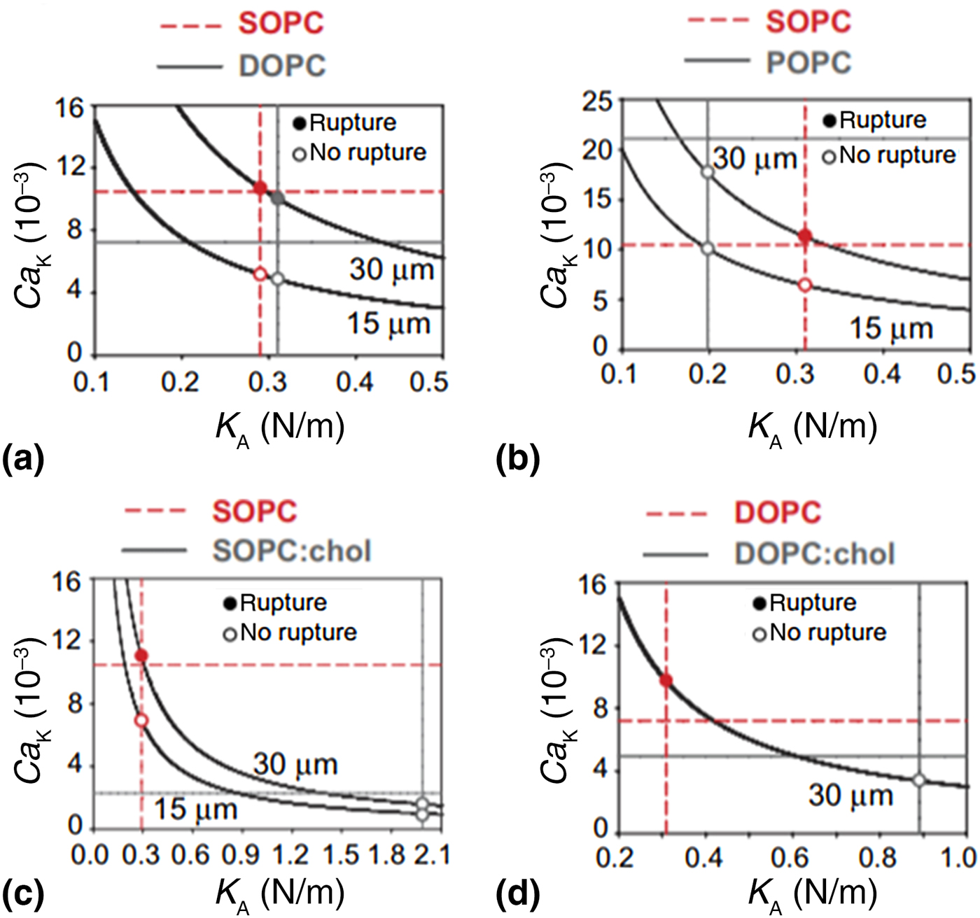
The effective capillary number was employed to evaluate the effective flow strength that is acting on the vesicles due to differences in their size. Vesicles with larger diameters were observed to rupture more easily under the same flow condition, in comparison with vesicles with smaller sizes. This is because a greater flow velocity gradient exerted larger shear stresses per surface area due to the lower curvature, resulting in higher effective capillary numbers (namely, Ca = ηG maxR/K A, where η is bulk viscosity, G max is the maximum strain rate of the vesicle, R is the vesicle radius, and K A is the stretching elasticity) for the vesicles with larger diameters. The co-authors of the study observed that the addition of 50% cholesterol to pure SOPC and DOPC vesicles, respectively, increased the stretching elasticity by factors of 3 and 5.[Reference Pommella, Brooks, Seddon and Garbin8] This observation has been corroborated by recent computational investigations of multicomponent phospholipid vesicles flowing in a channel.[Reference Chu, Yu, Greenstein, Aydin, Uppaladadium and Dutt3] Both these studies demonstrated that mixed or hybrid vesicles are more resistant to deformation. These results show that the presence of stiffer molecules in the hybrid vesicles can significantly decrease the effective capillary number (Fig. 1).
Relation between capillary number and stretching elasticity for (a) pure SOPC versus DOPC vesicle, (b) pure SOPC versus POPC vesicle, (c) pure SOPC versus SOPC:chol (50%) hybrid vesicle, and (d) pure DOPC versus DOPC:chol (50%) vesicle. In each panel, the two curves correspond to different vesicle sizes. Vertical lines denote theoretical calculations of critical capillary number and horizontal lines denotes literature values for bending elasticity for corresponding lipid species. Reproduced with permission from Ref. Reference Pommella, Brooks, Seddon and Garbin8. Licensed CC BY 4.0 https://creativecommons.org/licenses/by/4.0/ (Springer Nature, 2015).

The impact of channel characteristics on the response of phospholipid vesicles
The properties of the channel have been observed to impact the response of the vesicle to flow. This was observed to hold for both single and multiple vesicles flowing through a channel. Earlier studies have examined the effect of the channel diameter on the flow of a vesicle through a channel. For conditions where the channel diameter was comparable with the vesicle diameter, interesting effects have been reported. The apparent viscosity of blood flow through a capillary was observed to decrease when the channel diameter was decreased from 0.3 mm to 10 µm.[Reference Fahraeus and Lindqvist10] The observation was called the Fahraeus–Lindqvist effect.[Reference Fahraeus and Lindqvist10] For multiple vesicles flowing through a channel, the spacing between two consecutive vesicles along the direction of flow was observed to affect the interactions between the vesicles in narrow channels. Studies on red blood cells have shown that when the cell-to-cell distance becomes greater than the channel diameter, the cells become hydrodynamically independent or decoupled.[Reference McWhirter, Noguchi and Gompper9] It should be noted that membranes of red blood cells are constituted of a phospholipid bilayer, which is supported by a membrane skeleton comprised of spectrin (namely, a cytoskeletal protein).
The impact of flow conditions on the response of phospholipid vesicles
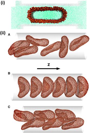
Flow conditions can also induce changes in the morphology, or shape reconfiguration, of the vesicles. A vesicle that is spherical under zero flow conditions will elongate as the flow rate in the channel is increased. To account for both the inertial and viscous effects of the flow on the vesicle, the flow can be characterized with the Reynold's number (Re). The Reynold's number is defined as Re = ρvd/μ, where ρ, v, d, and μ are the fluid density, fluid velocity, channel diameter, and fluid viscosity, respectively. Studies have shown vesicles to transition into several distinct shapes under increasing values of the Reynold's number. For example, the vesicles will transform from a sphere to a bullet-like shape and then into a parachute-like shape.[Reference Vitkova, Mader and Podgorski7] Further increasing the Reynold's number can result in the eventual rupture of the vesicle.[Reference Chu, Yu, Greenstein, Aydin, Uppaladadium and Dutt3] Even under small fluid flow velocities, where the shear forces are not sufficiently high to deform the vesicle shape, the flow conditions can affect the vesicle orientation in the channel. Studies have shown that the hydrodynamic interactions of small fluid flows can determine the orientation of the red blood cells in microcapillaries (Fig. 2).[Reference McWhirter, Noguchi and Gompper11] By tuning the channel length, diameter, and pressure gradient along the direction of flow, the system can be made to approximate an equilibrium Poiseuille flow, which corresponds to biological systems.[Reference Vitkova, Mader and Podgorski7]
(i) Deformation of a spherical vesicle into a spherocylinder during transit through a nanochannel. Reproduced with permission from Ref. Reference Bertrand and Joos14. (American Physical Society, 2012). (ii) The morphology and self-organization of red blood cells during flow through a channel. Snapshots of six red blood cells flowing in (A) disordered-discocyte phase, (B) aligned-parachute phase, and (C) zigzag-slipper phase. Reproduced with permission from Ref. Reference McWhirter, Noguchi and Gompper11. (National Academy of Sciences, 2009).

Role of other external conditions on the response of phospholipid vesicles to flow
Phospholipid vesicles under flow can be impacted by changes in the external conditions that impact the mechanical properties of the vesicle bilayer or the fluid shear stresses. These conditions include temperature, salt, and pH.
Temperature
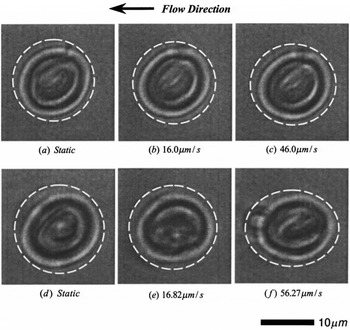
Temperature plays a critical role in the elastic properties of lipid bilayers, including the membrane of red blood cells.[Reference Artmann, Kelemen, Porst, Buldt and Chien26, Reference Mishima, Nakamae, Ohshima and Kondo27] As the elastic properties of bilayers can change the behavior of the vesicles under flow, it is important to understand the effect of temperature on the flow-induced shape deformation of vesicles. Foo et al. used optical tweezers and computational fluid dynamics to investigate the impact of temperature on the shape deformation of dipalmitoylphosphatidylcholine (DPPC) vesicles under different fluid flow velocities.[Reference Foo, Liu and Chan12] Their studies demonstrated that increasing the temperature (thereby, inducing a gel-to-liquid-crystalline transition in the vesicle bilayer) had an effect of reducing the viscous drag of the phospholipid vesicles. This was due to the vesicle bilayer becoming less rigid and highly deformable. As a consequence, the shape of a spherical vesicle transitioned into a more asymmetric stream-wise vesicle (Fig. 3).
Phase-contrast image showing deformations of DPPC vesicle at 23.0 °C in the gel phase (top row) and at 48.6 °C (bottom row) in the liquid-crystalline phase, under different flow velocities: (a) static, (b) 16 µm/s, (c) 46 µm/s, (d) static, (e) 16.82 µm/s, and (f) 56.27 µm/s. The vesicle is held by the optical trap. Reproduced with permission from Ref. Reference Foo, Liu and Chan12. (Springer, 2003).

Another study also used optical tweezers to examine the shape transformations of suddenly stopped vesicles by removing the flow-induced stress from vesicles moving at a constant velocity.[Reference Foo, Chan and Liu13] This study investigated the effect of temperature on the flow-induced deformations of vesicles composed of purely DPPC and a mixture of DPPC and cholesterol. The observations from this study revealed that the viscoelastic property of the vesicles is dominant when the temperature is above the phase transition temperature of the bilayer. Under these conditions, the vesicles take longer time to recover their initial spherical shape.
Salt and pH
Studies have examined the impact of salt on the morphology of vesicles.[Reference Renoncourt, Vlachy, Bauduin, Drechsler, Touraud, Verbavatz, Dubois, Kunz and Ninham28] These studies reported a transition from a micelle to a vesicle upon the addition of salt. However, these studies were performed under zero flow conditions. Furthermore, no studies on the impact of pH on the morphology of vesicles under flow have been examined till date.
Computational studies of phospholipid vesicles under flow
Particle-based coarse-grained simulation methods
All atom molecular dynamics simulations are not suitable for studying the effect of fluid flow on the morphological and structural properties of phospholipid vesicles. The underlying reason is that such studies will require using millions of atoms where most of the atoms will be part of the solvent molecules. Furthermore, these studies will be limited to a maximum of a few microseconds, even with the use of state-of-the-art high-performance distributing computing. In addition, all atom simulations that have been run for extended durations of time may not give desired results due to the rough energy landscape, which could cause a system to be trapped in a local energy minimum.[Reference Laio and Parrinello29] In contrast, coarse-grained simulations have smoother free energy surfaces as there are fewer degrees of freedom and this can accelerate sampling of the conformations so that experimentally comparable results can be obtained within a shorter time scale. Therefore, coarse-grained simulations can be used to study the characteristics of the vesicles under flow. Particle-based methods, such as coarse-grained molecular dynamics, dissipative particle dynamics, or multi-particle collision simulation techniques can resolve the structure and morphology of the vesicles along with the mechanisms and dynamical processes underlying the changes in the shape of vesicles under flow.
Deterministic particle-based methods such as the coarse-grained molecular dynamics simulation technique have been used to examine the extrusion of small vesicles through nanochannels[Reference Bertrand and Joos14] (see Fig. 2i). These studies used coarse-grained representations of both the solvent and the vesicle. The vesicle was pushed into a nanochannel, resulting in a change in its shape. The study demonstrated a spherical vesicle to transition into a spherocylindrical vesicle during flow through a nanochannel. Under sufficiently high pressures, the spherocylindrical vesicle was observed to rupture.
A hybrid simulation technique called multi-particle collision method combines a particle-based simulation technique for the fluid hydrodynamics with a triangulated membrane model to study the flow-induced morphology and collective behavior of vesicles or red blood cells. In particular, multi-particle collision technique has shown the flow-induced aggregation of suspensions of red blood cells (see Fig. 2ii).[Reference Noguchi and Gompper30] Furthermore, high-volume fractions of the red blood cells yield three phases, each characterized by a distinct morphology of the red blood cells and their relative organization. Under shear flow, the shape of the vesicles transitions from discocyte-to-prolate or prolate-to-discocyte.[Reference Noguchi and Gompper15] For low membrane viscosity and reduced volumes, the shear flow induces the shape of the vesicle to transition from a discocyte to a prolate.[Reference Noguchi and Gompper16] However, under high membrane viscosity, the shear flow induces the vesicle to transition from a prolate to a discocyte,[Reference Noguchi and Gompper16] or oscillations between the two states.
Dissipative particle dynamics is a mesoscopic simulation technique that uses soft-sphere coarse-grained models to capture both the molecular details of the nanoscopic building blocks and their supramolecular organization while simultaneously resolving the hydrodynamics of the system over extended time scales.[Reference Aydin, Ludford and Dutt23, Reference Groot and Warren31, Reference Dutt, Kuksenok, Nayhouse, Little and Balazs32] Dissipative particle dynamics has been extensively used to study the deformation of red blood cells and their dynamics during flow in a tube, as recently reviewed.[Reference Ye, Phan-Thien and Lim33] However, there are very few studies on the dynamics of vesicles under flow using the dissipative particle dynamics method.
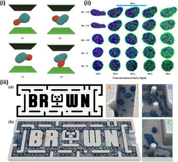
Smith and Uspal investigated the effect of shear flow on the budding dynamics of a multi-component vesicle by using the dissipative particle dynamics technique.[Reference Smith and Uspal17] The authors employed self-assembly of amphiphilic lipid molecules to create the vesicles. The single-component vesicles under shear experienced a tank treading motion, consistent with experimental observations. Budding was introduced by using two types of lipid species that phase separate within the bilayer, while shear was imposed by moving the planar walls at a constant velocity. The observations from the study demonstrated either the release of a bud through pinch off or the flattening of the budded domain via the shear forces (Fig. 4i). The study also determined the mathematical relation between the line tension and the shear rate in order for the pinch off to occur.
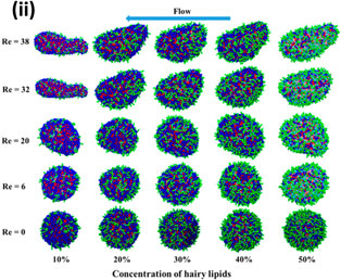
(i) Shear-driven bud flattening with time for reduced volume of 0.86, dimensionless line tension of 2.15, dimensionless shear rate of 6.52, and relative domain size of 0.23. The vesicle configuration was shown from a side view. Reproduced with permission from Ref. Reference Smith and Uspal17. (American Institute of Physics, 2007). (ii) Deformation of hairy vesicle as a function of the concentration of hairy lipids and Reynolds number. Reproduced with permission from Ref. Reference Chu, Yu, Greenstein, Aydin, Uppaladadium and Dutt3. (American Chemical Society, 2017). (iii) (a) The vector graphic image used to build the dissipative particle dynamics system of a microfluidic device. (b) Flow of surfactant solution through channels of a microfluidic device; (A–C) show the close-up views at different regions of simulation system given in (b). Reproduced with permission from Ref. Reference Li, Bian, Tang and Karniadakis36. (Elsevier, 2018).

Chu et al. studied the flow-induced response of bio-inspired multicomponent vesicles and hairy vesicles in a channel, under different flow conditions and molecular compositions.[Reference Chu, Yu, Greenstein, Aydin, Uppaladadium and Dutt3] The study examined the effect of the Reynolds number and the key molecular components on the behavior of the two types of vesicles under Poiseuille flow in a channel. The hairy vesicles were composed of phospholipid and polyethylene glycol-grafted phospholipid species. Whereas the bio-inspired multicomponent vesicles encompassed two different phospholipid species, a glycolipid and cholesterol. The channel was represented by a cylinder. The shear flow through the channel was implemented by imposing a constant body force on all solvent particles except those inside the vesicle (as opposed to using planar walls and moving them at a constant velocity[Reference Smith and Uspal17]). The vesicles were found to exhibit increased resistance to flow-induced deformation for higher concentration of cholesterol or polyethylene glycol-grafted lipids (Fig. 4ii) and longer polyethylene glycol chains. A similar behavior was observed in experimental studies showing mixed vesicles encompassing cholesterol and lipid molecules to deform less than pure lipid vesicles under flow.[Reference Needham and Rashmi34] Moreover, the structural integrity of the vesicle was found to improve with the phase separation of the polyethylene glycol-grafted lipids by preventing the opposition to the flow of the solvent.
One of the important problems in dissipative particle dynamics simulations of systems with fluid flow and wall boundaries is the penetration of the coarse-grained particles in to the wall due to the soft repulsive interactions. Some formulations were developed to alleviate this problem. For example, the two dissipative particle dynamics studies mentioned in this review used a bounce-back boundary condition at the wall surface to prevent the dissipative particle dynamics particles from penetrating into the solid walls.[Reference Smith and Uspal17] The Maxwellian reflection scheme is another commonly used method to prevent the penetration by bouncing back particles when they overlap with the wall.[Reference Revenga, Zúñiga and Español35] In order to use these methods, the position of the wall needs to be numerically defined; therefore, these methods are not applicable for walls with arbitrary shapes. A recent study solved this problem by developing a boundary method for describing arbitrarily shaped solid walls with the correct no-slip boundary conditions in dissipative particle dynamics simulations.[Reference Li, Bian, Tang and Karniadakis36] This method dynamically computes the position of the wall boundary by using information about the local particle configurations, which allow automatic detection of the wall boundaries by the particles. The dissipative particle dynamics simulations demonstrated correct no-slip boundary conditions for the walls that are stationary or moving with very low fluctuations in the density and temperature near the wall surface. Finally, this method was applied to a very complex microfluidic system by flowing surfactant solution through arbitrarily shaped small cylinders in dissipative particle dynamics simulations (Fig. 4iii). The geometry of the system was generated directly from a vector graphics image. The model was validated by examining the dynamics of surfactant micelles flowing through the microfluidic device, observing the formation of micellar structures, and correlating these results with experimental measurements.
Continuum-based simulation methods
Continuum-based methods neglect the structure and dynamics of individual lipid molecules in the vesicles bilayer, thereby significantly simplifying the lipid membrane mechanics. These methods treat the surface of a vesicle as a volume-incompressible and homogeneous material. The static topology of the vesicle surface is determined by the minimization of the sum of elastic and bending energies. In the presence of fluid flow, the velocity profile is usually described by the Stokes equation. The equilibrium deformation of the vesicle is an outcome of the force balance between the bending and elastic forces on the vesicle surface and the viscous force from the flow medium.[Reference Li, Vlahovska and Karniadakis37, Reference Abreu, Levant, Steinberg and Seifert38]
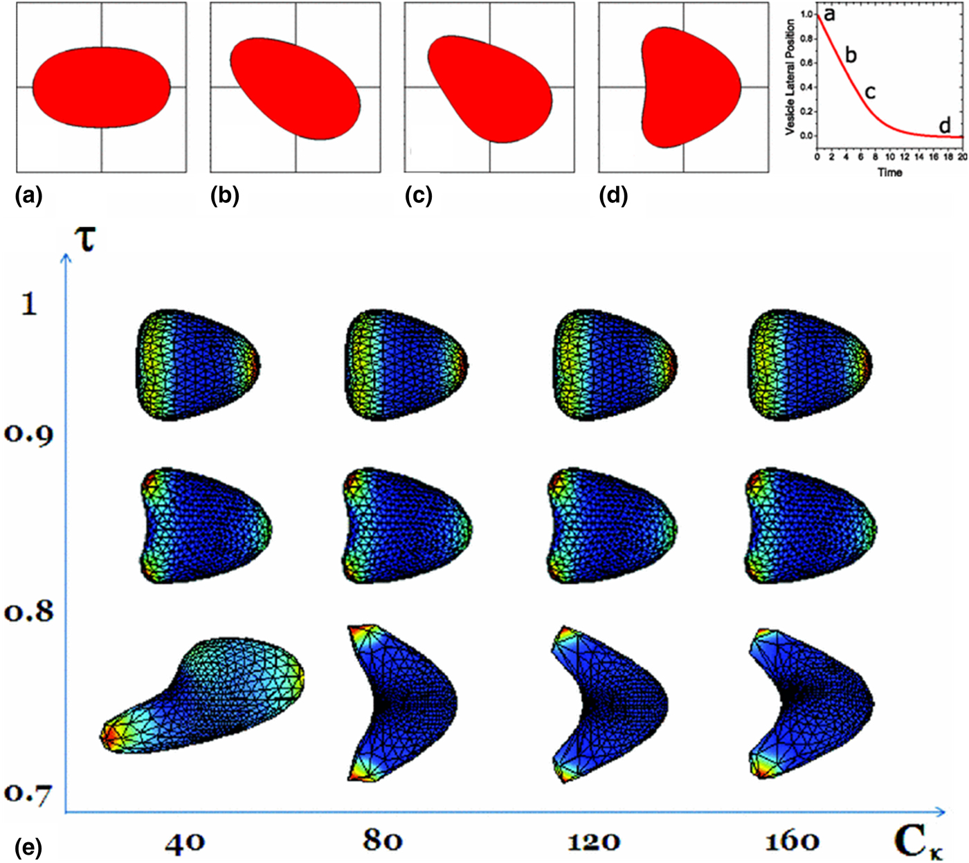
A straightforward continuum-level approach assumes the vesicle to be a second fluid phase immiscible to the surrounding flow. In this approach, numerical simplification can be applied by treating the vesicle as an incompressible droplet in a planar unbounded Poiseuille flow.[Reference Kaoui, Ristow, Cantat, Misbah and Zimmermann39] One of the insightful results obtained from this approach is that the vesicle gradually migrates vertically to the center of the flow if it is initially placed off the center. In addition, the shape of the vesicles changes from an ellipsoid to a parachute corresponding to different vertical positions during its migration.[Reference Kaoui, Ristow, Cantat, Misbah and Zimmermann39]
The boundary integral method is an efficient approach to treat the surface of the vesicles as mesh of elements whose velocities and positions are governed by a set of Green's functions. The Green's functions determine the force acting on the elements by the continuum representation of the flowing fluid. In this method, an enclosed vesicle surface was defined by an arbitrary reduced volume, which was critical for determining either the vesicle's static shape in the absence of flow or steady-state shape in the presence of flow. Via the use of such methods, the effect of the reduced volume of the initial shape of a vesicle was investigated. Figure 5 shows that different values of the reduced volume of the vesicles will result in diverse changes in their morphology under the same flow conditions. This explains why red blood cells with a biconcave structure exhibit a parachute shape that was rarely observed for a flowing vesicle that was initially given a reduced volume consistent with a spherical shape.[Reference Kaoui, Tahiri, Biben, Ez-Zahraouy, Benyoussef, Biros and Misbah18] The same applies for a red blood cell-like vesicle to adapt itself into a bullet-like structure in Poiseuille flow. Similar results regarding the reduced volume are obtained through the immersed boundary method where the surface tension was formulated as a spring-like function instead of the Lagrange's multiplier to capture the inextensibility of the surface boundary.[Reference Kaoui, Tahiri, Biben, Ez-Zahraouy, Benyoussef, Biros and Misbah18, Reference Hu, Kim and Lai40, Reference Fedosov, Peltomaki and Gompper41] In these studies, the effect of reducing the confinement was found to be equivalent to increasing the mean flow velocity as both avenues promote the shape transition from a bullet-like to a semi-parachute-like shape. In addition, the equilibrium shape remains unchanged for different bending rigidities for a constant capillary number.[Reference Fedosov, Peltomaki and Gompper41]
The shape transition of a vesicle as it migrates from off-center position to the central axis of cylindrical Poiseuille flow. (a–d) correspond to different vertical position as labelled in the curve. Reproduced with permission from Ref. Reference Kaoui, Ristow, Cantat, Misbah and Zimmermann39. (American Physical Society, 2008). (e) Phase diagram of shape transition among slipper, parachute, and bullet-like shape of a vesicle at different capillary number C k and reduced volume τ. Reproduced with permission from Ref. Reference Kaoui, Tahiri, Biben, Ez-Zahraouy, Benyoussef, Biros and Misbah18. (American Physical Society, 2011).

Differences in the viscosity of the interior and exterior fluid of the vesicle can be captured using phase field methods. Phase field methods impose a solid boundary to differentiate between the velocity field inside and outside the vesicle. Results from such methods demonstrate the existence of a critical value of the difference in the viscosity where the shape transitions from tank-treading to tumbling of the vesicles, at a certain flow velocity field. This critical value of the difference in the viscosity was shown to be positively correlated to the initial reduced volume.[Reference Biben and Misbah42] Also, vesicles became more inert to instability as the tank-treading angle significantly decreases as the difference in the viscosity increases.[Reference Beaucourt, Rioual, Seon, Biben and Misbah43]
The lattice Boltzmann method (LBM) is another continuum-based method to simulate fluid dynamics via solution of the discrete Boltzmann equation. LBM has been used to model shape changes of vesicle-like structures under shear flow.[Reference Kaoui and Harting44, Reference Li, Yi, Shan and Fang45] Li et al. used this method to study the deformation of two-dimensional vesicles (specifically, red blood cells) under flow for different Reynolds numbers by incorporating elastic modulus and bending rigidity in the LBM simulations.[Reference Li, Yi, Shan and Fang45] The results of the study showed that the physiologically relevant biconcave shape of the red blood cells is highly effective in resisting changes related to the bending rigidity of the membrane. In addition, the vesicles attain a highly flattened shape and undergo tank-tread like motion under shear flow.
The limitations of continuum-based methods yield bending and elastic energy calculations that are linearly dependent on the size of the system based upon the assumption of a homogenous distribution of material density. In other words, the prediction of in-flow topologic responses of lipid vesicles does not change with the size of the vesicles. This assumption allows continuum-based methods to work well in the microscale but lose accuracy in the mesoscale, where molecular behavior plays a more important role in determining the packing densities of lipid heads or tails at regions of different curvatures. In addition, thermal perturbation, which is critical for mesoscopic systems, is not considered by most continuum methods.[Reference Abreu46, Reference Fedosov, Dao, Karniadakis and Suresh47]
Conclusions
In summary, the transitions between different shapes of the vesicles under flow is the outcome of an interplay of multiple factors, including vesicle dimension, mechanical properties, differences in the viscosity between the interior and exterior fluid, and the reduced volume. The mechanical properties are determined by the molecular composition of the vesicles. Whereas, the reduced volume is a more complex characteristic that is controlled by membrane proteins, cholesterol concentrations, and the constraints from the cytoskeleton networks in biological cells.[Reference Goni48] Experimental studies are constrained to using a limited number of natural phospholipid molecules for synthesizing mixed vesicles, and lack precise molecular-level control for determining the reduced volume of the vesicles. Hence, it is difficult for experimental studies to contribute a systematic understanding to the relation between vesicle shape transition and the multiple factors mentioned earlier, including mechanical properties. It is also worthwhile to point out that low reduced volume shapes are not energetically favorable for vesicles with small diameters (<100 nm) to adapt to the fluid flow. This occurs as the high curvature regions of the vesicles often involve a rapid change of the orientations of the phospholipid molecules, resulting in a high exchange energy penalty. This means that a vesicle with a diameter of hundred nanometers shows less variation in its shape under flow than its counterparts whose diameters are in the micrometer scale. It is critical to consider this issue when using phospholipid vesicles to encapsulate, store, and transport active molecules.
Both continuum-based and particle-based coarse-grained simulation methods have been used to study flow-induced response and shape deformations of vesicles or red blood cells. Continuum approaches are computationally more efficient and can reach larger length scales than particle-based models as molecular scale features are not included in these approaches. On the other hand, particle-based models are useful for understanding underlying mechanism of the relation between response of the vesicles under flow and specific interactions between different vesicle components by resolving molecular scale features.
Future directions
Via the use of other classes of surfactant molecules, such as polymers or amphiphilic peptides, greater molecular level control of the vesicle characteristics can be achieved. Another route is to introduce variation in the surface texture at the vesicle–fluid interface through the grafting of solvophilic chains on to the polar groups of the surfactant molecules encompassing the vesicles. Whereas experimental studies[Reference Zhu, Yang, Chen and Du49, Reference Vauthey, Santoso, Gong, Watson and Zhang50] have developed the aforementioned vesicles, the response of the vesicles under flow has yet to be studied.
Another area of potential growth is the development of computational methods that can capture the multiscale characteristics of the vesicles and their response to fluid flow. The dissipative particle dynamics method is restricted to the values of the Schmidt number,[Reference Ahlrichs and Dunweg51, Reference Lobaskin and Dunweg52] which is considerably lower than that corresponding to water at standard temperature and pressure. Hence, the need arises for a multiscale computational method, which can capture the chemical specifics of the molecules and the correct hydrodynamic interactions. Recent efforts[Reference Ahlrichs and Dunweg51–Reference Chatterji and Horbach59] have interfaced particle dynamics techniques to discreet fluid dynamics techniques. Other efforts[Reference Ando and Skolnick60] have introduced hydrodynamic interactions into Brownian dynamics. Despite these advances, further development of these methods is required. The success of such classes of techniques would enable the representation of a range of chemical and physical interactions, coupled with long range electrostatics and hydrodynamic interactions.
Acknowledgement
The authors would like to acknowledge Xiang Yu and Geetartha Uppaladadium for their contributions to Ref. 3.