Introduction
Fully grown vertebrate oocytes are arrested at the prophase in the dictyate stage of the first meiotic division (germinal vesicle [GV] stage) until the start of oocyte maturation (Edwards, Reference Edwards1965). In amphibians, meiosis is resumed by the action of gonadotrophins, which act indirectly through the stimulation of the intrafollicular production of steroid hormones that act directly on the oocytes (Masui, Reference Masui1967; Schuetz, Reference Schuetz1974; Patiño et al., Reference Patiño, Yoshizakib, Thomasc and Kagawad2001).
The progression of the meiotic cell cycle is regulated by the M phase/maturation-promoting factor (MPF) (Masui & Markert, Reference Masui and Markert1971; Labbé et al., Reference Labbé, Capony, Caput, Cavadore, Derancourt, Kaghad, Lelias, Picard and Dorée1989) and mitogen-activated protein kinase (MAPK) (Sagata et al., Reference Sagata, Watanabe, Vande Woude and Ikawa1989; Nebreda et al., Reference Nebreda, Gannon and Hunt1995).
During meiosis resumption, oocytes undergo a series of nuclear and cytosolic changes that prepare them for fertilization and are referred to as oocyte maturation. These events are characterized by germinal vesicle breakdown (GVBD), chromatin condensation and spindle formation and, among cytosolic changes, organelle redistribution and maturation of Ca2+ release mechanisms (Fujiwara et al., Reference Fujiwara, Nakada, Shirakawa and Miyazaki1993; Gavin et al., Reference Gavin, Cavadore and Schorderet-Slatkinem1994). It is evident that the mechanism of calcium homeostasis is also modified during the maturation of oocytes from several different species (Duesbery & Masui, Reference Duesbery and Masui1996).
MPF, which is a Ser/Thr kinase composed of a regulatory cyclin B subunit and p34cdc2 subunits, displays a cyclical activity that peaks at the metaphase stage (Wu et al., Reference Wu, Ignotz, Currie and Yang1997; Kalous et al., Reference Kalous, Kubelkam, Rimkevicova, Guerrier and Motlik1993). Fully grown immature Rhinella arenarum oocytes contain a deposit of pre-MPF, located mainly in the perinuclear cytoplasm, which remains inactive by phosphorylations on the sites Thr-14/Tyr-15 of cdc2 by the Myt1 kinase. Although phosphorylation on Thr-161 is a prerequisite for the activation of MPF (Brizuela et al., Reference Brizuela, Draetta and Beach1989; Fesquet et al., Reference Fesquet, Labbé, Derancourt, Capony, Galas, Girard, Lorca, Shuttleworth, Dorée and Cavadore1993), it is activated after dephosphorylation on Thr-14 and Tyr-15 by cdc25 phosphatase (Russell & Nurse, Reference Russell and Nurse1987; Borgne & Meijer, Reference Borgne and Meijer1996). The mechanism by which MPF triggers maturation is poorly understood, although Ca2+ has been suggested as a possible second messenger. Early reports argued that a Ca2+ increase is sufficient to induce oocyte maturation (Wasserman & Masui, Reference Wasserman and Masui1975; Moreau et al., Reference Moreau, Guerrier and Dorêe1976; Schorderet-Slatkine et al., Reference Schorderet-Slatkine, Schorderet and Baulieu1976).
MAP kinase is part of a kinase cascade that is initiated probably by c-mos. This pathway seems to be involved in meiotic spindle organization, extrusion of the first polar body, and meiotic arrest at the metaphase II (MII) stage (Verlhac et al., Reference Verlhac, de Pennart, Maro, Cobb and Clarke1993; Colledge et al., Reference Colledge, Carlton, Udy and Evans1994; Choi et al., Reference Choi, Rulong, Resau, Fukasawa, Matten, Kuriyama, Mansour, Ahn and Vande Woude1996). c-mos activates MAP kinase, MEK and p90, the last step of the MAP kinase cascade, which inhibits the Mytl kinase, contributing to MPF activation. In Xenopus oocytes, the role of MAP kinase is more extensive, and its activation appears to be required for GVBD initiation (Kosako et al., Reference Kosako, Gotoh and Nishida1994).
There is a long standing debate in the literature as to whether Ca2+ signals are required for Xenopus oocyte maturation/meiosis (Duesbery & Masui, Reference Duesbery and Masui1996). Ca2+ transients induced by agonists also trigger Ca2+ waves in immature Xenopus oocytes (Lechleiter et al., Reference Lechleiter, Girard, Peralta and Clapham1991) and this factor causes GVBD and the development of the oocytes to the metaphase of the second maturation division. However, oocytes injected with high concentrations of Ca2+ buffers were unable to mature (Moreau et al, Reference Moreau, Guerrier and Dorêe1976; Duesbery & Masui, Reference Duesbery and Masui1996).
In mammalian oocytes, spontaneously occurring Ca2+ oscillations mark the acquisitions of meiotic competence and may also play a role in driving the final stage of oocyte maturation (Carrol & Swann, Reference Swann1992; Homa et al., Reference Homa, Carroll and Swann1993; Carroll et al., Reference Carroll, Swann, Whittingham and Whitaker1994). The Ca2+ signalling mechanisms of the human oocytes are known to evolve during the final stages of maturation (Herbert et al., Reference Herbert, Gillespie and Murdoch1997).
The role of Ca2+ as a mediator or trigger of GVBD during mammalian oocyte maturation (Tsafriri & Bar-Ami, Reference Tsafriri and Bar-Ami1978; Jagiello et al., Reference Jagiello, Ducayen, Downey and Jonassen1982; Powers & Paleos, Reference Powers and Paleos1982) is in conflict with reports of its Ca2+ independence (Paleos & Powers, Reference Paleos and Powers1981). In hamster oocytes (Fujiwara et al., Reference Fujiwara, Nakada, Shirakawa and Miyazaki1993) and mouse oocytes (Boni et al., Reference Boni, Tosti, Roviello and Dale1999), a series of spontaneous Ca2+ oscillations occur in the oocytes after isolation from the follicle up to the GVBD stage, although they do not seem to be required for its completion (Carrol & Swann, Reference Swann1992; Tombes et al., Reference Tombes, Simerly, Borisy and Schatten1992). In several other groups of animals, calcium ions have been implicated as regulators of oocyte maturation (Schuetz, Reference Schuetz1975).
In bovine (Homa, Reference Homa1991) and porcine (Kaufman & Homa, Reference Kaufman and Homa1993), it is well known that the Ca2+ rise during the germinal vesicle stage is essential for meiosis resumption because GVBD has been shown to be dependent on Ca2+ mobilization and frequently accompanied by transient rises in Ca 2+ (Schuetz, Reference Schuetz1975; Poenie et al., Reference Poenie, Alderton, Tsien and Steinhardt1985; Steinhardt & Alderton, Reference Steinhardt and Alderton1988; Silver, Reference Silver1989; Kao et al., Reference Kao, Alderton, Tsien and Steinhardt1990).
Similarly, there are reports that GVBD in invertebrates and amphibians are either induced by Ca2+ (Moreau et al., Reference Moreau, Guerrier and Dorêe1978), accompanied by a Ca2+ transient (Wasserman & Masui, Reference Wasserman and Masui1975), or dependent on internal Ca2+ mobilization (Masui et al., 1977). Changes in the level of intracellular free Ca2+ ions have also been strongly implicated in the triggering of the initiation of the M phase (Whitaker & Patel, Reference Whitaker and Patel1990). Due to this vital role played by Ca2+ oscillations during oocyte maturation, the study of their molecular mechanisms is of great importance.
The endoplasmic reticulum (ER) is the main Ca2+ store in the oocytes. During oocyte maturation, the ER undergoes a profound reorganization and this process seems to play an important role in the establishment of the ability of the oocytes to generate long-lasting [Ca2+]i oscillations (Shiraishi et al., Reference Shiraishi, Okada, Shirakawa, Nakanishi, Mikoshiba and Miyazaki1995).
Two types of intracellular Ca2+ receptor/channels are known to mediate intracellular Ca2+ release from the ER lumen (Miyazaki et al., Reference Miyazaki, Shirakawa, Nakada and Honda1993; Whitaker, Reference Whitaker1993). The most abundant, the inositol 1,4,5-trisphosphate receptor (IP3R), and the other Ca2+ channel, the ryanodine receptor (RyR), have also been reported to mediate Ca2+ release in several oocytes (Swann, Reference Swann1992; Berridge, Reference Berridge1993; Coronado et al., Reference Coronado, Morrissette, Sukhareva and Vaughan1994; Macháty et al., Reference Macháty, Wang, Day and Prather1997).
IP3R is known to play a central role in triggering Ca2+ release at fertilization in a number of different mammalian species (Miyazaki et al., Reference Miyazaki, Shirakawa, Nakada, Honda, Yuzaki, Nakade and Mikoshiba1992; Fissore et al., Reference Fissore, Pinto-Correia and Robl1995).
The importance of IP3 is evident through its ability to induce calcium oscillations when injected or perfused into Xenopus oocytes (Oron et al., Reference Oron, Dascal, Nadler and Lupu1985; DeLisle et al., Reference DeLisle, Krause, Denning, Potter and Welsh1990). However, Ca2+ alone was insufficient to induce GVBD in Xenopus oocytes (Duesbery & Masui, Reference Duesbery and Masui1996). Combinations of ionophore A23187 with IP3 or nocodazole-induced microtubule depolymerization not only accelerated progesterone-induced GVBD, but also induced GVBD in the absence of progesterone (Bellé et al., Reference Bellé, Ozon and Stinnakre1977).
The participation of IP3R in the regulation of intracellular calcium may also be studied by incubating inhibitors known to interfere with cellular metabolism. For example, heparin prevents calcium mobilization by its interaction with the IP3 gated-receptor in intracellular stores (Ajduk et al., Reference Ajduk, Małagocki and Maleszewski2008). In contrast, thimerosal, an artificial activator to IP3R, is a sulphydryl reagent that sensitizes calcium-induced calcium release mechanisms in cells (Swann, Reference Swann1991) and induces multiple intracellular Ca2+ transients in pig oocytes (Macháty et al., Reference Macháty, Wang, Day and Prather1997).
RyRs have not been previously detected in Xenopus laevis oocytes (Erickson et al., Reference Erickson, Mooren, Moore-Nichols and Dunn2004), although their presence in oocytes from both lower and higher order species has been reported. Functional RyR/channel complexes seem to be present in the eggs of some species such as sea urchins, mice, bovines and humans (Swann, Reference Swann1992; Galione et al., Reference Galione, McDougall, Busa, Willmott, Gillot and Whitaker1993; Ayabe et al., Reference Ayabe, Kopf and Schultz1995; Yue et al., Reference Yue, White, Reed and Bunch1995; Herbert et al., Reference Herbert, Gillespie and Murdoch1997), but their level of expression is comparatively much lower than that of the IP3R and the contribution of RyR to Ca2+ release during oocyte maturation and fertilization is as yet speculative (Ayabe et al., Reference Ayabe, Kopf and Schultz1995; He et al., Reference He, Damiani, Parys and Fissore1997; Macháty et al., Reference Macháty, Wang, Day and Prather1997; Yue et al., Reference Yue, White, Reed and King1998).
RyR agonists include exogenous ryanodine and caffeine, and endogenous cyclic ADP ribose (c-ADP), a CICR modulator that has been proposed as second messenger (Berridge, Reference Berridge1993; Galione et al., Reference Galione, McDougall, Busa, Willmott, Gillot and Whitaker1993). RyR antagonists include ruthenium red and procaine.
These channels form the pathways for the Ca2+ release that amplifies signals transmitted via plasma membrane receptor. The amplitude, frequency and subcellular localization of Ca2+ signals play an important role in determining cellular responses.
Ca2+ signals can be generated by Ca2+ release from intracellular Ca2+ stores (ER) or by Ca2+ influx from the extracellular space. In this sense, the L-type Ca2+ channel plays an important role in the incorporation of Ca2+ from the extracellular space. Upon entry into the intracellular space, Ca2+ is bound by a number of calcium-binding proteins that include calmodulin (Sun et al., Reference Sun, Hodeify, Haun, Charlesworth, MacNicol, Ponnappan, Ponnappan, Prigent and Machaca2008). Calmodulin is a ubiquitous protein involved in the regulation of several cell processes such as oocyte maturation (Cartaud et al., Reference Cartaud, Ozon, Walsh, Haiech and Demaille1980).
The aim of this work is the study of the participation of IP3R, RyR and L-type Ca2+ channel in the spontaneous and progesterone-induced maturation of Rhinella arenarum oocytes.
Materials and methods
Animals
Adult specimens of Rhinella arenarum were collected in northwestern Argentina from May to August (non-reproductive period) and from September to December (reproductive period) and kept at 15°C until use, generally 15 days after collection.
Obtainment and culture of denuded oocytes
Experimental manipulation and culture were performed at room temperature (22–25°C) in amphibian Ringer solution (AR) (6.6 g NaCl/l, 0.15 g CaCl2/l and 0.15 g KCl/l) containing penicillin G-sodium (30 mg/l) and streptomycin sulphate (50 mg/l), pH 7.4. Fully grown follicles (1.7–1.8 mm in diameter) were isolated from other ovarian tissues using watchmaker's forceps. Denuded oocytes were obtained by manually pulling off the follicle epithelium and theca layer using fine forceps under a dissecting microscope (Lin & Schuetz, Reference Lin and Schuetz1985). Follicle cells were removed by incubation of defolliculated oocytes on AR for 5 min with gentle shaking (100 oscillations/min). Denuded oocytes were kept in AR until use.
Routine in vitro cultures were carried out using plastic multiwell culture dishes (Costar 3524, Cambridge, MA, USA). Randomized samples of 20 oocytes were distributed into separate wells containing 2ml of AR. The reagents were added (5 μl) directly to the culture medium. Two well duplicates were routinely run in each determination. Oocyte maturation was assessed by detecting GVBD 24 h after hormone or reagent addition. Meiosis resumption was scored both by the presence of a transient white spot in the animal pole and by the absence of a nuclear envelope after subsequent dissection of the oocytes fixed in trichloroacetic acid (TCA).
Denuded oocytes obtained during the reproductive period (September–December) were considered competent to mature spontaneously, in contrast with those oocytes obtained during the no reproductive period (May–August), which were considered incompetent (Zelarayán et al., Reference Zelarayán, Oterino and Bühler1995).
The removal of the follicle cells in the oocytes competent to mature spontaneously (in the reproductive period) and the addition of progesterone (2.5 μM) to those incompetent to mature spontaneously (in the non-reproductive period) were considered as time 0 of the treatment. Only viable oocytes were taken into consideration at the end of the incubation period
Hormones and reagents
All hormones and reagents were purchased from Sigma. Progesterone (5 μl) was dissolved in ethanol and added directly to the culture medium to give a final concentration of 2.5 μM.
Calcium ionophore A23187 was prepared in stock solutions in dimethyl sulphoxide (DMSO).
Results
Participation of intracellular Ca2+in nuclear maturation
Effect of a calcium chelator
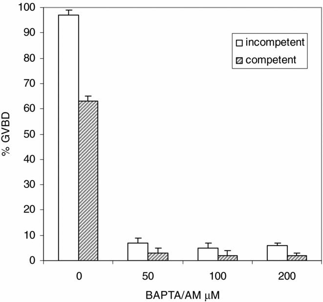
In order to evaluate the participation of intracellular Ca2+ in nuclear maturation, competent and incompetent oocytes were treated with the permeable calcium chelator BAPTA/AM (50–200 μM) for 30 min before the addition of progesterone (2.5 μM) or the removal of follicular cells. GVBD was scored after 24 h of incubation.
The results obtained (Fig. 1) indicate that a significant decrease in the ability of oocytes to resume meiosis was observed in both types of oocytes with 50 μM BAPTA/AM. A further increase in chelator concentration did not modify GVBD percentages. These results suggest that the presence of free intracellular calcium is critical to induce meiosis resumption in either type of oocytes.
Effect of BAPTA/AM on nuclear maturation. Competent and incompetent oocytes were treated with the permeable calcium chelator BAPTA/AM (50–200 μM) for 30 min before the addition of progesterone (2.5 μM) or the removal of follicular cells. Germinal vesicle breakdown (GVBD) was scored after 24 h of incubation. Values are the mean ± standard error of the mean (SEM) (n = 4). Each experiment was performed on a different animal.

Effect of the ionophore A23187
It has been shown that the oocytes of some species can be induced to reinitiate meiosis by a great variety of signals that trigger a transient intracellular calcium increase. In this case we used the ionophore A23187 to induce the translocation of Ca2+ across the membrane with the concomitant increase in free intracellular calcium.
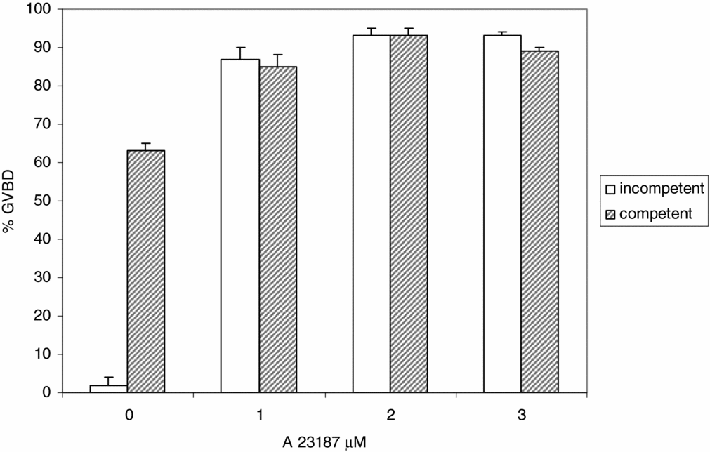
Denuded oocytes competent and incompetent to mature spontaneously were incubated in AR containing A23187 (1–3 μg/ml) in order to stimulate both external calcium influx and internal calcium release. GVBD was scored after 24 h of culture at 25°C.
The results in Fig. 2 show that after treatment with the ionophore at concentrations above 1 μg/ml the percentage of GVBD increased abruptly in both types of oocytes. Oocytes induced to mature by A23187 were similar in appearance to oocytes that had undergone GVBD after hormone treatment.
Effect of the ionophore A23187 on nuclear maturation. Denuded oocytes competent and incompetent to mature spontaneously were incubated in AR containing A23187 (1–3 μg/ml) in order to stimulate both external calcium influx and internal calcium release. Germinal vesicle breakdown (GVBD) was scored after 24 h of culture at 25°C. Values are the mean ± standard error of the mean (SEM) (n = 3). Each experiment was performed on a different animal.
Participation of extracellular Ca2+ in progesterone-induced nuclear maturation
Effect of antagonist of Ca2+ L-type channel
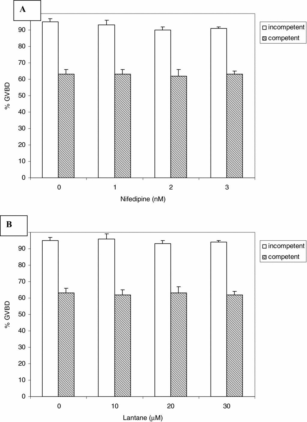
To determine whether the presence of calcium in the incubation medium was necessary for meiosis resumption, denuded oocytes competent and incompetent to mature spontaneously were incubated in AR in the presence of Ca2+ L-type channel blockers, nifedipine or lanthane, capable of inhibiting Ca2+ influx through the plasma membrane.
Oocytes competent and incompetent to mature spontaneously were pre-incubated in the presence of nifedipine (1–10 nM) or lanthane (10–30 μM) for 30 min before addition of progesterone (2.5 μM) or removal of the follicle cells. The percentage of GVBD was scored after 24 h of culture. The results shown in Fig. 3a, b show that no significant differences were found between the test groups and those of the control group.
Effect of antagonist of Ca2+ L-type channel. (A) Oocytes competent and incompetent to mature spontaneously were pre-incubated in the presence of nifedipine (1–10 nM) for 30 min before the addition of progesterone (2.5 μM) or the removal of the follicle cells. (B) Oocytes competent and incompetent to mature spontaneously were pre-incubated in the presence of lanthane (10–30 μM). The percentage of germinal vesicle breakdown (GVBD) was scored after 24 h of culture. Values are the mean ± standard error of the mean (SEM) (n = 3). Each experiment was performed on a different animal.
Participation of IP3R in nuclear maturation
Effect of thimerosal
We investigated whether immature oocytes competent and incompetent to mature spontaneously possessed IP3R capable of mediating Ca2+ release from intracellular stores and inducing GVBD.
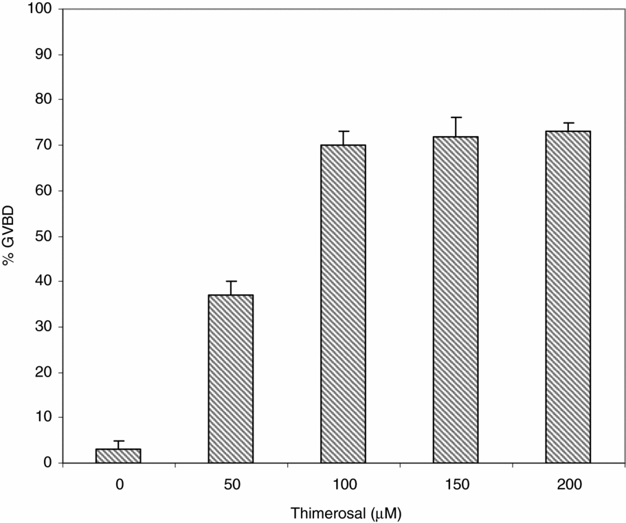
The effect on nuclear maturation of thimerosal, an agonist of IP3R, was studied by incubating incompetent denuded oocytes in AR in the presence of different doses of thimerosal (50–200 μM). GVBD was scored after 24 h of culture at 25°C.
Results (Fig. 4) indicated that thimerosal was able to induce GVBD in a dose-dependent manner. The increase in thimerosal concentration above 100 μM did not change the percentages of GVBD.
Effect of thimerosal on oocyte maturation. Incompetent denuded oocytes were culture in AR in the presence of different doses of thimerosal (50–200 μM). Germinal vesicle breakdown (GVBD) was scored after 24 h of culture at 25°C. Values are the mean ± standard error of the mean (SEM) (n = 4). Each experiment was performed on a different animal.
Effect of heparin
In order to determine if the effect of thimerosal was mediated by the activation of IP3R, the competitive IP3R antagonist heparin was used.
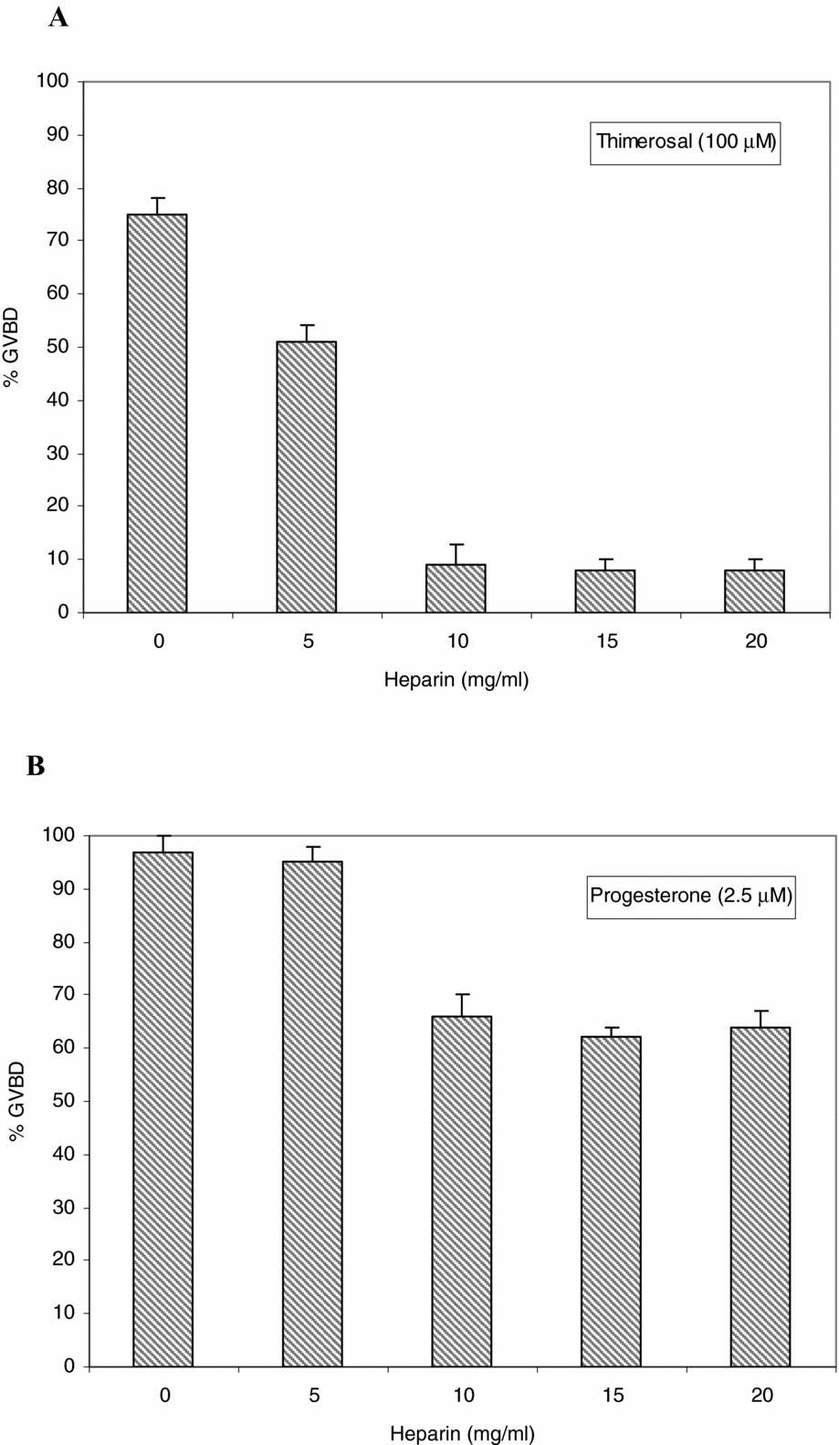
Oocytes incompetent to mature spontaneously were pre-incubated for 60 min in AR in the presence of different doses of low-molecular-weight sodium heparin (5–20 mg/ml) before the addition of thimerosal (100 μM) or progesterone (2.5 μM).
The results shown in Fig. 5 indicate that treatment with heparin significantly inhibited thimerosal-induced GVBD. This finding suggests that this agent mobilizes Ca2+ from the intracellular stores by activating the IP3 receptor. However, preincubations in heparin did not completely block progesterone-induced maturation and, at concentrations above 10 μM, GVBD rates were not modified.
Effect of heparin on oocyte maturation. Oocytes incompetent to mature spontaneously were pre-incubated for 60 min in AR in the presence of different doses of low-molecular-weight sodium heparin (5–20 mg/ml) before the addition of thimerosal (100 μM) (A) or progesterone (2.5 μM) (B). Values are the mean ± standard error of the mean (SEM) (n = 4). Each experiment was performed on a different animal.
These results suggest that the inhibitory effect of heparin is more effective in thimerosal than in progesterone-induced maturation.
Participation of RyR in nuclear maturation
Effect of caffeine
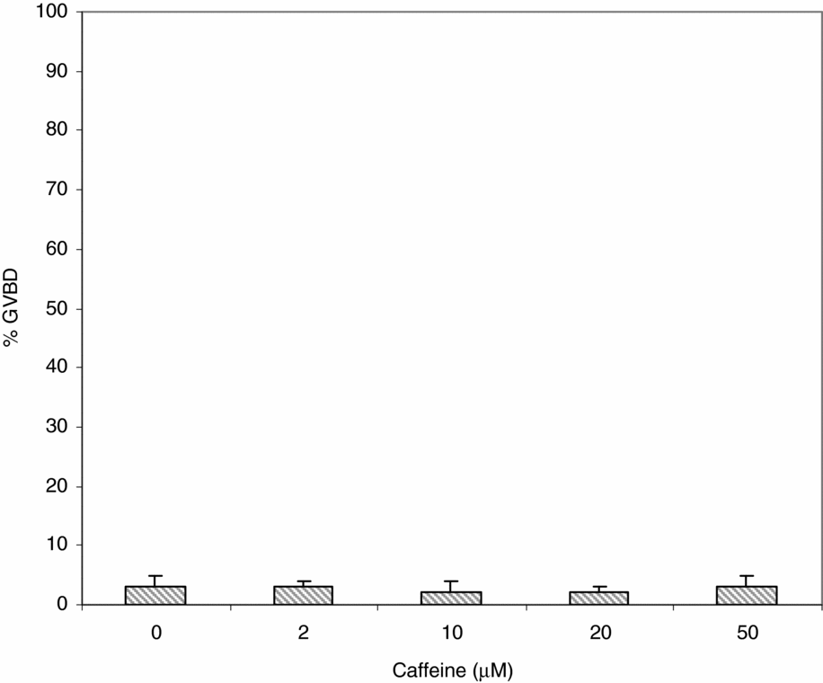
In order to determine whether ryanodine Ca2+ channel receptors are active in oocytes during maturation, we used caffeine, an agonist of the receptor, which potentiates Ca2+ release from a ryanodine-sensitive store. Incompetent denuded oocytes were treated with caffeine (5–50 μM). GVBD was scored after 24 h of culture at 25°C.
As shown in Fig. 6, treatment with caffeine did not induce GVBD, suggesting that RyR stimulation did not induce Ca2+ release.
Effect of caffeine on oocyte maturation. Incompetent denuded oocytes were treated with caffeine (5–50 μM). Germinal vesicle breakdown (GVBD) was scored after 24 h of culture at 25°C. Values are the mean ± standard error of the mean (SEM) (n = 3). Each experiment was performed on a different animal.
When incompetent oocytes were treated with low doses of thimerosal and caffeine, the percentage of GVBD was similar to the one obtained with thimerosal alone (data not shown). These results provide additional data suggesting that ryanodine receptors are no present in immature oocytes, or are not functional, or are involved to a lesser degree in Rhinella arenarum oocyte maturation.
Effect of MPF inhibition on thimerosal-induced GVBD
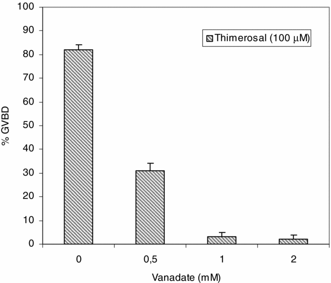
During hormone-induced maturation, MPF activity increases to complete GVBD. In order to verify if the increase in Ca2+ induced by thimerosal could bypass MPF activation, we used sodium metavanadate (NaVO3), an MPF activation inhibitor. Incompetent denuded oocytes of Rhinella were pre-incubated in vanadate (0.5–2 mM) for 30 min before treatment with thimerosal (100 μM). Results (Fig. 7) indicated that the inhibition of MPF activity by vanadate blocked thimerosal-induced GVBD.
Effect of inhibition of M phase/maturation-promoting factor (MPF) on thimerosal-induced germinal vesicle breakdown (GVBD). Incompetent denuded oocytes of Rhinella were pre-incubated in vanadate (0.5–2 mM) for 30 min before treatment with thimerosal (100 μM). GVBD was scored after 24 h of culture at 25°C. Values are the mean ± standard error of the mean (SEM) (n = 4). Each experiment was performed on a different animal.
Effect of the MAPK inhibitors on thimerosal-induced GVBD
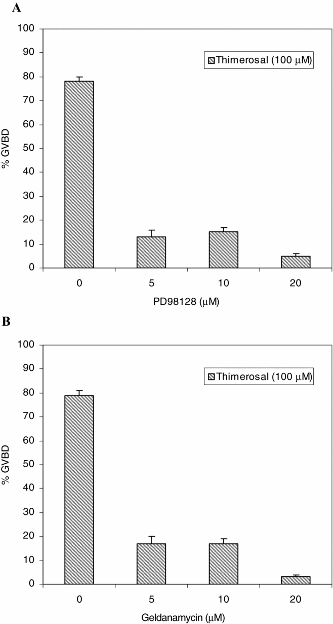
In order to determine the importance of MAPK in the GVBD induced by thimerosal we used PD98128, an inhibitor of MEK, and geldanamycin, an inhibitor of P90. Incompetent oocytes were pretreated with different doses of PD98128 (5–20 μM) or geldanamycin (5–20 μM) for 30 min before the addition of thimerosal (100 μM). GVBD was scored after 24 h of culture at 25°C.
Results (Fig. 8) showed that the inhibition of MEK or P90 inhibited the GVBD induced by thimerosal, suggesting that the activation of IP3R is not enough to bypass the lack of activity of the MAPK.
Effect of the mitogen-activated protein kinase (MAPK) inhibitors on thimerosal-induced germinal vesicle breakdown (GVBD). Incompetent oocytes were pretreated with different doses of PD98128 (5–20 μM) (A) or geldanamycin (5–20 μM) (B) for 30 min before the addition of thimerosal (100 μM). GVBD was scored after 24 h of culture at 25°C. Values are the mean ± standard error of the mean (SEM) (n = 4). Each experiment was performed on a different animal.
Discussion
The question of whether Ca2+ increases are a signal for causing GVBD is controversial in different species.
We investigated the participation of Ca2+ in the spontaneous and progesterone-induced nuclear maturation in Rhinella arenarum oocytes and the effect of different pharmacological agents known to produce modifications in Ca2+ channels.
It has been proposed that the intracellular increase in Ca2+ in amphibian oocytes is a consequence of progesterone action and that calcium is the most necessary second message for meiosis resumption (Kleis-San Francisco et al., Reference Kleis-San Francisco and Schuetz1987; Schorderet-Slatkine et al., 1984).
In agreement with these studies, we found that meiosis resumption was sensitive to Ca2+ deprivation and that a Ca2+ increase in the cytoplasm was able to trigger entry into meiosis in Rhinella arenarum oocytes. We demonstrated that loading competent and incompetent oocytes with the intracellular calcium chelator BAPTA/AM caused suppression of spontaneous and progesterone-induced GVBD. This finding is in agreement with results in cow (Homa, Reference Homa1991) and pig (Kaufman & Homa, Reference Kaufman and Homa1993) oocytes in which BAPTA/AM inhibited GVBD. However, the resumption of meiosis in mouse oocytes was not affected by chelation of intracellular calcium (Carroll & Swann, Reference Carroll and Swann1992) or depletion of extracellular calcium (Tombes et al., Reference Tombes, Simerly, Borisy and Schatten1992).
Ca2+ could diffuse by ion flow through the oocyte plasma membrane or from the ER directly from intracellular reservoirs. It is difficult to estimate the relative importance of both Ca2+ sources in the progesterone-induce GVBD.
However, in our results, the capacity of progesterone to trigger meiosis reinitiation in Rhinella in the presence of L-type Ca2+ channel blockers (nifedipine and lanthane) indicated that spontaneous and progesterone-induced maturation would be independent of extracellular calcium influx. The same result was reported by Zelarayán et al. (Reference Zelarayán, Sánchez Toranzo, Oterino and Bühler2004) using Ni and Cd. In contrast, previous findings in mouse (Lee et al., Reference Lee, Yoon and Bae2004), invertebrate (Cuomo et al., Reference Cuomo, Silvestre, De Santis and Tosti2006) and Pleurodeles (Ouadid-Ahidouch, Reference Ouadid-Ahidouch1998) oocytes demonstrated that L-type Ca2+ channels participate in meiosis resumption.
In Rhinella oocytes maturation, external Ca2+ seems to play a minor role, while intracellular Ca2+ mobilization mechanisms appear to be more important. However, the specific way in which Ca2+ affects meiosis progression is unclear.
The Ca2+ release induced by IP3 has been suggested as a primary mechanism for the maturation of bovine oocytes because the cytoplasmic IP3 receptors increase in number during meiotic progression (He et al., Reference He, Damiani, Parys and Fissore1997). In immature bovine oocytes the distribution of IP3R throughout the oocytes is similar to that in Xenopus, mouse and hamster oocytes (Kume et al., Reference Kume, Muto, Aruga, Nakagawa, Michikawa, Furuichi, Nakade and Okano1993, Reference Kume, Muto, Inoue, Suga, Okano and Mikoshiba1997; Shiraishi et al., Reference Shiraishi, Okada, Shirakawa, Nakanishi, Mikoshiba and Miyazaki1995; Mehlmann et al., Reference Mehlmann, Mikoshiba and Kline1996). Our results indicated that in Rhinella arenarum the intracellular increase in Ca2+ during maturation is also mediated mainly by IP3R, as demonstrated by the effect of thimerosal and heparine (Figs. 4 and 5). The ability of thimerosal to induce maturation in mouse oocytes was also reported by Carroll & Swann (Reference Swann1992).
Experiments performed on Xenopus oocytes suggest that IP3R sensitivity to Ca2+ closely depends on the cytoplasmic level of IP3. Consequently, when IP3 concentration increases, Ca2+ release from the ER lumen occurs through the IP3 receptors (IP3R) and lasts longer, causing more pronounced changes in Ca2+ levels (Mak & Foskett, Reference Mak and Foskett1998).
In Rhinella the response to thimerosal was similar but not identical to those occurring to the response(s) to progesterone. These observations indicate that progesterone may release intracellular Ca2+ directly or indirectly, possibly by triggering the production of IP3 via the activation of phospholipase C (Morrison et al., Reference Morrison, Waggoner, Whitworth-Langley and Stith2000; Zelarayán et al., Reference Zelarayán, Sánchez Toranzo, Oterino and Bühler2004).
In our work conditions we found that both the hormone-induced maturation and the response induced by thimerosal were blocked by heparin, indicating that thimerosal acted on the IP3-induced Ca2+ release mechanism in the oocytes.
Further studies are therefore needed to clarify whether the increased Ca2+ release is due to increased IP3 receptor sensitivity or to the sensitization of other elements of the pathway.
A second class of Ca2+ release channel, the ryanodine receptor, was first characterized as the major Ca2+ release channel of the muscle sarcoplasmic reticulum. This receptor has marked similarities with the IP3 receptor in that they are both transmembrane proteins. The presence of the ryanodine receptor is not clear among different species. There is agreement that in sea urchin eggs both IP3 and ryanodine-sensitive stores are involved in generating Ca2+ release (Lee et al., Reference Lee, Aarhus and Walseth1993; Buck et al., Reference Buck, Hoffmann, Rakow and Shen1994).
In pig oocytes, ryanodine receptors already operate in the GV stage and, like IP3 receptors, show redistribution during oocyte maturation. However, a minor role or null has been attributed to Ca2+ release through ryanodine receptors in bovine oocytes, in which they are poorly expressed (He et al., Reference He, Damiani, Parys and Fissore1997).
Although the presence of RyRs in oocytes from both lower and higher species has been reported, it has not been detected in Xenopus laevis oocytes (Erickson et al., Reference Erickson, Mooren, Moore-Nichols and Dunn2004). Our results using caffeine, an agonist of the RyR, could suggest that in Rhinella Ca2+ release from ryanodine-sensitive stores is not essential for oocyte maturation. This result may be explained by differences in the organization of the ER or by the lower sensitivity of ryanodine receptors in this species. In addition, it was reported that caffeine may modify the activity of phosphodiesterase to induce an increase in cAMP levels (Kren et al., Reference Kren, Ogushi and Miyano2004; Mishra & Joy, Reference Mishra and Joy2006; Minelli & Bellezza, Reference Minelli and Bellezza2011) that could inhibit meiosis resumption. Our experiments cannot rule out the possibility that the inhibitory effect of cAMP may be more important than the stimulatory effect of the Ca2+ released by RyR activation.
In amphibians, MPF and MAPK play a central role during oocyte maturation, controlling several events such as chromosome condensation, MTOC formation and GV migration toward the animal pole (Sánchez Toranzo et al., Reference Sánchez Toranzo, Bonilla, Bühler and Bühler2010). However, no definitive relationships have been identified between Ca2+ and MPF or MAPK.
Previous works had shown that MPF regulated the IP3-dependent Ca2+ oscillatory signals in ascidian oocytes (Levasseur & McDougall, Reference Levasseur and McDougall2000). In starfish oocytes, treatment with 1-MA to induce meiosis resumption affected Ca2+ increase induced by IP3 injection. This option would provide additional support to the suggestion that MPF is involved in the regulation of the IP3R function. Our results indicated that the decrease in MPF activity with NaVO3 negatively affected the percentage of thimerosal-induced GVBD. This finding suggests that Ca2+ release through IP3R could be involved in the signalling pathway that induces MPF activation.
MAPK is an important component of many signal transduction pathways that transmit various stimuli and regulate cell cycle progression and cell proliferation in eukaryotic cells, but the function of this kinase in mediation of the hormone-induced meiotic resumption of oocytes is unclear.
MAP kinase activation has been reported to be essential in Xenopus oocyte maturation (Tung & Jackson, Reference Tung and Jackson2005; Sellier et al., Reference Sellier, Bodart, Flament, Baert, Gannon and Vilain2006; Git et al., Reference Git, Allison, Perdiguero, Nebreda, Houliston and Standart2009) because it is apparently activated simultaneously with MPF activation and displays a switch-like behaviour (Ferrell & Machleder, Reference Ferrell and Machleder1998). Palmer et al. (Reference Palmer, Gavin and Nebreda1998) proposed that a kinase, the p90, acting downstream of MAPK, inactivates the Cdc2 inhibitory kinase Myt1 by phosphorylation and thereby links MAP kinase activation to MPF activation.
In our experiment with Rhinella oocytes, the inhibition of MEK by PD98128 or P90 by geldanamycin produced a significant decrease in the percentages of GVBD induced by thimerosal (Fig. 8a, b). This result suggests that Ca2+ release per se can not bypass the inhibition of the MAPK activity.
Conclusions
In Rhinella arenarum oocytes spontaneous and progesterone-induced maturation would be independent of extracellular calcium influx as indicated by the experiments with L-type Ca2+ channel blockers (nifedipine and lanthane) but would be sensitive to intracellular Ca2+ deprivation.
The stimulation of IP3R by thimerosal-induced GVBD in a way similar to progesterone. However, in this species, the Ca2+ release from ryanodine-sensitive stores may not be essential for oocyte maturation.
The Ca2+ release induced by thimerosal was not sufficient to induce GVBD when MPF activity was inhibited by vanadate. In the same way, the inhibition of MEK and P90 decreased significantly the GVBD induced by thimerosal.
Acknowledgments
This work was supported by a grant from the Science Council of the National University of Tucumán (CIUNT) and the National Agency for the Promotion of Science and Technology (FONCYT), Argentina.