


Introduction
The use of antiseptic products including chlorhexidine gluconate (CHG) as part of a multimodal approach has been associated with a reduction in healthcare-associated infections (HAIs). CHG body washes, oral rinses, skin antisepsis and central line care have been shown to decrease rates of HAIs in adults Reference Bleasdale, Trick, Gonzalez, Lyles, Hayden and Weinstein1–Reference Lee, Bartsch and Wong4 as well as rates of bacteremia and surgical site infections in children. Reference Milstone, Elward and Song5–Reference Raulji, Clay, Velasco and Yu8 Such practices help decrease the colonization burden with bacterial pathogens as well as minimize the likelihood of exogenous pathogens coming in contact with patients. Moreover, these strategies for the prevention of HAIs are supported by the Infectious Diseases Society of America (IDSA) and the Centers for Disease Control and Prevention. Reference Mangram, Horan, Pearson, Silver and Jarvis9–Reference Yokoe, Anderson and Berenholtz11
Gram-positive pathogens as a group are the most common cause of HAIs in children with Staphylococcus aureus being among the pathogens of highest clinical impact. Reference Blackburn, Henderson, Minaji, Meuller-Pedobdy, Johnson and Sharland12,Reference Wisplinghoff, Seifert, Tallent, Bischoff, Wenzel and Edmond13 Notably, a subset of S. aureus possess genes encoding inducible multidrug resistance efflux pumps, including qacA/B and smr, which may confer higher MICs to CHG and other antiseptics. In prior work, up 44% of hospital-acquired S. aureus isolates carried either qacA/B or smr. Reference McNeil, Kok and Vallejo14 While CHG MIC elevations in these strains are modest, ex vivo models of S. aureus venous catheter infection demonstrated that the presence of qacA/B or smr are associated with significant reduction in the effectiveness of CHG decontamination procedures at concentrations far exceeding the MIC. Reference McNeil, Sommer, Vallejo, Hulten and Kaplan15 While the clinical impact of these phenomena are uncertain, a number of centers have reported decolonization failure as well as breakthrough infections in patients colonized with S. aureus carrying these genes. Reference Lee, Macedo-Vinas and Francois16,Reference Batra, Cooper, Whiteley, Patel, Wyncoll and Edgeworth17
Notably, children with cancer are at unique risk for antimicrobial resistant infections given their frequent hospitalizations, exposure to antibiotics and antiseptics as well as immunosuppression. In a study from St. Jude’s Children’s Research Hospital from the mid-2000s, S. aureus was the second most common pathogen of bloodstream infection in children with malignancy. Reference McCullers, Williams and Wu18 We previously reported an increase in the presence of antiseptic tolerance (AT) genes among S. aureus infections in pediatric oncology patients following an escalation of CHG use at Texas Children’s Hospital (TCH). Reference McNeil, Hulten, Kaplan, Mahoney and Mason19 It is conceivable, that regular exposure to antiseptics as well as the healthcare system in general could be selecting for colonization with antiseptic tolerant organisms among high-risk children. Little data are available, however, regarding the frequency or clinical impact of asymptomatic colonization with antiseptic tolerant pathogens in children. Moreover, children have been reported to have higher rates of S. aureus colonization than adults in some studies, Reference Kuehnert, Kruszon-Moran and Hill20 potentially making them an ideal population to investigate the relationship between healthcare exposure and antiseptic tolerant S. aureus (ATSA) colonization. We conducted a longitudinal evaluation of nasal and axillary colonization with antiseptic tolerant ATSA in children with cancer relative to a cohort of healthy children.
Methods
Subjects and study procedures
Subjects were enrolled into high- and low-risk cohorts in a 1:2 ratio with each cohort having similar but distinct eligibility criteria. High-risk subjects comprised children 1 months –<18-years old with a recent diagnosis (< 3 months) of malignancy (including hematologic malignancy and solid tumors) recruited from the Texas Children’s Cancer Center (TCCC) clinic in Houston, TX. Subjects with expected survival > 12 months and without the anticipated need for hematopoietic stem cell transplant were eligible. Enrollment of subjects began in November 2019 but was temporarily halted in late March 2020 due to restrictions imposed by the SARS-CoV-2 pandemic; enrollment was reinitiated in Oct 2020 and was ultimately completed in November 2021. Pediatric oncology patients were chosen as the study population because of 1) previous studies demonstrating high-levels of qacA/B-positive S. aureus infections, Reference McNeil, Hulten, Kaplan, Mahoney and Mason19 2) high-levels of healthcare, antibiotic and antiseptic exposure and 3) the need for regular medical follow-up encounters in the TCCC, enhancing subject retention.
The low-risk cohort was comprised of healthy children from 1 months– <18 years of age recruited from either of two Houston, TX area primary care practices affiliated with Texas Children’s Hospital (TCH). Low-risk subjects with a major medical comorbidity or ongoing use of mupirocin or chlorhexidine gluconate products were excluded. Only one child per household was eligible. From both cohorts, subjects with an active known or suspected S. aureus infection (e.g., skin abscess, folliculitis, etc.) were excluded. Additionally, subjects with parents/guardians unable to read/write English or Spanish were excluded. A subset of low-risk subjects was reported in a prior publication. Reference McNeil, Flores, Kaplan and Hulten21
Following informed consent procedures, subjects had anterior nares and axillary cultures obtained and guardians were asked to complete a questionnaire focusing on general health, antimicrobial/antiseptic use, healthcare exposure and staphylococcal infections. Additional questionnaires were administered, and colonization cultures collected every 3 months (±30 days) for a 12-month period. At the time of enrollment, guardians of low-risk subjects were instructed in culture collection. Reference Lautenbach, Nachamkin and Hu22 Follow-up questionnaires, culture collection supplies and written instructions were then shipped to low-risk subjects; questionnaires and culture swabs were returned promptly to study personnel. For the high-risk cohort, study follow-up encounters occurred every 3-months and coincided with standard of care visits in the TCCC. For high-risk subjects, nares cultures were rescheduled if the absolute neutrophil count was < 500 cells/mm3 or platelet count < 50,000 cells/mm3 at the time of study visit. Likewise, for patients in either cohort actively receiving antibiotics, culture collections were deferred until the subject was off anti-staphylococcal therapy for ≥48 hours. All subjects had follow-up telephone encounters at 18-months post enrollment to assess for incident infections as well as general health. The primary outcome was colonization with ATSA at either body site at least once during study follow-up. This study was approved by the IRB of Baylor College of Medicine.
All culture samples were examined for the presence of S. aureus; identified S. aureus underwent PCR for the detection of qacA/B and smr. Reference McNeil, Kok and Vallejo14 ATSA were collectively regarded as those isolates bearing either qacA/B and/or smr. Detailed microbiology methods as well as sample size considerations are provided in the Supplemental Appendix.
Infection prevention measures
CHG with or without alcohol is the skin cleanser of choice prior to central venous line (CVL) insertion and maintenance procedures at our institution as well as for cleansing of the catheter hub prior to accessing the CVL. Oncology patients with a CVL are advised to take a weekly bath with CHG wipes during hospital admissions. Colonization status determined as part of this study was not disclosed to subjects or providers; children with malignancy are not routinely screened for S. aureus colonization at our center and as such, the non-disclosure of colonization status did not impact the standard of care.
Statistical analyses
Subjects with samples submitted for ≥ 3 study encounters were eligible for inclusion in the final analysis. Summary statistics were stratified by study arm (high- vs. low-risk) and compared using independent, two-sample t-tests, Wilcoxon rank sum test, chi-square, and Fisher exact test. A similar analysis compared baseline characteristics between subjects who had an ATSA colonization with those who did not. Baseline characteristics that were significantly different between groups at the p < 0.05 level were subsequently included in a multiple logistic regression model. The frequency of S. aureus/ATSA colonization was compared at each observation period and presented as the proportion of subjects colonized at either body site. Subanalyses included an assessment of risk factors for ATSA colonization within the high-risk cohort. Statistical analyses were performed using SAS for Windows ver. 9.4 (Cary, NC).
Results
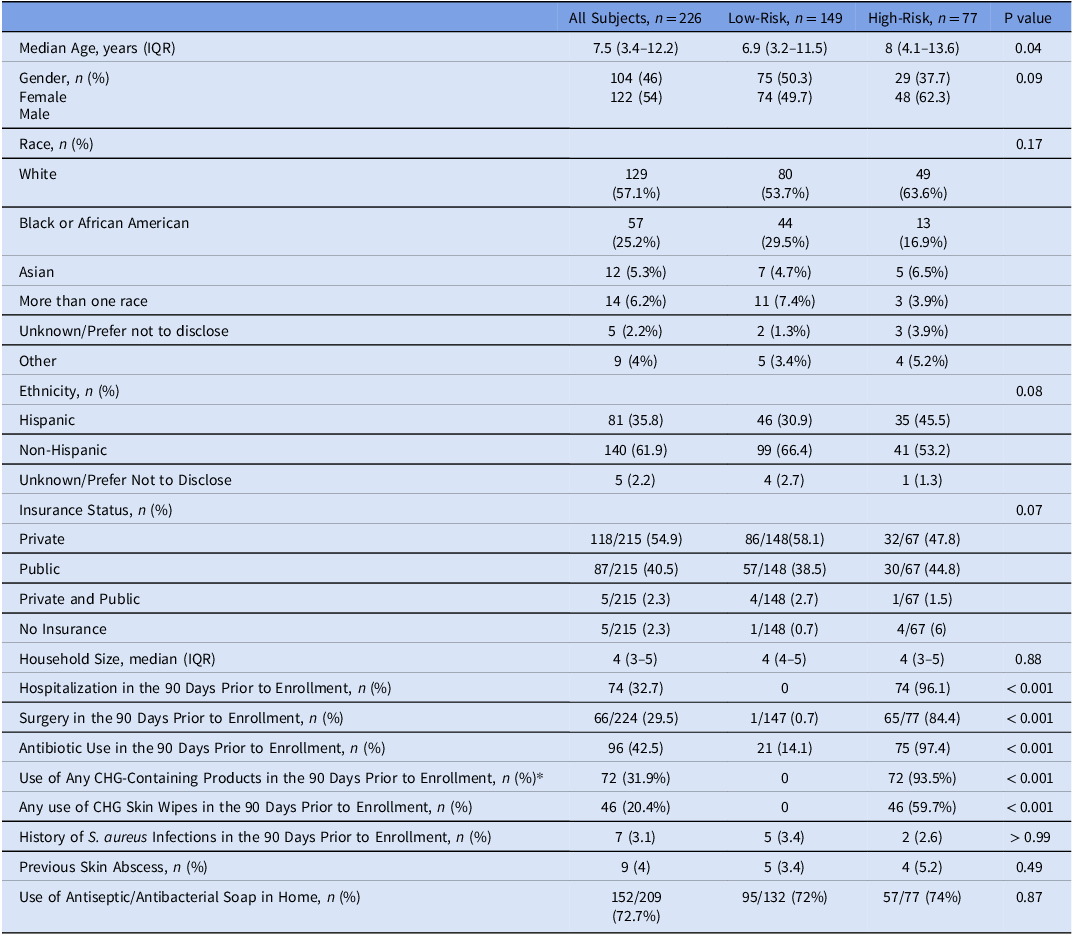
During the study period, 272 subjects were enrolled (188 low-risk and 84 high-risk subjects) with 226 (149 low-risk and 77 high-risk) being evaluable for the primary outcome. There were 1926 culture swabs included in the final analysis. Subjects in the high-risk cohort were older than those in the low-risk cohort (median 8 years [Interquartile range (IQR): 4.1–13.6 years] vs. 6.9 years [IQR: 3.2–11.5 years], p = 0.04, Table 1). Other demographic characteristics were similar between low- and high-risk cohorts. Among the high-risk cohort, the median time from oncologic diagnosis to enrollment was 88 days. As expected, a higher rate of overall health care exposure at enrollment was present in the high-risk cohort, including higher rates of surgery, hospital admission and antibiotic exposure in the 90-days prior to enrollment. Rates of prior S. aureus infection were comparable between groups.
Baseline characteristics at enrollment

* Includes CHG prior to CVL access, CHG skin wipes and mouthwash.
Among the high-risk cohort, the most common malignancy was acute lymphocytic leukemia (ALL, n = 41) followed by sarcomas as a group (n = 12), Hodgkin’s lymphoma (n = 9) and Wilm’s tumor (n = 5, Supplemental Table 1 ). Seventy-five high-risk subjects (97.4%) had a CVL in place at the time of study enrollment and 72 (93.5%) reported regular use of any CHG-containing product (e.g., CVL care, CHG-wipes, mouthwash). Over the course of one-year follow-up, the frequency of high-risk subjects with CVL present and/or reported CHG use as well as recent hospitalization at a given time point declined (Supplemental Figure 1 ).
Overall S. aureus colonization
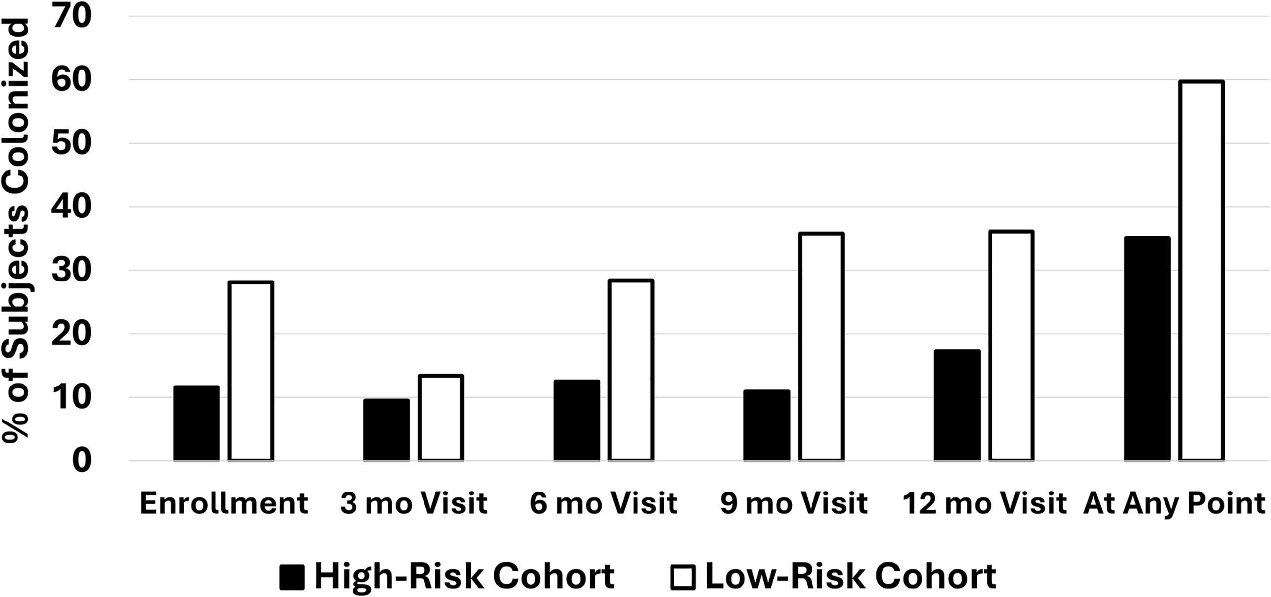
From subjects evaluable for the primary outcome, 275 S. aureus colonizing isolates were obtained. One-hundred sixteen subjects (51.3%) were colonized with S. aureus at least once during the study period and 15 (6.6%) had MRSA colonization. S. aureus colonization occurred in 27/77 (35.1%) high-risk subjects and 89/149 (59.7%) low-risk subjects (p < 0.001, Figure 1). MRSA colonization occurred in 2/77 (2.6%) of high-risk subjects and 13/149 (8.7%, p = 0.09) low-risk subjects. In 70 subjects (25.7%), S. aureus was recovered at more than one time point; low-risk subjects more often had S. aureus recovered at more than one-time point (60/89, 67.4%, vs. 10/27, 37%, p = 0.007). Four subjects were colonized with MRSA more than once. A negative relationship was observed at numerous time points between overall recovery of S. aureus and any antibiotic use in the 90 days prior to sample collection (Supplemental Figure 2 ). Higher rates of antibiotic use were noted among the high-risk cohort at all time-points (Supplemental Figure 3).
S. aureus Colonization Over Time. The depicted values represent the proportion of subjects in each risk cohort with colonization at the respective time point. Overall, a greater proportion of low-risk subjects were colonized with S. aureus at least once (p < 0.001).

ATSA colonization
Thirty-five subjects (15.5%) were colonized with ATSA at least once during follow-up. Twenty-three subjects (10.2%) were colonized at least once with a qacA/B-positive strain and 22 with an smr-positive strain (9.7%). Two subjects were colonized at more than one time point with ATSA with identical antimicrobial susceptibility patterns.
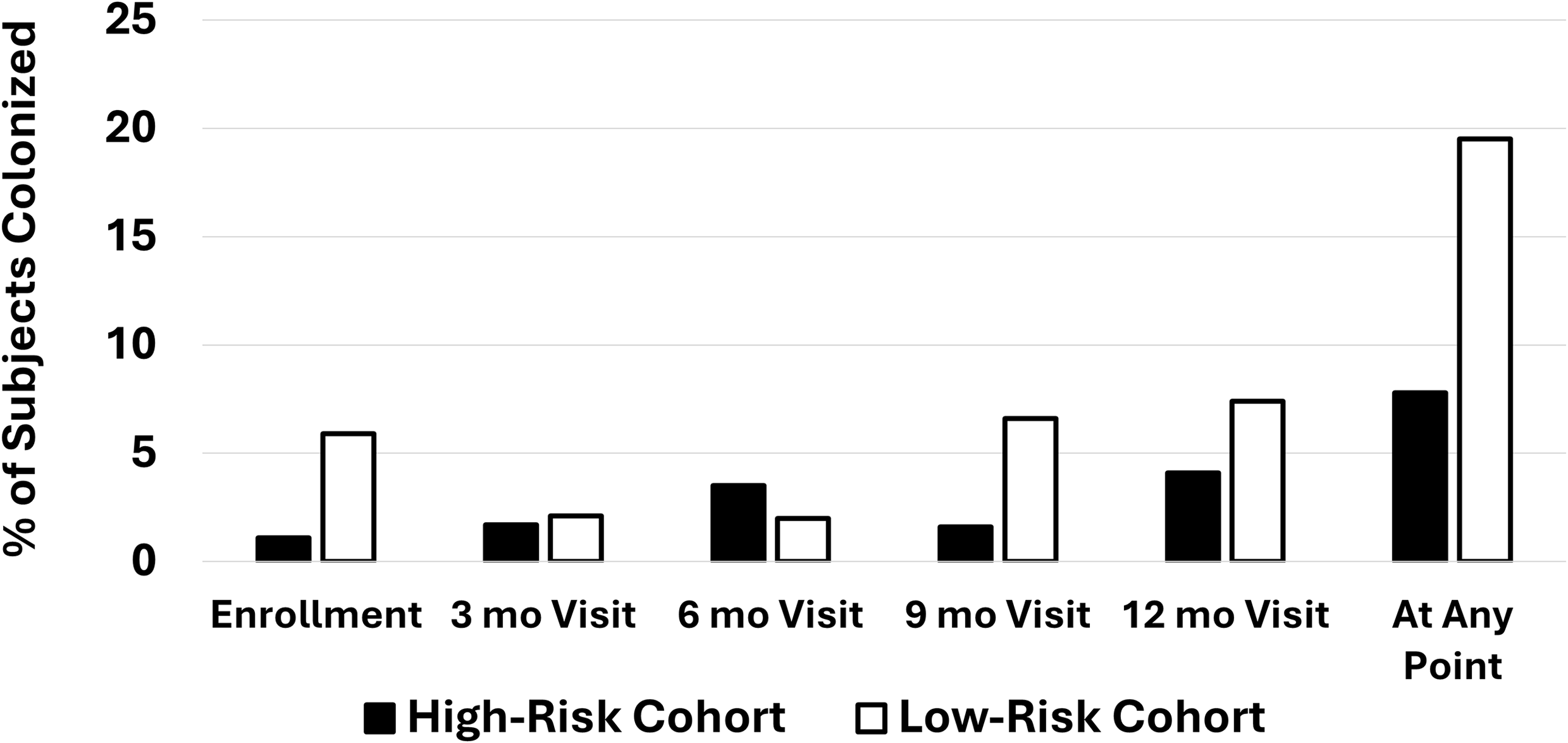
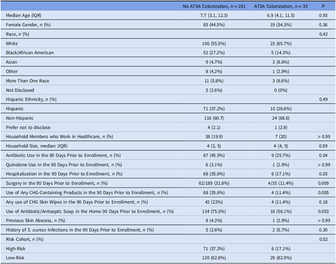
The proportion of subjects colonized with ATSA at a given encounter ranged from 2-7.4% among the low-risk cohort and 1.1–4.1% in the high-risk cohort (Figure 2). A greater proportion of subjects with ATSA colonization at any point during the study belonged to the low-risk cohort compared to those without ATSA colonization (29/35, 82.9%, vs. 120/191, 62.8%, p = 0.02, Table 2). Other associations observed with ATSA colonization included lower rates of antibiotics use (25.7% vs. 45.5%, p = 0.04), hospitalization (17.1% vs. 35.6%, p = 0.03), surgery (11.4% vs. 32.8%, p = 0.009), and use of CHG (11.4% vs. 35.6%, p = 0.002). After adjustment for confounding variables, there was no association between risk-cohort and ATSA colonization status at a statistically significant level (p = 0.97). No predictors were significantly associated with ATSA colonization in the full model.
ATSA Colonization Over time. The depicted values represent the proportion of subjects in each risk cohort with colonization at the respective time point. A larger proportion of low-risk subjects were colonized with ATSA at least once (p = 0.02).

Comparison of subjects with and without ATSA colonization

Subjects with and without qacA/B-positive S. aureus colonization were compared in additional sub-analyses. Subjects with qacA/B-positive S. aureus colonization reported less use of antiseptic soap in the home (9/23, 39.1%, vs. 143/203, 70.4%, p = 0.004) and more frequently had a history of documented S. aureus infection prior to enrollment (4/23, 17.4% vs. 6/203, 2.9%, p = 0.01).
Within the high-risk cohort, most subjects with ATSA colonization had a hematologic malignancy (5/6, 83.3% vs. 50/71, 70.4%, p = 0.6, Supplemental Table 2 ). Additionally, high-risk subjects with ATSA colonization more often had a history of quinolone use (33.3% vs. 14%, p = 0.2) and less often reported use of antibacterial soap in the home (33.3% vs. 73.5%, p = 0.03). In the high-risk cohort, the number of hospital days as well as antibiotic days from enrollment to last follow-up were similar among those with and without ATSA colonization. Among high-risk subjects with ATSA colonization, one developed an incident S. aureus skin infection diagnosed at an outside facility- the isolate was unavailable for comparison to the colonizing strain. Another ATSA colonized high-risk subject developed cellulitis that was presumed staphylococcal in nature.
Antimicrobial Susceptibility
CHG MICs were determined for 275 S. aureus colonizing isolates. Overall, qacA/B-positive strains were associated with modest elevations in CHG MIC compared to strains lacking this gene (p = 0.02, Supplemental Table 3 ).
qacA/B-positive isolates were more often MRSA (9/27, 33.3% vs. 14/248, 5.6%, p < 0.001); qacA/B-positive isolates were also associated with a numerically higher rate of ciprofloxacin resistance (4/27, 14.8%, vs. 16/248, 6.5%, p = 0.1, Supplemental Table 4 ). smr-positive and -negative isolates had comparable rates of resistance to systemic antibiotics.
Discussion
The use of antiseptic preparations, including CHG, when implemented as part of a bundled approach are associated with reductions in HAIs. Despite these potential benefits, several studies have reported the emergence of infections due to S. aureus with reduced susceptibility to CHG in populations with high levels of exposure. Reference McNeil, Hulten, Kaplan, Mahoney and Mason19,Reference Suwantarat, Carroll and Tekle23,Reference Warren, Prager and Munigala24 We sought to evaluate the frequency of colonization with ATSA in children with cancer.
In a prior study of neonatal intensive care unit patients colonized with MRSA, 8% of isolates had reduced CHG susceptibility. Reference Reich, Boyle and Hogan25 By contrast, other studies by these same investigators noted that <1% of community-acquired S. aureus skin infections were due to qacA/B-positive isolates. Reference Fritz, Hogan and Camins26 Taken together these findings suggest that healthcare exposure may predispose to colonization with these organisms. Notably, in a large study utilizing decolonization approaches to reduce skin-infections in military recruits, prolonged exposure to CHG did not increase rates of colonization with ATSA. Reference Schlett, Millar and Crawford27 We unexpectedly found that rates of colonization with ATSA in children with cancer were comparable to those in healthy children after adjustment for confounding variables. The reasons for these findings in our population are uncertain but could conceivably be related to antibiotic exposure. We observed a strong negative correlation between preceding antibiotic exposure and S. aureus colonization at multiple time points as well as higher rates of antibiotic use in the high-risk cohort. Despite efforts to avoid sample collection in subjects actively receiving anti-staphylococcal therapy, it is conceivable that antibiotic use may have suppressed the S. aureus/ATSA colonization burden in the high-risk cohort, impacting our observations. Additionally, location of recruitment may have influenced our findings. As high-risk subjects were recruited from the oncology clinic our sample may have been biased towards those subjects who were well enough to spend significant time in an ambulatory setting. The frequency of hospitalization among the oncology cohort, however, remained high throughout the study minimizing the impact of this limitation. Interestingly, the overall rate of ATSA colonization in our study population was 15.5%, suggesting that our region may represent an area of relatively high ATSA endemicity. While recognizing the limitations, our prospective observations suggest that, despite the high rate of healthcare exposure experienced by ambulatory pediatric oncology patients, such children are not at increased risk for colonization with antiseptic tolerant staphylococci. Notably, in a recent clinical trial of CHG bathing in children with hematologic malignancy, regular CHG exposure did not increase the risk of bloodstream infection with antimicrobial resistant organisms, Reference Zerr, Milstone and Dvorak28 adding credence to our findings. These authors noted, however, a higher rate of colonization with coagulase-negative staphylococci with elevated CHG MIC in subjects receiving CHG baths, suggesting a potential unintended impact of antiseptics.
In previous work, 60–>80% of children have been reported to be colonized with S. aureus over the course of a year. Reference Kuehnert, Kruszon-Moran and Hill20,Reference Fritz, Krauss and Epplin29,Reference Fritz, Garbutt, Elward, Shannon and Storch30 In our study, the overall S. aureus colonization rate of 51.3% was somewhat lower than these prior reports. Notably, our longitudinal study spanned the pre- and post-SARS-CoV-2 eras. A number of studies have reported declines in bacterial upper respiratory infections, including Streptococcus pneumoniae and S. pyogenes in the early SARS-CoV-2 pandemic, followed by a surge in disease activity as the pandemic abated. Reference Engstrom, Plattner and McNeil31 Notably, we previously reported a decline in MRSA colonization in healthy children during the first year of the pandemic. Reference McNeil, Joseph, Sommer and Flores32 Moreover, we observed an association with reduced S. aureus colonization and the engagement in nonpharmaceutical interventions (masking, social distancing, etc.) intended to curb the pandemic. Thus, it is at least conceivable that these interventions may have impacted the findings regarding S. aureus colonization in the present study.
Previous studies have reported associations between carriage of antiseptic tolerance genes in S. aureus and the presence of resistance to other antibiotics including methicillin, fluoroquinolones, and mupirocin as well higher vancomycin MICs. Reference Noguchi, Hase, Kitta, Sasatsu, Deguchi and Kono33–Reference McNeil, Hulten, Mason and Kaplan35 We observed a significantly higher rate of methicillin-resistance among qacA/B-positive colonizing strains highlighting the potential clinical impact of this genotype. Interestingly, only two subjects (0.7%) had ATSA colonization with concordant strains at more than one time point. Similarly, previous longitudinal studies of healthy children suggest that MRSA colonization is more transient than that due to MSSA and that the introduction of staphylococcal colonization within household units is more common with MSSA. Reference Fritz, Krauss and Epplin29,Reference Mork, Hogan and Muenks36 Such findings raise questions regarding the trade-off between antimicrobial/antiseptic resistance and strain fitness in microbial physiology. Reference Touati, Bellil, Barache and Mairi37
There are limitations to this work which should be acknowledged. Our findings represent that of a single center, albeit one with a previously reported high incidence of ATSA infection, Reference McNeil, Hulten, Mason and Kaplan35 and thus the results may not be generalizable. Moreover, institutional variability in CHG use may impact microbial ecology and carriage of ATSA. Likewise, our focus on a pediatric population may limit the ability to extrapolate our findings to adults. Given work demonstrating a higher prevalence of S. aureus colonization in children, Reference Kuehnert, Kruszon-Moran and Hill20 a much larger sample size may be needed to replicate this study in adults. The small number of high-risk children with ATSA colonization limits the degree to which any clinical variables can be associated with ATSA. The reliance on culture for organism identification rather than purely molecular diagnostics, may have underestimated colonization in the high-risk cohort given the very high frequency of antibiotic exposure in this group. While PCR-based methods are increasingly utilized for the identification of MRSA colonized patients, such approaches are not validated for the detection of ATSA. Although qacA/B and smr are the most common genes associated with antiseptic tolerance in S. aureus in North American and European studies, Reference Rosa, de Paula Baptista and Tran38,Reference Vali, Davies, Lai, Dave and Amyes39 we did not screen isolates for other more rare mechanisms of antiseptic resistance, Reference Liu, Zhao, Han, Shu, Wu and Ni40 potentially underestimating ATSA burden. Finally, adherence to the prescribed use of CHG products was not rigorously quantified in our study, potentially underestimating the impact of these interventions on ATSA colonization. Given the complex needs of pediatric oncology patients, in addition to routine childcare, requiring detailed subject/guardian documentation of CHG use (such as through diaries) would have placed high-burdens on caregivers and was not believed to be feasible for this study.
In summation, children with cancer do not experience higher rates of colonization with ATSA than healthy community-dwelling children. These findings suggest that repeated use of infection prevention strategies including CHG do not predispose to colonization with antiseptic tolerant staphylococci in the ambulatory setting. While ATSA are frequently resistant to systemic antimicrobials including methicillin, colonization with these organisms is transient. Further work should focus on the impact of frequent exposure to the healthcare system on the overall microbiota.
Supplementary material
To view supplementary material for this article, please visit https://doi.org/10.1017/ice.2025.10243
Acknowledgements
JCM receives grant support from Merck for investigator-initiated research unrelated to this manuscript; JCM receives royalties from UpToDate. JCM was the site investigator on a clinical trial sponsored by Nabriva Therapeutics unrelated to this manuscript.
Financial support
This study was supported by AHRQ R01HS026896