I. INTRODUCTION
The energy demand in the past decades has significantly increased due to the industrial development in many countries throughout the world. This has led to the increase in consumption of conventional energy resources including fossil fuel and oil. Additionally, this consumption has posed many environmental and health related threats. To overcome these problems, researchers have seriously invested in creating a clean and sustainable energy system. The production of hydrogen via photoelectrochemical (PEC) water splitting is considered as a clean and environmentally friendly fuel, which is formed with the help of a renewable source like sunlight. Here the natural resource, which is water, is also widely available. Thus PEC water splitting for the production of hydrogen has a promising future. Reference Khaselev and Turner1–Reference Li, Yu and Jaroniec13
Copper(I) oxide (Cu2O) is a p-type semiconductor with a direct band gap of 2 eV. The energy band levels of Cu2O are favorably positioned to effectively split water under solar irradiation. The conduction band level of Cu2O lies 0.7 V negative to the hydrogen evolution potential and the valence band position lies positive to the oxygen evolution potential. Due to the position of the energy band levels, a small overpotential is required to drive the half-cell reaction for water oxidation. Additionally, Cu2O has a theoretical solar-to-fuel conversion efficiency of 18%. Reference Paracchino, Laporte, Sivula, Grätzel and Thimsen14 Moreover, Cu2O is an inexpensive, abundantly available material and can be fabricated by simple, inexpensive, and scalable methods including electrodeposition and sputtering. Reference Paracchino, Laporte, Sivula, Grätzel and Thimsen14–Reference Shi, Zhang, Li, Zhang, Wang and Shen22 Paracchino and co-workers fabricated Cu2O via electrodeposition. They produced high quality photocathodes with overlayers of aluminum doped zinc oxide and titanium oxide followed by platinum nanoparticles as catalyst. The overlayers served as a protective layer for the Cu2O photocathode, which increased the stability of the photocathode as high as 1 h. The photocathode geometry and design yielded high photocurrent density for a Cu2O based photocathode. Reference Paracchino, Laporte, Sivula, Grätzel and Thimsen14 In another study, Cu2O nanowire photocathodes have been used for the efficient water splitting process. Here, Cu2O was prepared by the sputtering of a Cu film on indium doped tin oxide (ITO) on a glass substrate, followed by anodization to form an oxide. In this study, it was demonstrated that the formation of oxide was dependent on the initial thickness of the sputtered Cu film. Additionally, atomic layer deposition was used to obtain a conformal coating of titanium oxide (TiO2) followed by a ruthenium oxide (RuO x ) catalyst. These photocathodes were highly efficient and very durable lasting for as long as 50 h. Reference Luo, Steier, Son, Schreier, Mayer and Grätzel17
Along with Cu2O, plasmonic structures like gold (Au) nanostructures have often been utilized for solar energy related applications. The plasmonic structures like Au nanostructures contribute to enhanced light absorption in solar devices including photovoltaics, photoelectrodes, etc. The increased light absorption is mainly attributed to the scattering events and localized surface plasmon resonance contributions due to the presence of Au nanostructures in any solar device. Thus the Au nanostructures have been utilized greatly to improve efficiency of the solar devices. Reference Atwater and Polman23–Reference Zhang, Wang and Gong29 Zhang and co-workers fabricated a photoelectrode comprising of TiO2, decorated with 20 nm Au nanoparticles. The design of the photoelectrode was such that the surface plasmonic resonance matched with the photonic band gap of TiO2. The photoelectrode was then tested for solar water splitting. It demonstrated the highest photocurrent density recorded for such a combined system of Au and TiO2. Reference Zhang, Zhang, Hedhili, Zhang and Wang30 The evidence of Au enhancing the catalytic activity was further demonstrated by Primo and co-workers. Reference Primo, Marino, Corma, Molinari and García31 Here, ceria nanoparticles were utilized for solar water splitting. These ceria nanoparticles were decorated with 1 wt% Au. The evolution of oxygen from water was high for Au decorated ceria nanoparticles, when a minute amount of Au was utilized in this process. In another example, Gao and co-workers deposited Au nanostructures initially as a thin film followed by nanoimprinting using PMMA and then deposited Au in the form of nanopillars on a silicon substrate. Reference Gao, Liu, Jeong and Yang32 The Au nanostructures were coated with hematite to form a photoanode. The presence of the Au nanopillars greatly enhanced the photocurrent density compared to the hematite control sample. From these examples, it is clear that the presence of plasmonic structures like gold is beneficial and has the potential to significantly influence the efficiency of the solar devices.
Here, in our present investigation we explore the use of an Au underlayer for a Cu2O photocathode in PEC water splitting. The Au nanostructures were electrodeposited in varying morphologies by changing the process variables, followed by electrodeposition of the Cu2O photocathode. The PEC performance of an Au underlayer Cu2O photocathode was significantly higher, ∼81%, than the Cu2O control sample. The samples were characterized by scanning electron microscopy, X-ray diffraction (XRD), and electrochemical impedance spectroscopy (EIS).
II. EXPERIMENTAL
The chemicals used for Au nanostructure deposition were gold(III) chloride trihydrate (HAuCl4·3H2O, ≥99.9%) purchased from Sigma Aldrich (Milwaukee, WI, USA), sodium hydroxide (NaOH, ≥99.1%) and hydrochloric acid (HCl, ≥37.0%) from Fisher Scientific (Hanover Park, IL, USA), and L-cysteine (C3H7NO2S, ≥98%) from EMD Millipore (Billerica, MA, USA). The chemicals used for Cu2O electrodeposition were cupric sulfate pentahydrate (CuSO4·5H2O, ≥98%), lactic acid (C3H6O3, ≥88.5%), and potassium hydroxide (KOH, ≥85.8%). These chemicals were purchased from Fisher Scientific (Hanover Park, IL, USA). All chemicals were used without any further purification. The aqueous solutions were prepared using deionized water. The substrate used for the electrodeposition was ITO coated on glass (size: 25 × 25 × 1.1 mm, conductivity: 7 Ω/sq) purchased from University Wafer, Inc. (Boston, MA, USA). The chemicals used for cleaning the ITO substrate were acetone (100%, 200 proof), HCl, and HNO3.
The electrodeposition was carried out using an electrochemical cell, consisting of a reference electrode (RE), counter electrode (CE), and working electrode (WE). For the present investigation, an Ag/AgCl wire was used as the RE. A 2 mm diameter platinum wire served as the CE and ITO coated on a glass substrate was the WE. Prior to electrodeposition, the ITO substrate was sonicated in a bath of acetone for 10 min. The next step consisted of cleaning the ITO substrate by using HCl and HNO3 consecutively for 2 min. The ITO substrate was rinsed with deionized water between every cleaning step.
For the electrodeposition of Au nanostructures, the electrolyte solution was prepared by dissolving the Au precursor in deionized water. The solution was placed in a 100 mL beaker. The electrolyte pH was varied by HCl and NaOH. The chemical additive L-cysteine was added to the solution after a particular pH value was obtained. The experiments were performed under ambient conditions with a reaction time of 30 min. To obtain the Au nanostructures with varying size, density, and morphology, several process parameters were varied. Table I provides a summary of the process parameters and their variations.
Important process parameters and their variations for electrodeposition of Au nanostructures.

For the electrodeposition of the Cu2O overlayer, the Cu precursor was dissolved in deionized water. The aqueous solution of Cu was stabilized by the addition of C3H6O3 to the solution. The pH of the final solution/electrolyte was adjusted by using KOH. The temperature during electrodeposition was maintained at 50 °C, by placing the electrochemical cell on a hot plate. The electrodeposition duration was 50 min and a constant current density of 0.5 mA/cm2 was applied during this deposition.
The morphology of the nanostructures was evaluated by scanning electron microscopy (SEM), which was a FEI Quanta-250 SEM instrument (Thermo Fisher Scientific, Waltham, Massachusetts), operating at 10 kV accelerating voltage. To investigate the composition and crystallinity of the nanostructures, XRD was performed, which was a Siemens D500 instrument (Siemens, New York, New York). The X-ray diffractometer used a Cu Kα radiation (λ = 1.5406 Å) at 45 kV and 30 mA, using a scan range of 20°–80°. For the PEC measurements, a PEC cell, a solar simulator, and a potentiostat were used. An ABET solar simulator with 100 mW/cm2 was utilized for all the measurements. The electrodeposition, PEC measurements, and PEC impedance spectroscopy measurements were performed using a CHI601E potentiostat from CH Instruments (Austin, Texas).
III. RESULTS AND DISCUSSIONS
The Au nanostructures were electrodeposited according to the above mentioned process conditions. The variations in the applied potential, electrolyte pH, and presence of L-cysteine has produced the following outcomes.
A. Effects of the applied potential during electrodeposition on Au nanostructures
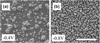
The applied potential during electrodeposition strongly affects the size and density of the deposited Au nanostructures. The deposition occurs via nucleation and growth, and has been documented in the literature. Reference Sakai, Arai, Yu and Tatsuma33 Figure 1 shows SEM images of Au nanostructures deposited at varying applied potentials of −0.4 and −0.8 V. These depositions were carried out at an electrolyte pH of 3, a deposition time of 30 min, and under ambient conditions. From the SEM images, it can be seen that the size of the Au nanostructures decreases with an increase in the applied negative potential. At an applied potential of −0.4 V, the Au nanostructures appeared slightly elongated and facetted. The increased applied potential, which is −0.8 V, changed the Au nanostructures from being elongated to quasi-spherical. Additionally, the morphology of the Au nanostructures changed from irregular to smooth. The density of the Au nanostructures increased with the varying applied potential from −0.4 V to −0.8 V. The Au nanostructures deposited at an applied potential of −0.8 V appeared denser with additional deposition over the initial deposited layer.
SEM images of Au nanostructures deposited, at a pH of 3, at (a) −0.4 V and (b) −0.8 V. The scale bar is 4 µm.

The observations can be explained with the effects of the applied potential on the formation mechanism of the Au nanostructures. It has been observed that when the applied potential is varied to a more negative value, the current density increases in the electrodeposition process. This is accompanied by an increase in the rate of deposition. Reference Sakai, Arai, Yu and Tatsuma33 Due to these conditions, there is an increase in the initial number of nuclei formed during electrodeposition. The increased number of nuclei results in smaller and denser nanostructures. The dense nanostructures also appear smooth. Further, the electrolyte pH was varied from 3 to 8 and their effects have been discussed in the following section.
B. Effects of electrolyte pH variation on Au nanostructures
The electrolyte pH has a significant effect on the morphology of the Au nanostructures. Figure 2 represents the SEM images of Au nanostructures at a varying applied potential from −0.4 V to −0.8 V with an electrolyte pH of 8. These SEM images in Fig. 2 were compared with their corresponding samples having similar applied potentials in Fig. 1. From this comparison, it was observed that the size, density, and morphology were varied with changes in the electrolyte pH. It was observed that the Au nanostructures appeared more spherical and facetted at an electrolyte pH of 8. Further, the nanostructure size and distribution appeared to be larger for samples prepared with a basic electrolyte. The density of the Au nanostructures was sparse at pH 8, compared to samples fabricated at pH 3, thus exposing the underlying substrate.
SEM images of Au nanostructures deposited, at a pH of 8, at (a) −0.4 V and (b) −0.8 V. The scale bar is 4 µm.

These changes in size, distribution, density, and morphology can be explained by the Au complex formation and their reactivity and stability. The Au precursor rapidly hydrolyzes to form Au complexes, when dissolved in water. It is known from the literature that at an electrolyte pH of 3, the Au complexes may include AuCl4 −, [AuCl3OH]−, and [AuCl2(OH)2]−, whereas at an electrolyte pH of 8, the Au complexes may include [AuCl(OH)3]− and [Au(OH)4]−. Reference Wang, Qian, Bi and Huang34,Reference Yannopoulos35
During hydrolysis, when chlorine ligands were replaced by hydroxyl ligands, the resulting Au complexes were less reactive. With the increase in the electrolyte pH to 8, the Au complexes become more stable, thus leading to a decrease in the deposition rate of the Au nanostructures. The lower deposition rate results in a decrease in the number of nuclei and subsequent spare density of the Au nanostructures. Due to the low deposition rate, the nucleation process occurs during the entire deposition process, and thus polydispersed nanostructures are formed. Reference Depestel and Strubbe36–Reference Ye, Yan, Ye and Zhou39 Furthermore, the preferential nucleation of AuCl4 − occurs on existing Au nanostructures rather than on the ITO substrate, leading to sparse deposition of large nanostructures. Reference Sakai, Arai, Yu and Tatsuma33 The next step in this investigation was to study the effect of L-cysteine on the Au nanostructure morphology.
C. Effects of L-cysteine on the Au nanostructures
A change in morphology was observed for the Au nanostructures fabricated in the presence of L-cysteine. The chemical additive L-cysteine is often used in the synthesis of Au nanoparticles as a morphology controlling agent. This is because it has the ability to adsorb on certain crystallographic planes with the help of the thiol (−SH) group. Reference Sakai, Arai, Yu and Tatsuma33 The L-cysteine adsorbs on low index planes of Au nanostructures and can be selectively desorbed at particular potentials during electrodeposition. Reference Sakai, Arai, Yu and Tatsuma33
The SEM images in Fig. 3 represent Au nanostructures fabricated in the presence of L-cysteine, at varying electrolyte pH and applied potentials. The SEM image in Fig. 3(a) showed some dendritic structures formed in the presence of 0.3 mM L-cysteine, whereas the SEM image in Fig. 3(b) did not exhibit any dendritic structures. When the pH was increased to 8, both samples shown in Figs. 3(c) and 3(d) exhibited a large particle size with rough, needlelike structures covering the fabricated nanostructures.
SEM images of Au nanostructures deposited, in the presence of 0.3 mM L-cysteine in the electrolyte, at varying potentials of −0.4 and −0.8 V at (a, b) pH 3 and at (c, d) pH 8. The scale bar is 4 μm.

The SEM images in Figs. 1–3 indicate a strong relationship between the applied potential, the electrolyte pH, and the L-cysteine during electrodeposition on the final outcome of the Au nanostructures. It is evident from Fig. 3 that the morphology of the Au nanostructures was affected by the presence of L-cysteine. From the literature, it is known that L-cysteine adsorbs on low index planes and plays an important role in controlling the morphology of the fabricated Au nanostructure. Reference Sakai, Arai, Yu and Tatsuma33 In Fig. 3, when the applied potential is changed from −0.4 V to −0.8 V at pH 3, the morphology appeared dense and quasi-spherical. This change in morphology from dendritic to quasi-spherical is due to the very high deposition rate at −0.8 V. Furthermore, when the pH of the electrolyte is changed from 3 to 8, there was an additional change in the morphology leading to the formation of needlelike structures. The formation of these rough structures was attributed to the adsorption L-cysteine on the Au nanostructures. Furthermore, the very slow deposition process at basic pH contributed to the needle formation as the gold ions were able to deposit on preferred crystallographic planes in a timely manner. Reference Sakai, Arai, Yu and Tatsuma33
The fabricated Au nanostructures were characterized using X-ray diffraction. Figure 4 shows the XRD plots for the samples prepared with an applied potential of −0.8 V, electrolyte pH of 3 and 8, and in the presence of L-cysteine. The XRD plot indicates a face-centered cubic crystal structure for the deposited Au. The strong peak at 38.3 was indexed as a (111) crystal plane of Au. Thus it is clear from the XRD data that the (111) crystal plane of Au was the dominant plane in all the samples. Reference Lin, Lin, Liu, Sheu and Hung40 The next step in the investigation was to deposit Cu2O films via electrodeposition on the Au nanostructures and test these composite photocathodes for their PEC performance.
XRD data of Au nanostructures deposited at a potential of −0.8 V with pH of 3 and 8 in the presence and absence of L-cysteine.

D. PEC performance of Cu2O photocathode and Au underlayer Cu2O photocathodes
In this phase of the investigation, the experimental scheme was to perform and compare the PEC performance of the composite Au–Cu2O photocathodes with the Cu2O control sample. The control sample of Cu2O photocathode was fabricated by electrodeposition of Cu2O films on ITO on a glass substrate. These Cu2O films exhibited a cubic crystal structure with (111) crystallographic plane being the dominant plane (Fig. S1). The next set of samples was prepared by the electrodeposition of Cu2O on the Au nanostructures forming a composite Au–Cu2O photocathode.
The PEC measurements were performed using a PEC cell. The PEC cell was made of quartz, which was covered with a black tape on three sides of the cell to avoid any parasitic light from entering the cell. It contained a Teflon lid that accommodated the RE, WE, and CE. A reaction area of 0.5 cm2 was defined with the help of epoxy that seals the remaining area of the sample. Silver paint was used to connect the copper wire to the photocathode. The silver paint was placed on the front side of the WE and was dried in air for four hours after which the epoxy (HYSOL 9462) was used to define the reaction area. A sufficient thickness of epoxy was required to minimize the formation of pinholes that can cause undesirable effects on the PEC measurements. The epoxy was cured for at least 12 h under ambient conditions.
The PEC measurements were carried out on the Cu2O control photocathode and on the composite Au–Cu2O photocathode. An ABET solar simulator was utilized to illuminate the defined reaction area on the photocathodes. The electrolyte used for PEC measurements was 0.5 M sodium sulfate at a pH of 5. A linear sweep voltammetry (LSV) with a scan rate of 20 mV/s was utilized. The measured potential versus Ag/AgCl was converted to reversible hydrogen electrode (RHE) by Nernst’s equation (E RHE = E Ag/AgCl + 0.059 pH + 0.197). Figure 5 shows a typical applied potential versus photocurrent density plot of the control Cu2O photocathode and the Au–Cu2O composite photocathode. A photocurrent density of the control Cu2O photocathode was measured, at 0 V versus RHE, to be −1.6 mA/cm2 (Fig. 5). Although this photocurrent value appears to be low, it is important to note that these PEC measurements were a proof of concept experiments. Further, the photocurrent values for the Au–Cu2O composite photocathode was measured to be −2.9 mA/cm2 (Fig. 5). It is clear from these measurements that the presence of an Au underlayer enhances the photocurrent density of the photocathode by 81%. The remaining photocurrents for all the fabricated Au–Cu2O composite photocathodes are presented, in the supporting information, in Figs. S3 and S4.
Enhanced photocurrent response in the presence of Au nanostructure underlayer.

The enhanced photocurrent density in the presence of Au underlayer can be explained with the help of Fig. 6. The Au nanostructures, in the Au–Cu2O composite photocathodes, act as scattering centers for the in-coming light [Fig. 6(a)]. This increases the optical path length of light in the Cu2O overlayer, which leads to trapping of light in the Cu2O layer. The trapped light is absorbed by the Cu2O layer and thus results in enhanced photocurrent density for the composite photocathode. The Au nanostructures also exhibit plasmonic properties. At particular in-coming wavelengths, the Au nanostructures have the ability to resonate creating a high electromagnetic field in the surrounding medium, thus leading to the concentration of light in the Cu2O overlayer [Fig. 6(b)]. This light concentration also results in the increased light absorption in Cu2O layer, which in turn increases the photocurrent density. Thus the scattering and resonance contribution from the Au nanostructures help enhance the photocurrent density of the composite photocathode. It is important to note that all the Au–Cu2O composite photocathodes exhibited similar photocurrent enhancement. This could be due to the difficulty in achieving strictly monodispersed Au nanostructures during the electrodeposition process that could resonate at particular in-coming wave length. Thus, our future work will further investigate to deconvolve the scattering and resonance contribution from the Au nanostructures by performing systematic studies on varying size, density, and morphology of the plasmonic nanostructures.
Schematic representation of the (a) scattering and (b) resonance contribution from Au nanostructures in enhancing PEC performance.

Along with the PEC measurements, the PEC response of Au–Cu2O composite photocathode was studied over a prolonged period. Here, the photostability measurements were performed at an applied potential of −0.5 V versus Ag/AgCl, which was equivalent to 0 V versus RHE for 1 h, in 0.5 M Na2SO4 electrolyte. The working electrode was illuminated by using a chopped AM 1.5 light from an ABET solar simulator at 100 mW/cm2. Figure 7 shows a typical photostability plot over a time of 1 h for the Au–Cu2O composite photocathode sample. The inset shows initial photocurrent density values during the experiment, when light was turned on and off. During the initial illumination, the photocurrent density was ∼2.73 mA/cm2, which matched well with the LSV data in Fig. 5 for the Au–Cu2O photocathode sample. When the illumination was turned off, the dark current approached close to 0 mA/cm2 and remained constant throughout the experiment. The dark current for the entire experiment is shown in Fig. S4. As the experiment progressed, the photocurrent density reduced from 2.73 mA/cm2 to 0.5 mA/cm2. This reduction occurred rapidly and was attributed to the corrosion of Cu2O film on the Au–Cu2O composite photocathode.
Photostability plot of Au–Cu2O composite photocathode. The inset shows the initial photocurrent density of the photocathode.

Additionally, EIS was performed to study the carrier transport in different samples including Au, Cu2O, and Au–Cu2O. A frequency range of 0.01–1 kHz was chosen for this study. For the EIS measurements, the carrier transfer resistance between the electrode and the electrolyte was studied. The resistance of the aforementioned samples was compared. This resistance was evaluated by measuring the diameter of the semicircle that is a characteristic of each EIS plot. Figure 8 shows the EIS plots for Au (also shown in the inset), Cu2O, and Au–Cu2O samples. The electrodeposited Au on ITO shows an EIS plot with a very small resistance of 150 Ω, as shown in the inset. This indicates an easy carrier transfer between the Au layer and the electrolyte, whereas the Cu2O sample on ITO shows a larger carrier resistance of approximately 2500 Ω. Further, the EIS plot for the Au–Cu2O composite photocathode also shows a high carrier resistance between the electrode and the electrolyte. This high resistance in Cu2O and Au–Cu2O samples was attributed to the Cu2O crystalline quality, grain boundaries, large film thickness of (4 µm) Cu2O, and the rough interface between Au and Cu2O films. The SEM image in Fig. 8(b) exhibits a relatively rough interface between the Au and Cu2O films. It is important to note that the nature and quality of the samples can be further improved by systematically optimizing the electrodeposition conditions, followed by an appropriate thermal treatment.
(a) EIS plots of Au, Cu2O, and Au–Cu2O samples. (b) SEM image of the Au–Cu2O sample edge exhibiting the Au and Cu2O films. Scale bar is 500 nm.

Along with EIS plots, admittance plots were also analyzed for Au, Cu2O, and Au–Cu2O samples. The admittance plots are presented in the supporting information (Fig. S5). From the EIS and admittance data, it is clear that Au samples demonstrated higher conductance for carrier transport, whereas the Cu2O samples exhibited higher resistance or lower conductance to carrier transport. The Au–Cu2O composite photocathodes exhibited slightly greater conductance than the Cu2O samples, indicating that the presence of Au underlayer was beneficial for carrier transport. However, the performance of the Au–Cu2O samples can be enhanced by improving the sample quality. The absolute quantum efficiency (QE) was obtained for Cu2O and Au–Cu2O photocathodes (Fig. S6). The absolute QE of Au–Cu2O was slightly higher than the Cu2O photocathode, through the wave length range of 300–900 nm. The enhancement was attributed to the presence of the Au underlayer. However, the magnitude of enhancement was not very large, and this was attributed to the Cu2O crystalline quality and grain boundaries and the rough interface between the Au layer and Cu2O film.
IV. CONCLUSION
In summary, Au nanostructures of varying size, density, and morphology were successfully electrodeposited by controlling the process parameters including applied potential, electrolyte pH, and presence of L-cysteine. An overlayer of Cu2O was successfully electrodeposited on the Au nanostructures. The PEC measurements of the Cu2O control photocathode and the Au–Cu2O composite photocathodes were performed. A photocurrent enhancement was observed for Au–Cu2O composite photocathodes. The enhancement was attributed to the increased light absorption in the Cu2O overlayer in the composite photocathodes. The improved light absorption was due to the scattering and resonance contribution of the Au nanostructures. Additionally, all the Au–Cu2O photocathodes demonstrated similar enhancement in photocurrent density which could be due to the difficulty of achieving truly monodispersed Au nanostructures. Further, the EIS and admittance data indicate that the presence of Au underlayer was beneficial for carrier transport between the electrode and the electrolyte.
Supplementary Material
To view supplementary material for this article, please visit https://doi.org/10.1557/jmr.2017.130.
ACKNOWLEDGMENTS
Authors acknowledge funding support from Iowa State University Startup Fund. S.P. thanks the Catron Fellowship. The authors would like to thank Dr. Warren Straszheim for help with SEM characterization. The authors declare no competing financial interest.