




No CrossRef data available.
Article contents
- Abstract
- Where (and When) to Start Looking for the Neurobiological Bases of Social Dysfunction in Autism?
- White and Gray Matter Growth Pathology in the First Years of Life in Autism: MRI Evidence
- Microstructural Defects in Autism: Prominent Cerebral and Cerebellar Abnormalities
- Summary and Conclusions
- References
Autism at the beginning: Microstructural and growth abnormalities underlying the cognitive and behavioral phenotype of autism
Published online by Cambridge University Press: 01 November 2005
Article contents
- Abstract
- Where (and When) to Start Looking for the Neurobiological Bases of Social Dysfunction in Autism?
- White and Gray Matter Growth Pathology in the First Years of Life in Autism: MRI Evidence
- Microstructural Defects in Autism: Prominent Cerebral and Cerebellar Abnormalities
- Summary and Conclusions
- References
Abstract
Autistic symptoms begin in the first years of life, and recent magnetic resonance imaging studies have discovered brain growth abnormalities that precede and overlap with the onset of these symptoms. Recent postmortem studies of the autistic brain provide evidence of cellular abnormalities and processes that may underlie the recently discovered early brain overgrowth and arrest of growth that marks the first years of life in autism. Alternative origins and time tables for these cellular defects and processes are discussed. These cellular and growth abnormalities are most pronounced in frontal, cerebellar, and temporal structures that normally mediate the development of those same higher order social, emotional, speech, language, speech, attention, and cognitive functions that characterize autism. Cellular and growth pathologies are milder and perhaps nonexistent in other structures (e.g., occipital cortex), which are known to mediate functions that are often either mildly affected or entirely unaffected in autistic patients. It is argued that in autism, higher order functions largely fail to develop normally in the first place because frontal, cerebellar, and temporal cellular and growth pathologies occur prior to and during the critical period when these higher order neural systems first begin to form their circuitry. It is hypothesized that microstructural maldevelopment results in local and short distance overconnectivity in frontal cortex that is largely ineffective and in a failure of long-distance cortical–cortical coupling, and thus a reduction in frontal–posterior reciprocal connectivity. This altered circuitry impairs the essential role of frontal cortex in integrating information from diverse functional systems (emotional, sensory, autonomic, memory, etc.) and providing context-based and goal-directed feedback to lower level systems.
The authors were supported by funds from the National Institute of Mental Health (2-ROI-MH36840) and National Institute of Neurological Disorders and Stroke (2-ROI-NS19855) awarded to Eric Courchesne.
Information
- Type
- Research Article
- Information
- Copyright
- © 2005 Cambridge University Press
Autism begins in many ways. In the first weeks of life, a mother notices something is not right about her newborn baby boy: he has marked difficulty coordinating his sucking and swallowing and sometimes he seems floppy and then at other times strangely rigid. As the first months go by, he appears to have good visual attention, perhaps too good, because sometimes his attention seems stuck. He is also unexpectedly sensitive to touch or sounds at one moment, but at others he seems almost completely oblivious to them. However, he is cuddly and he smiles and coos and seems to be socially connected. Then, during the second half of his first year of life, his vocalizations do not continue to develop and mother becomes truly concerned because now it is not just his motor, attention, and sensory responses that are awry; he takes a reduced interest in her and rarely smiles or looks at her or anyone else. Instead, he seems to regard objects with as much interest as people, whom he regards with disinterest or even avoidance. By his first birthday, a frightening and sad thought occurs to the mother: perhaps my baby has autism.
Another mother and father swell with joy because their newborn baby girl is so perfect, and as months come and go, she grows more delightful. Her interest in others grows and her skills at interacting and engaging others brings a sense of wonder to mother and father, and their dreams of what a special person she is to become expand. Each day, the little toddler's face is filled with different emotions as she expands her social, emotional, speech, and language skills. She seems as filled with love for her parents as they are for her. In the months before her second birthday, though, mother notices that she has not been herself lately. What is it? Perhaps a little quieter, perhaps a little bit less emotional. Worse, as her mother watches, each day she seems to fade a bit more until there is no doubt: her little girl is not herself anymore. She has gradually become more and more remote; her face no longer shows much emotion and she no longer delights in others. In fact, she no longer seeks out mom and dad. She has slowly faded away from them.
Thus, autism begins. In one case described by Dawson, Osterling, Meltzoff, and Kuhl (2000), the beginning was early, rapid, and unmistakable, but in another case described by Lord (C. Lord, personal communication, NAAR Conference, November 12, 2004), signs of autism did not appear until later in the second year of life. New research has identified key behavioral red flags for autism during the first 2 years of life (see Table 1; Wetherby, Woods, Allen, Cleary, Dickinson, & Lord, 2004), and many of them involve a lack of normal socioemotional behavior and an apparent lack of the normal desire to make socioemotional contact.
Nine red flags for autism

How could the desire for social connection not be there in an infant? Or, even more mysteriously, how could that desire appear strongly for a time, only to slowly dwindle away, leaving a strange void? What could do this to a quality so essentially human and so essential for human togetherness? What could snuff out social drive, a drive that ought to be as strong as the drive to breath, eat, or survive? What goes wrong in how the brain is organized and operates, and how might the impact of these processes be mitigated or prevented?
Where (and When) to Start Looking for the Neurobiological Bases of Social Dysfunction in Autism?
Over the past 40 years, cognitive psychologists have attempted to identify cognitive deficits underlying the myriad of behavioral symptoms seen in autism. Contemporary research has identified deficits in complex processing (Minshew, Goldstein, & Siegel, 1997), weak central coherence (Frith & Happe, 1994), impairment in the dynamic control of orienting, disengaging, and switching attention (Courchesne, Townsend, Akshoomoff, Saitoh, Yeung–Courchesne, Lincoln, James, Haas, Schreibman, & Lau, 1994; Townsend, Courchesne, Covington, Westerfield, Harris, Lyden, Lowry, & Press, 1999), deficits in executive functions (Rogers & Pennington, 1991), deficits in theory of mind (Baron–Cohen, Leslie, & Frith, 1985), deficits in imitation (Rogers & Pennington, 1991), impairments in social and affective relations (Hobson, 1993), and impairments in joint social attention (Mundy, 1995). However, the majority of this research was not aimed at identifying the specific neural bases of this disorder. With the use of cognitive neuroscience techniques (i.e., functional magnetic resonance imaging [fMRI], positron emission tomography [PET], magnetic encephalography [MEG], event-related potential [ERP], and EEG) the brain bases of these behavioral deficits can be elucidated. By utilizing the growing basic and clinical literature of cognitive neuroscience, evidence of cognitive and behavioral dysfunctions in autism can point to candidate brain regions of abnormality (e.g., see Mundy, 2003; Williams, Whiten, Suddendorf, & Perrett, 2001).
Cognitive neuroscience studies of autism have identified altered patterns of neurofunctional activity, specifically reduced activity in higher order frontal, temporal, and cerebellar regions but normal to increased activity in lower order posterior visual regions (for reviews, see Belmonte et al., 2004; Courchesne, Redcay, & Kennedy, 2004; Frith, 2003). These higher order regions are critical to the initiation, perception, and interpretation of socioemotional and communicative functions, which are strikingly impaired in autism, as well as other higher order cognitive, attention, and memory functions that are also abnormal in this disorder. Reduced frontal activation has been reported in dorsal or medial frontal cortices in a theory of mind task (Castelli, Frith, Happe, & Frith, 2002), in response to socially familiar faces (Pierce, Haist, Sedaghat, & Courchesne, 2004), during a gender or emotion decision task (Hubl, Bolte, Feineis–Matthews, Lanfermann, Federspiel, Strik, Poustka, & Dierks, 2003), in working memory tasks (Luna, Minshew, Garver, Lazar, Thulborn, Eddy, & Sweeney, 2002), in an embedded figures task (Ring, Baron–Cohen, Wheelwright, Williams, Brammer, Andrew, & Bullmore, 1999), in an emotion Stroop task (Kennedy, Redcay, & Courchesne, 2004), in visual spatial attention tasks (Belmonte & Yurgelun–Todd, 2003), and during sentence comprehension (Just, Cherkassky, Keller, & Minshew, 2004; Muller, Behen, Rothermel, Chugani, Muzik, Mangner, & Chugani, 1999; Muller, Chugani, Behen, Rothermel, Muzik, Chakraborty, & Chugani, 1998). ERP studies have consistently found reduced or absent physiological responses from frontal cortex during a variety of auditory and visual attention and orienting tasks (Ciesielski, Courchesne, & Elmasian, 1990; Courchesne, Kilman, Galambos, & Lincoln, 1984; Townsend, Courchesne, Covington, Westerfield, Harris, Lyden, Lowry, & Press, 1999). Reduced temporal lobe activity has been reported in higher order lateral temporal regions during processing of vocal sounds (Gervais, Belin, Boddaert, Leboyer, Coez, Sfaello, Barthelemy, Brunelle, Samson, & Zilbovicius, 2004), speech sounds (Boddaert, Belin, Chabane, Poline, Barthelemy, Mouren–Simeoni, Brunelle, Samson, & Zilbovicius, 2003; Boddaert et al., 2004), and faces (Pierce, Müller, Ambrose, Allen, & Courchesne, 2001), and during a mentalizing task (Castelli et al., 2002). Further, ERP and MEG studies have identified reduced or abnormal temporal lobe responses to a variety of speech and nonspeech sounds (Bruneau, Bonnet–Brilhault, Gomot, Adrien, & Barthelemy, 2003; Bruneau, Roux, Adrien, & Barthelemy, 1999; Ceponiene, Lepisto, Shestakova, Vanhala, Alku, Naatanen, & Yaguchi, 2003; Dawson, Finley, Phillips, Galpert, & Lewy, 1988; Gage, Siegel, Callen, & Roberts, 2003) and during spatial tuning of attention to selective auditory sound sources (Teder–Salejarvi, Pierce, Courchesne, & Hillyard, 2005). In contrast to these reductions of activity, recent fMRI studies report normal activation in visual cortex in response to basic visual stimuli (Hadjikhani, Chabris, Joseph, Clark, McGrath, Aharon, Feczko, Tager–Flusberg, & Harris, 2004). Furthermore, several of the above-mentioned fMRI studies that show reductions in frontal and/or temporal regions show increased activity in occipital regions, including lateral extrastriate regions (Ring et al., 1999), medial occipital cortex (Hubl et al., 2003), and ventral occipital cortex (Belmonte & Yurgelun–Todd, 2003). These findings suggest that posterior, lower order regions may be relatively spared while anterior, higher order regions may be more severely affected. Furthermore, functional connectivity studies have revealed reduced functional connectivity between lower order and higher order brain regions (e.g., occipital to frontal, Castelli et al., 2002; superior temporal to inferior frontal, Just et al., 2004; and parietal to frontal, Horwitz, Rumsey, Grady, & Rapoport, 1988).
Findings from the cerebellar cortex in autism appear to parallel those of the cerebral cortex in that a similar dissociation between higher order and lower order processes and regions is seen. Although only a handful of functional neuroimaging studies of the cerebellum in autism exist, these studies reveal reduced cerebellar activity during higher order tasks, including attention (Allen & Courchesne, 2003), listening to and generating sentences (Muller et al., 1998, 1999), and judgement of facial expression (Critchley, Daly, Phillips, Brammer, Bullmore, Williams, Van Amelsvoort, Robertson, David, & Murphy, 2000) with normal to increased activity during lower order motor tasks (Allen & Courchesne, 2003; Allen, Muller, & Courchesne, 2004; Muller, Pierce, Ambrose, Allen, & Courchesne, 2001). The cerebellum has reciprocal projections to both prefrontal (Middleton & Strick, 2001) and temporal (Schmahmann & Pandya, 1991) lobes, and thus, not surprisingly, reductions of activity in the cerebellum during higher order tasks are often seen with concurrent reductions in prefrontal or temporal lobe activity (e.g., Muller et al., 1999). These functional findings suggest a system of neural dysfunction including higher order frontal, temporal, and cerebellar regions may underlie many of the social and speech abnormalities seen in older children and adults with autism.
An important question is what structural developmental abnormalities lead to these outcome frontal, temporal, and cerebellar neurofunctional abnormalities. It is argued here that useful models and knowledge of the neural developmental biology that precedes and produces the initial autistic behavioral dysfunctions are necessary on three fronts: they can provide explanations for outcome neurobehavioral deficits that have been carefully documented by behavioral and neuroimaging research. They can provide information that constrains and directs candidate causes that can plausibly lead to these more specific early neural defects. Last, they can provide specific neurobiological processes and defects for targeting in animal models; autism is a disorder of higher order social, emotional, speech, language, and cognitive functions that are not convincingly modelable in rodents, which are the animals of choice for genetic and environmental factors research on autism.
Although much data exist to document the behavioral and cognitive impairments in autism, comparatively little is known about the underlying microstructural neural defects. Existent postmortem neuropathology studies that have relied on visual qualitative inspection in autism have shown abnormalities in microanatomy within frontal, temporal (Bailey, Luthert, Dean, Harding, Janota, Montgomery, Rutter, & Lantos, 1998), limbic (Bauman & Kemper, 2005; Kemper & Bauman, 1998), and cerebellar regions (Bailey et al., 1998; Kemper & Bauman, 1998; Ritvo, Freeman, Scheibel, Duong, Robinson, Guthrie, & Ritvo, 1986). However, these qualitative studies have been few in number, and none of these abnormalities have been rigorously quantified. Further, the nature of observed abnormalities revealed is diverse, from increased cell packing density to laminar abnormalities to ectopic neurons in white matter (Bailey et al., 1998). Indeed, numerous microstructural abnormalities likely underlie the diverse behavioral phenotype of autism. Systematic and quantitative neuropathological investigations require such an investment of time and effort that an uninformed search for potentially subtle, but important, neuropathological differences would be inefficient and impractical. Thus, knowledge of candidate brain regions of abnormality in autism, as gained through cognitive neuroscience, allows for an efficient hypothesis-driven analysis of neurobiological abnormalities in autism. Further, knowledge of regional neurofunctional abnormalities provides an essential framework for interpreting the potential functional significance of postmortem neuronal and molecular findings. For example, the neuroimaging literature would predict neuronal defects that are more apparent in prefrontal and higher order temporal regions but less apparent in occipital cortex. In sum, to understand the emergence of autism, a multilevel analysis approach is useful wherein behavior informs cognitive neuroscience, which informs basic neurobiology, and vice versa.
In the following paper, we will focus on findings from macroscopic (e.g., MRI) and microscopic structural investigations of autism, highlighting findings from very young children with autism. Further, we will attempt to relate this new microstructural evidence with findings of macroscropic structural and functional abnormalities in autism.
White and Gray Matter Growth Pathology in the First Years of Life in Autism: MRI Evidence
The first years of life are uniquely important for brain development
Figure 1 is an example of the sparse neural connections in the frontal cortex of a newborn. The circuitry necessary for complex information processing and complex behavior does not yet exist. Instead, it will be created in the first postnatal years by neuronal differentiation and growth, dendritic and axonal growth, axonal myelination, and a tremendous increase in synapse numbers. Higher order social, emotional, cognitive, attention, speech, and language functions are all mediated by slowly maturing regions such as frontal cortex, while basic sensory and perceptual functions are mediated by relatively more rapidly and earlier maturing systems.

Golgi-stained sections showing the growth of pyramidal neuron soma and dendrites in the middle frontal gyrus. The normal newborn has sparse neural circuitry; then, with increasing age, there is a tremendous increase in the complexity of dendritic arborizations. In this frontal cortical area, the dendrite arbors for layer 3 pyramidal neurons, which are only 3% of mature size in the newborn, are still only about 50% by 2 years of age and do not reach 100% until the end of childhood (see text). From The Human Brain (3rd ed), by J. Nolte, 1993, St. Louis, MO: Mosby Year Book/Elsevier. Copyright 1993 by Mosby Year Book/Elsevier. Reprinted with permission from Elsevier.
In the frontal cortex at birth, the length of the pyramidal cell dendritic arbors in layer 3 is only 3% of full mature size, and by 2 years of age it is only 48% (see example in Figure 1); many additional years will pass before full size is reached. By comparison, in primary visual cortex pyramidal dendritic arbors are already 33% of full size at birth, and by 2 years they have reached mature size (table 1.3 in Huttenlocher, 2002). Myelination follows the same functional hierarchical pattern, with basic-level systems developing earlier than higher order association systems (Kinney, Brody, Kloman, & Gilles, 1988). Compared to more posterior cortices, frontal cortex undergoes synapse formation later, and for a longer period of time, and develops far larger pyramidal neurons with far more synapses (about 100,000 vs. about 20,000) and far larger dendritic (e.g., in layer 3, 6836 vs. 2900 μm) and axonal arbors and axonal projections (Huttenlocher, 2002).
It has long been recognized that this unique period of neural differentiation and circuit formation is also a time when the brain is particularly vulnerable to abnormal events that disrupt the formation of cortical connectivity, producing aberrant circuits and functions and behavioral deficits (Dobbing, 1981; Huttenlocher, 2002; Kinney et al., 1988). More slowly maturing brain regions such as frontal cortex have a longer window of vulnerability than more rapidly maturing ones, such as the occipital cortex.
Findings in young autistic children
Recent evidence indicates that autism may involve brain growth pathology during this very window of vulnerability; it is early, brief, and age delimited (for reviews, see Courchesne, 2004; Courchesne et al., 2004; Courchesne & Pierce, 2005a; Dementieva, Vance, Donnelly, Elston, Wolpert, Ravan, DeLong, Abramson, Wright, & Cuccaro, 2005). At birth, head circumference in autism, and therefore brain size (Bartholomeusz, Courchesne, & Karns, 2002), were found to be either equivalent to the normal average or slightly smaller than normal (Courchesne, Carper, & Akshoomoff, 2003; Courchesne et al., 2001; Dementieva et al., 2005; Gillberg & de Souza, 2002; Lainhart, Piven, Wzorek, Landa, Santangelo, Coon, & Folstein, 1997).1
There is excellent agreement among reports of birth head circumference in autism: it was 34.7 cm in Gillberg and de Souza (2002), 34.17 cm in Lainhart et al. (1997), and 34.65 cm in Courchesne et al. (2003). Normal average birth head circumference is between 34.5 and 35.9 cm, depending on the clinical norms used for reference (Bartholomeusz et al., 2002).
Both cerebral and cerebellar white matter volumes in autistic toddlers have been found to be abnormally enlarged (18 and 39%, respectively); cerebral gray matter is enlarged by 11% (Courchesne et al., 2001). Most importantly, there were striking regional differences in this overgrowth pathology: in 2- to 4-year-old autistic toddlers, frontal cortex and frontal white matter volumes were the most abnormally enlarged, but the occipital lobes did not differ significantly from normal (Carper, Moses, Tigue, & Courchesne, 2002). Within the frontal cortex, dorsolateral and mesial prefrontal cortices were most abnormal, but the precentral gyrus, like the occipital lobe, was not significantly different from normal (Carper & Courchesne, 2005). Temporal gray matter and parietal white matter were also enlarged but not to the magnitude of frontal gray and white volumes (Carper et al., 2002); in the limbic system, the amygdala was also enlarged by 4 years of age (Sparks et al., 2002). A PET study of 3- to 4-year-old autistic children reported hypoperfusion in frontal cortex in autism which was interpreted as evidence of delayed frontal maturation (Zilbovicius, Garreau, Samson, Remy, Barthelemy, Syrota, & Lelord, 1995).
An equally striking and important second phase of growth pathology in autism follows on the heels of the early overgrowth: abnormally slow or arrested growth. A recent meta-analysis of 12 MRI brain volume studies showed an autism brain size difference from normal of about 11% between 1 and 3 years of age, which declined through childhood (when the normal, but not autistic, brain continues to grow); by adolescence the autism brain size difference from normal is only about 1–2% (Redcay & Courchesne, 2005). Between 2–4 and 6–8 years of age, frontal and temporal cortical gray matter increase by 20 and 17% in normal children but change by only 1 and −1%, respectively, in autism (Carper et al., 2002). The dorsolateral subregion of frontal cortex increases by 27% from 2–5 years to 5–9 years of age in normals, but by only 7% in autistic children (Carper & Courchesne, 2005). White matter growth is likewise retarded; for example, between 2 and 4 years and 7 and 11 years of age, frontal white matter volume increases by 45% in normal children, but by only 13% in autistic children (Carper et al., 2002), and between 2 and 3 years of age and adolescence, cerebellar white matter volume increases by 50% in normal children, but by only 7% in autistic children (Courchesne et al., 2001). Thus, regions that show the greatest early overgrowth in autism, frontal lobe and cerebellar white matter, also show sharply reduced or arrested growth thereafter.
Last, in contrast to the cerebellar white matter changes just described, during early development in autism there appears to be a reduction in the size of one or another region of the cerebellar vermis, which is largely gray matter (review in Courchesne, 2004). In one MRI study with over 200 autistic and control subjects (making this the largest autism MRI study ever done), Hashimoto, Tayama, Murakawa, Yoshimoto, Miyazaki, Harada, and Kuroda (1995, p. 229) reported underdevelopment of the cerebellar vermis in autistic individuals ranging from infants to adolescents. In another MRI study of 3- to 9-year-old children, reduction in the size of cerebellar vermis lobules VI–VII was found to be specific to autistic children compared with normal, fragile X, fragile X with autism, Down syndrome, and Down syndrome with autism children (Kaufmann, Cooper, Mostofsky, Capone, Kates, Newschaffer, Bukelis, Stump, Jann, & Lanham, 2003). In a third recent MRI study of autistic children, the posterior portion of the cerebellar vermis was found to be significantly reduced in size (Levitt, Blanton, Capetillo–Cunliffe, Guthrie, Toga, & McCracken, 1999). These findings are consistent with some previous developmental MRI studies reporting hypoplasia of the one or another region of the cerebellar vermis (e.g., Ciesielski, Harris, Hart, & Pabst, 1997; Courchesne, Yeung–Courchesne, Press, Hesselink, & Jernigan, 1988).
Findings in older autistic children and adults
Recent structural imaging results on older autistic children are compatible with this evidence of gray and white matter abnormality in autistic toddlers. For example, Herbert et al. (2003) reported greater white matter volume in 7- to 11-year-old autistic children and in a second study found that this deviation from normal was greatest in frontal white matter underlying cortex (so-called “radiate white matter”) and least in occipital lobe (Herbert, Ziegler, Makris, Filipek, Kemper, Normandin, Sanders, Kennedy, & Caviness, 2004). Hardan, Jou, Keshavan, Varma, and Minshew (2004) found abnormally increased gyrification in frontal cortex in an autistic sample ranging in age from 8 to 50 years. Levitt, Blanton, Smalley, Thompson, Guthrie, McCracken, Sadoun, Heinichen, and Toga (2003) reported that some frontal and temporal sulci are abnormally shifted superiorly and posteriorly in autistic children. Altered serotonin synthesis in the cerebello–thalamo–frontal pathway has been described in autistic children (Chugani, Muzik, Rothermel, Behen, Chakraborty, Mangner, da Silva, & Chugani, 1997) and a theory has been proposed linking serotonin abnormality during prenatal development to minicolumn and other neural abnormalities in autism (Chugani, 2000).
Microstructural Defects in Autism: Prominent Cerebral and Cerebellar Abnormalities
According to in vivo MRIs of autistic toddlers and young children, the frontal lobe, which would be predicted to be the most vulnerable during postnatal neuronal growth, differentiation, and circuit formation, is indeed the structure with the greatest early growth pathology and later functional pathology. Conversely, occipital lobes would be predicted to be the least vulnerable, and in fact, they show a nonsignificant difference from normal both structurally and functionally. Temporal and parietal cortices fall between these two extremes. This raises the hypothesis proposed by Courchesne and Pierce (2005a, 2005b) that fine quantitative mapping of neural microstructure and white matter in autism will show that the greatest pathology occurs in cortical association regions that have the latest and most protracted neuronal and functional developmental time tables.
Similarly, the cerebellum is vulnerable during the first 2 years of postnatal life because the genesis of cerebellar cells continues well into the first and perhaps second year of life. It is unique in this regard because neurogenesis during brain development is prenatal for all other major brain structures; whether this final phase of cytogenesis in the cerebellum generates neurons, glia or both is uncertain. This vulnerable structure also develops abnormally in autism in the first years of life according to the in vivo MRI data reviewed above, and of all MRI abnormalities, the most striking and pronounced is overgrowth of cerebellar white matter (Courchesne et al., 2001).
Neuron numbers: Excess in cerebrum, reduction in cerebellum
In collaborative and as yet unpublished research projects, neuron numbers in the cerebrum, cerebellum, and subcortical structures in a set of autistic and control postmortem cases aged 4–67 years were counted (Schmitz, 2004; Wegiel, 2004). These were among the first studies to utilize modern stereological procedures in a study of the autistic brain. There appeared to be a tendency toward an excess number of cerebral cortical neurons was found in autistic cases as compared to controls (Schmitz, 2004). Interestingly, the magnitude of this excess showed an age-related decline across child to adult autism cases. It is not yet known whether there are regional differences in the excess neuron numbers or in the rate and magnitude of the age-related decline in numbers.
In contrast, decreased neuron numbers were reported for the cerebellum and basal ganglia (Wegiel, 2004). Cerebellar Purkinje neuron numbers were decreased by about 30% and the volume of the cerebellum was reduced by about 19%. This loss of Purkinje neurons was seen at all ages in the autism sample from 4- to 67-year-olds. Large decreases in neuron numbers were also reported for the nucleus accumbens and basal ganglia, but neuron numbers in the hippocampus were remarkably similar in autistic and control cases. This quantitative and stereological study adds to a large number of observational reports that cerebellar Purkinje neuron numbers are abnormally reduced in autism (see reviews in Courchesne, 1997, 2004). Cerebellar Purkinje neuron loss may be the single most commonly reported neuronal abnormality in the autism literature to date.
Implications of increased cortical neurons and decreased Purkinje neurons
Many fundamental questions remain before the implications of an increase in cortical neurons and axons can be fully interpreted. Are there regional increases in neurons and axons that parallel regional increases in MRI gray and white matter volume? The only existent stereological count of cerebral neurons was global, not regional (Schmitz, 2004). In our pilot study, we reported what appeared to be an increased number of neurons in layer 3 throughout frontal cortical regions but not in primary visual cortex. Although this fits the reports of regional MRI volumetric differences, it was not a stereologically conducted count. It will be valuable for future studies to perform regional stereological neuron counts. Because cerebral neurons in different layers are generated at different but somewhat overlapping developmental times, it will also be important to determine whether or not the increases are layer specific. There have been hypotheses that autism involves deficient cortical inhibitory control, and so it will be interesting to know if there is an imbalance in the numbers of excitatory pyramidal neurons and inhibitory interneurons such as chandelier cells. Because roughly 75–80% of cortical neurons are pyramidal cells (Jones, 1984), an aberrant increase in their numbers could have a significant overall effect on both total neuron counts and gray matter volume even if the number of inhibitory neurons remained constant or even decreased slightly.
An excess number of cerebral neurons could be because of any one of several possibilities: a failure to correctly regulate the number of neurons produced during the neurogenesis stage of prenatal development in autism, a deficit or delay in apoptosis so that too many survive into postnatal life, or a compensatory neural genesis during perinatal or postnatal life that is triggered by adverse events such as those that ignite the neuroinflammatory reaction reported by Vargas, Nascimbene, Krishnan, Zimmerman, and Pardo (2005).
There is no experimental data as yet that could speak to the abnormal neurogenesis possibility. However, if the excess resulted from an early prenatal failure to regulate genesis, then it might be expected that this early excess would cause the brain to appear to be enlarged at birth as well as after birth, but in fact, in the majority of cases, head circumference at birth in autism is normal to slightly smaller than normal. On the other hand, if the excess in neuron numbers is regionally limited to only late maturing frontal and other higher order association cortices, then perhaps at birth its presence might not be apparent and only becomes so across the first months and years of postnatal life.
The second possibility involving abnormal deficits or delays in apoptosis is compatible with observations of abnormally increased levels of molecules that should promote apoptosis molecules in frontal and cerebellar cortices in adult autistic postmortem cases (Araghi–Niknam & Fatemi, 2003) and of an age-related decline in the magnitude of the excess numbers from childhood to adult years in autism (Schmitz, 2004).
The third possibility involving an aberrant late compensatory genesis could help explain why the brain at birth in autism is not abnormally enlarged in the great majority of cases. It is now appreciated that such a “compensatory” late genesis of neural and glial cells can occur in response to some adverse conditions (Vaccarino & Ment, 2004).
Abnormally slowed or arrested growth that follows the brief period of overgrowth in the autistic toddler could be because of a late onset of apoptosis as just discussed above, because of the conclusion of the period of late compensatory neural and glial genesis also just discussed, or because the excess of neurons and connections produces dysfunctional neural activity that causes elimination or retarded growth of ineffective neurons, axons, and dendritic and axonal arbors. Certainly there are a number of clear microstructural signs of arrest of growth, loss, and insufficient development in the older child or adult with autism, each of which could underlie the arrested or slowed growth of gray and white matter that has been observed in MRI studies of older autistic children and adults. Already mentioned were abnormal age-related declines in cerebral neuron numbers, the presence of abnormal levels of molecules that might promote apoptosis, and abnormally narrow minicolumns. Small cell size has been noted in the cingulate cortex (Kemper & Bauman, 1998) and in other frontal regions (Buxhoeveden, Semendeferi, Schenkar, Switzer, & Courchesne, 2005) in older autistic postmortem cases.
Increased numbers of cerebral neurons could underlie the volume increases in cerebral gray matter that have been reported in MRI studies. In addition, an excess of neurons likely means an excess of axons, and this could underlie volume increases in white matter that have also been reported in MRI studies of autism. These excesses mean cortical connectivity must be abnormal. Some have speculated that autism involves cerebral anatomical overconnectivity (Casanova, 2004), while others have argued in favor of cerebral functional underconnectivity (Belmonte & Yurgelun–Todd, 2003; Horwitz et al., 1988; Just et al., 2004). It has also been suggested that the key is an abnormally increased ratio of excitation to inhibition (Rubenstein & Merzenich, 2003). Here it is proposed that each of these defects underlies autism: there is an abnormal ratio of excitation to inhibition, there is anatomical overconnectivity but primarily within frontal cortical regions, and there is functional underconnectivity but principally in long-distance frontoposterior reciprocal pathways. Moreover, it is proposed that connectivity patterns and defects change with development.
Minicolumns are abnormally narrow in frontal and temporal cortex but not occipital cortex
The minicolumn is a fundamental unit of information processing. Figure 2 shows schematics of a minicolumn. The minicolumn is a roughly columnar vertical assembly of pyramidal neurons and interneurons, their interconnections, and input and output axons that extend from layer 6 up to the cortical surface. Each is 30–60 μm in diameter with perhaps 80–100 pyramidal neurons per minicolumn. In humans, minicolumns in frontal association cortex are nearly twice the diameter and several times the volume of those in primary sensory cortices such as the primary visual cortex (Buxhoeveden & Casanova, 2002). These 80–100 pyramidal neurons are thought to have originated from a common precursor cell during neurogenesis in the second and third trimesters. Thus, the number of minicolumns in cerebral cortex reflects the number of precursor cells that gave rise to each minicolumn. Expansion of cerebral cortex and increase in its processing power across evolution up to humans is due to an increase in the number of prenatally generated minicolumns. The vertical array of pyramidal cells is surrounded by neuropil space; in humans, this neuropil space that contains synapses and dendrites has expanded greatly, allowing for more complex microcircuitry and refined and powerful neural computation within each minicolumn (Buxhoeveden & Casanova, 2002). These vertical assemblies of interconnected pyramidal neurons have large inhibitory interneurons (e.g., chandelier cells) that modulate the synchronous output of clusters of minicolumns. Synchronous processing and signaling locally in minicolumns provide more powerful coherence of output to other cortical regions and cortico–cortical coupling. The function of minicolumns is to provide fine tuning of information processing and learning within a cortical column.

The minicolumn is a fundamental unit of information processing. (a) It is a roughly columnar vertical assembly of pyramidal neurons and interneurons, their interconnections, and input and output axons that extend from layer 6 up to the cortical surface. Each is 30–60 μm in diameter with perhaps 80–100 pyramidal neurons per minicolumn. (b) A schematic closeup of cortical layer 3 within a minicolumn. The shaded area represents the neuropil space that surrounds the vertical assembly of neurons. This area contains mostly synapses, dendrites, and unmyelinated axons. The unshaded section is where the majority of the cell soma reside and it contains myelinated axons. From “Morphological Differences Between Minicolumns in Human and Nonhuman Primate Cortex,” by D. P. Buxhoeveden, A. E. Switala, E. Roy, M. Litaker, and M. F. Casanova, 2001, American Journal of Physical Anthropology, 115. Copyright 2001 by American Journal of Physical Anthropology. Adapted with permission from Wiley–Liss, a subsidiary of John Wiley & Sons, Inc.
The first quantitative study of minicolumn size in autism examined layer 3 and found minicolumns to be abnormally narrow in one frontal and two temporal cortical areas (BA 9, 21, and 22; Casanova, Buxhoeveden, Switala, & Roy, 2002). In a pilot study, we aimed to determine which frontal regions have this minicolumn abnormality, and whether this abnormality is present at the time of clinical onset of autism. We measured minicolumn size in layer 3 throughout dorsal, mesial, and orbital frontal cortex in a postmortem 3-year-old autistic case; minicolumn size in primary visual cortex was measured as a contrast site (Buxhoeveden et al., 2005). To test whether there is arrest of minicolumn growth in frontal cortex in autism, we compared results from this 3-year-old to those from a postmortem 41-year-old adult with autism. We measured between 1,600 and 2,000 individual minicolumns per case, and we also measured controls. Minicolumn and neuropil sizes were significantly reduced throughout dorsal, mesial, and orbital frontal cortices in the 41-year-old adult with autism, being almost half the volume of normal adults. In addition, minicolumns and their surrounding neuropil space in the 3-year-old autistic case were nearly the same size as this 41-year-old autistic adult, which suggests the hypothesis that in autism minicolumn growth is arrested sometime during early childhood. In contrast to findings in frontal cortices, in both the 3-year-old and 41-year-old autistic cases, minicolumn size was normal in primary visual cortex. In addition, in the frontal cortex in these autistic cases, there appeared to be an excess number of neurons in each minicolumn, but this was not stereologically confirmed. The reduction in frontal minicolumn size in our small pilot study was greater than that reported for minicolumns in temporal cortex in the Casanova et al. (2002) study. Together, the Casanova study and our pilot study reveal regional differences in minicolumn size in autism, with maximum maldevelopment in the dorsal and orbital frontal cortex, somewhat less in the temporal cortex, and none detectable in the primary visual cortex.
Implications of underdeveloped minicolumns
An interesting question is whether the increase in cerebral neuron numbers indicates too many minicolumns or too many neurons per minicolumn. The number of minicolumns in cortex in autism has not been quantified. Certainly, a modest increase in the number of otherwise normal minicolumns cannot explain the emergence of autistic behavior in the first 2 years of life. On the contrary, one can imagine the opposite view: a modest increase in the number of otherwise normal minicolumns could produce a modest improvement in information processing power.
In contrast, an excess number of neurons per minicolumn would likely disrupt the normal intrinsic microcircuitry and physiological functioning within each minicolumn, especially if there is an imbalance between the number of excitatory pyramidal cells per column and the number of inhibitory cells. Because normally more neurons are generated than are later retained, an excess of neurons per minicolumn would be indicative of a failure of the normal neuron selection and pruning process of apoptosis. The retention of an abnormal number of neurons and their connections would alter minicolumn circuit design and function. Because minicolumn cell assemblies are thought to be the most basic unit of refined information processing in cortex, their defect could be part of the explanation of information processing deficiencies in autism.
A more profound disturbance of minicolumn circuit design and function would result from migration abnormality. Migration defects have been observed in several postmortem autism cases (Bailey et al., 1998), and Kennedy, Semendeferi, and Courchesne (unpublished) have also seen rafts of cells in aberrant locations and disoriented pyramidal neurons in the frontal cortex. Migration abnormality could fracture or interrupt the fundamental vertical integrative feature of the minicolumn organizational design, cause an excess of neurons in some minicolumns but reduction in others, and/or produce an excess of neurons settling in certain layers within minicolumns and a deficit in other layers. Such migration-based defects would lead to fractionated and incompletely or aberrantly formed minicolumn vertical circuitry, as well as an imbalance between excitation and inhibition within and between minicolumns. This could explain why minicolumns are “narrow” or “underdeveloped” and an excess of neuron numbers could explain why cortical gray matter volumes are larger than normal.
The presence of normal minicolumns in primary sensory cortex may signal that the autistic brain retains the capacity for detailed and refined processing of lower level visual information, and in fact, normal visual activation in autism has been found by recent fMRI studies (Hadjikhani et al., 2004). Thus, cortical areas of impaired and spared minicolumns parallel areas of impaired and spared function in autism. To our knowledge, this is the first evidence of such a regionally specific cerebral cytoarchitectonic–cerebral function parallel in autism. Major strides in understanding the fundamental neural bases of autistic behavior may ensue from combined investigations of regional differences in minicolumns, MRI volumetric growth patterns, and neurofunctional activity.
The double defect of aberrant migration and an excess of neurons due to failure of normal apoptosis would be devastating to minicolumn circuit formation and function. Although a simple increase in minicolumn numbers does not by itself explain abnormal function and behavior in the first 2 years of life in autism, dysfunctional minicolumn assembly could explain numerous findings of dysfunctional frontal activity. The underdevelopment of minicolumns in frontal cortex likely signals the failure of the normal emergence of a diversity of highly specialized vertical functional units that are necessary for the refined processing of and learning about information critical to higher order functions. Indeed, abnormalities in a variety of higher order social (Castelli et al., 2002; Pierce et al., 2004), emotional (Kennedy et al., 2004), and cognitive functions (Belmonte & Yurgelun–Todd, 2003; Just et al., 2004; Luna, Minshew, Garver, Lazar, Thulborn, Eddy, & Sweeney, 2002) have been demonstrated by neuroimaging in many studies of older children and adults with autism.
Abnormal glia activation and neuroinflammation are present in frontal lobes and cerebellum
Vargas et al. (2005) found evidence of astroglial and microglial activation and neuroinflammation in both white and gray matter samples taken from posterior cerebellar hemispheres, the middle frontal gyrus and anterior cinguate gyrus in 5- to 44-year-old autistic postmortem cases. Macrophage chemoattractant protein (MCP-1) and tumor growth factor-beta-1 (TGF-β-1) derived from glia were the most prevalent cytokines. CSF taken from living autistic children also showed a marked increase in MCP-1. In all three regions, there was enlargement of astroglial cell bodies and their processes. Microglial activation was present in the cerebellum and cerebral cortex and its underlying white matter; the cerebellum had the most pronounced microglial activation. In the cerebellum, glial activation was associated with degenerating Purkinje neurons, granule cells and axons, and in nearly all autistic brain examined, there was Purkinje and granule cell loss. Degenerating Purkinje cells were strongly immunoreactive for TGF-β-1. In the anterior cingulate and middle frontal gyrus, microglial activation was prominent at the junction of cortex and underlying white matter. There was no evidence of an adaptive immune reaction (e.g., T-cell infiltration or deposition of immunoglobulin) in the autistic brains, but evidence of deposition of complement membrane attack complexes was observed in the cerebellum, apparently associated with Purkinje cells. Interestingly, compared to the cerebellum and middle frontal gyrus, the anterior cingulate gyrus had a more complex array of increased proinflammatory and modulatory cytokines.
Implications of neuroinflammation and glial activation
Glial activation in cerebellar and frontal white matter reported by Vargas et al. (2005) might contribute to the significant increases in cerebellar and frontal lobe white matter volumes documented by in vivo MRI in 2- to 4-year-old autistic children (Carper et al., 2002; Courchesne et al., 2001), as well as to the retarded growth in white matter after this early overgrowth period. The reported glial activation involved enlargement of glial cell bodies in the autism postmortem cases (Vargas et al., 2005); although in that study the number of glial cells was not counted, it is possible that in autism there might also be an increase in glial cell numbers as part of the neuroinflammation reaction. Glial activation in white matter that increases glial cell size and number could be part of the explanation for the volume increases seen in structural MRI of young autistic children. It might also be speculated that when glial activation reaches a plateau or steady state, it would no longer drive volume increases; if so, this might explain the second phase of growth pathology in autism: abnormally slow or arrested growth.
In the anterior cingulate gyrus and middle frontal gyrus in the Vargas et al. (2005) study, microglial activation was prominent at the junction of cortex and underlying white matter. In vivo diffusion tensor imaging also reports white matter abnormality in these same two frontal zones in older autistic children (Barnea–Goraly, Kwon, Menon, Eliez, Lotspeich, & Reiss, 2004) and an in vivo MRI study reports greater volume in autism in the white matter underlying cortex, the zone of so-called radiate white matter tracts (Herbert et al., 2004). Because brain size is normal to smaller than normal at birth in autism and the abnormal acceleration of brain size begins in the first postnatal months, if glial activation does play a role in the overgrowth then it must begin to do so sometime between late in the third trimester and the first postnatal months of life.
It is uncertain how the neuroinflammation described by Vargas et al. (2005) might be related to the recent report of an increase in the number of cerebral neurons (Schmitz, 2004), but there are several possibilities. One is that the neuroinflammatory activity signals compensatory reactions that limit the process of apoptosis in cerebral cortex thus sparing too many neurons. Another is that the neuroinflammatory activity signals a late onset, compensatory proliferation of glia and cererbral cortical neurons (see review in Vaccarino & Ment, 2004). Still another is that genetic factors lead to overproduction of both neural and glial cells as in the example of the p27 gene knock-out model mentioned below.
In either event, the overproduction of neurons, and probably glial cells, may itself trigger a still later compensatory response; in the Schmitz (2004) study there was an age-related decline in the amount of excess cerebral neuron numbers; another study (Araghi–Niknam & Fatemi, 2003) found evidence in postmortem autism tissue of reduced anti-apoptotic and increased pro-apoptotic molecules in the cerebral cortex. The age-related decline in excess cerebral neuron numbers is compatible with the in vivo MRI studies of the young autistic brain reviewed earlier. Namely, by 2–4 years of age, frontal and temporal gray matter volumes are abnormally enlarged, but thereafter they fail to increase in volume, whereas normal cortex grows by about 20% in size across childhood (Carper et al., 2002; Courchesne et al., 2001).
Glial cells play key roles in brain organization during development as well as in neuroinflammatory reactions. They are involved in neural migration, minicolumn structural formation, and minicolumn function, as well as in apoptosis, including that of Purkinje cells (Marin–Teva, Dusart, Colin, Gervais, van Rooijen, & Mallat, 2004). For example, they normally provide crucial signaling to migrating neurons and growing axons, and disruption of glial development and activity would disrupt neural development and axon connectivity patterns. They are hypothesized to play an essential role in minicolumn structural and functional development (Colombo & Reisin, 2004). Disruption of this role may prevent the emergence of functionally discrete minicolumns. Glial developmental abnormalities, which apparently occur in autism, may play a part in the genesis of microstructural abnormalities in autism including migration defects, minicolumn abnormality, connectivity defects and abnormal neuron numbers. Further research is needed to determine whether the glial and molecular abnormalities described by Vargas et al. (2005) are fundamentally neuroinflammatory reactions that begin prenatal or early postnatal or reflect aberrations in genetic mechanisms that regulate the normal role of glia during neural development and organization.
Excess glial production and/or activation have the potential to produce any or all of the previously discussed microstructural findings, including frontal minicolumn abnormalities and increased neuron counts. Abnormal glial activation might also help explain some macrostructural findings in autism such as cerebellar and frontal white matter overgrowth and gray matter reduction in the cerebellum. In addition to its potential explanatory capacity for micro- and macrostructural findings, the increase in glial activity also could underlie theories of autism based on functional imaging studies, such as local overconnectivity and long-distance underconnectivity, based on the capacity of glia to alter synaptic strength (Ullian, Christopherson, & Barres, 2004). Excess glial activation in the cerebellum is also in accord with previously discussed findings of abnormal cerebellar function (Allen & Courchesne, 2003; Allen et al., 2004).
Summary and Conclusions
Evidence now supports the hypothesis that growth pathology throughout the first years of postnatal life prevents the developmental formation of neural circuitry in frontal, temporal, and cerebellar cortices that is essential for higher order social, emotional, language, speech, and cognitive functions (Courchesne & Pierce, 2005a, 2005b). In the autistic infant or toddler, the reason these higher order functions do not appear when they should or appear only in a nascent form and then regress, is that neural maldevelopment in these particular cortices precedes and prevents these essential circuits from forming in the first place. The reason that it is not until the second and more commonly the third year of life before it is realized that a toddler has autism, is because these frontal, temporal, and cerebellar circuits normally have a late and protracted development and do not normally “come online” until the second and third years of life. Thus, in the first year of life, the infant with autism and the normal infant are not easily distinguished from each other: that is, both the autistic and the normal infant lack many of these higher order functions (e.g., see Table 1; most of the red flags of autism are behaviors that are also undeveloped in the normal infant during the first month of life).
At the macroscopic structural level, this process of neural maldevelopment is signaled by an abnormally accelerated rate of brain growth after birth that is not sustained beyond early childhood. By 2 to 4 years of age, the structures most abnormally enlarged are frontal and temporal gray and white matter, the amygdala, and cerebellar white matter. Structural data from older children and adults with autism shows an abnormal shift in the relative location of several frontal and temporal sulci and increased folding of frontal cortex; white matter underlying frontal and temporal cortices may be especially abnormally increased in volume and may have abnormal diffusion patterns that are indicative of either abnormally oriented axons or other white matter pathology. Neurofunctional abnormalities in frontal, temporal, and cerebellar cortices have been demonstrated in children and adults with autism via a large number of fMRI, PET, and ERP experiments (Belmonte et al., 2004; Belmonte & Yurgelun–Todd, 2003; Chugani et al., 1997; Dawson, Osterling, Rinaldi, Carver, & McPartland, 2001; Friedman, Shaw, Artru, Richards, Gardner, Dawson, Posse, & Dager, 2003; Hughes, Russell, & Robbins, 1994; Luna et al., 2002; McEvoy, Rogers, & Pennington, 1993; Minshew et al., 1997; Pennington & Ozonoff, 1996; Pierce et al., 2004; Rumsey & Hamburger, 1990; Townsend et al., 2001; Zilbovicius et al., 1995).
At the microstructural level, both neuronal and glial abnormalities are present in cerebral and cerebellar structures. In the cerebrum, neuron numbers may be increased, but in the cerebellum they are significantly decreased. Although cerebral neuron numbers may be increased, in some frontal regions neurons may be too small and too densely packed, signs indicating underdevelopment. Minicolumns in frontal and temporal association cortices (but not in primary visual cortex) are too narrow, a finding suggestive of deviant development of this fundamental unit of neural microcircuitry. In frontal and cerebellar gray and white matter, astroglia and microglia are activated and molecular signals of a neuroinflammatory reaction are present, both findings suggestive of either an on-going but delayed developmental stage of apoptosis or an on-going innate inflammatory reaction to some yet to be identified trigger (e.g., a chemical or pathogen exposure, a genetic defect, etc.). Finally, according to visual inspection (and not yet verified by quantitative experiments), in some autistic postmortem cases neural migration defects and abnormally oriented pyramidal cells are both present in frontal and temporal cortices and migration defects are present in the cerebellum.
These microstructural defects are present in autistic cases as young as 3 and 4 years, ages during which in vivo MRI studies find abnormal enlargement of these same cerebral and cerebellar structures. Thus, these defects might play a role in the genesis of enlargement of those structures. Certainly an excess of cerebral neurons with its attendant excess of axons and activated glial cells (with perhaps an increase in the number of glial cells) might each contribute to the gray and white matter volume increases seen in the first years of life in autism. There is no evidence of an increase in the number of minicolumns, although it has been speculated that there must be an excess of them because cerebral gray matter volumes are increased. However, this is not the only explanation. Gray matter volumes might be increased because cortex is thicker due to an excess of neurons and panlaminar glial activation. Minicolumns might be thin but taller than normal. Another possibility is that minicolumns are initially generated in normal numbers but become structurally and functionally fractionated due to migration abnormalities (perhaps because glial guidance signals are abnormal), the failure of apoptosis to establish a normal number of neurons within a column, or the pathological glial activation, which might disturb the ability of astroglia to perform its developmental role in minicolumn functional organization. Together, an excess of neurons and fractionated minicolumns might give the misimpression of an increase in the number of columns that were originally generated in early prenatal life.
Brain enlargement is absent at birth in at least 94% or more of autistic infants (review in Courchesne & Pierce, 2005a). This means either that the environmental and/or genetic defects and processes that generate accelerated growth during the first years of life begin in perinatal or early postnatal periods, or that they remain occult in some way until then. Several scenarios can be conceptualized that would produce the observed microstructural and macrostructural outcomes. Adverse events (e.g., Glasson, Bower, Petterson, de Klerk, Chaney, & Hallmayer, 2004) during late prenatal, perinatal, or early postnatal life might trigger a neuroinflammatory reaction in frontal cortex including glial activation and genesis of new neural cells. Alternately, the number of cortical neurons generated might be normal, but for some reason there is a delay or failure in apoptosis in frontal and possibly other association cortices. In that event, glial activation reflects a delayed, deviant, or ineffective apoptotic process. Another possibility is that perhaps the number of cortical neurons generated far exceeds normal, and this triggers a prolonged, on-going “corrective” apoptotic process that abnormally extends into postnatal life. In addition to scenarios in which an inflammation-inducing insult or proliferative error causes multiple further disruptions, neuronal overproliferation and glial activation could share a common genetic root. One example of a potential genetic base for many of the observed micro and macrostructural changes is the p27 gene. Its loss causes dysregulation of cell proliferation cycles, resulting in a 250% increase in glia cell numbers in the cerebellum and a 30% increase in hippocampal neurons (Casaccia–Bonnefil, Tikoo, Kiyokawa, Friedrich, Chao, & Koff, 1997).
A common theme across these and other plausible scenarios is that in autism at birth, there is an imbalance of neuron numbers with an excess in the frontal cortex and perhaps other association cortical regions but not in basic-level systems, and there is concurrent glial activation. These abnormalities could set in motion a cascade of maldevelopment in the frontal cortex via several pathways (Courchesne & Pierce, 2005b). First, early development normally involves not only progressive growth processes, but also regressive, selection, and elimination processes (Huttenlocher, 2002; Quartz & Sejnowski, 1997). Thus, abnormal cellular processes that create the overgrowth and then arrested growth during this vulnerable developmental time could in turn accelerate, retard, or disrupt selection and regressive processes that would normally be occurring. Second, the activation of astroglia might disrupt the ability of astroglia to play their normal postnatal role in frontal minicolumn functional development. Third, the excess of frontal cortical neurons after the normal stage of apoptosis (which is normally largely completed prenatal) might impede the refinement of within-minicolumn circuits, tip the excitatory–inhibitory balance in minicolumns towards excess excitation, and abnormally increase the target size for long-distance axons from posterior lower level systems which would effectively dilute their impact on frontal neural functioning. Further, following the simple principle that neurons that fire together wire together, the abnormal excess of frontal neurons, in the absence of normal local inhibitory modulation, might be predicted to create local and very short distance eddies of excitation that develop into excessively overconnected but dysfunctional local and short-distance circuits. Conversely, long-distance cortical–cortical connectivity would be decreased because its development depends on spatiotemporally coherent bursts of activity. The net functional result is diminished impact of low-level information on frontal activity and diminished impact of frontal activity on posterior systems. In effect, then, frontal cortex is, relative to normal, “disconnected” from other cortical and subcortical structures, and instead frontal cortex mainly “talks with itself” (Courchesne & Pierce, 2005b). The central function of frontal cortex, to integrate diverse information from multiple systems and provide directive and adaptive feedback, does not develop in autism.
In the beginning in autism, we hypothesize that frontal functions fail to develop normally in the first years of life and so the first signs of autism are absent or deviant frontal-mediated behaviors. Underlying frontal dysfunction may be defective minicolumn microcircuitry, excessive but disorganized local and short-distance connectivity, and deficient long-distance reciprocal cortical–cortical connectivity. Low-level, basic information processing may be relatively spared but it may fail to be utilized in the serve of high-order context-based, goal-directed behavior. Thus, aberrant cortical long-distance and local circuitry would impair the essential role of frontal cortex in integrating information from diverse and distant functional systems (emotional, sensory, autonomic, memory, etc.) and in providing context-based and goal-directed feedback to lower level systems.
This hypothesis of neural information processing is corroborated by neurofunctional findings of reduced activity in higher order frontal and temporal regions but normal to increased activity in lower order posterior regions, as well as reduced functional connectivity between regions as described above. A lack of input from higher order association regions would lead to a cognitive processing style that is fragmented and incomplete. Rather, sensory cortices may receive input normally but fail to send and receive inputs from higher order association regions, due to deficient long distance connectivity. This may account for the behavioral findings of decreased contextual and social processing but enhanced pitch and visual–perceptual processing.
It is important that these findings of reduced neural activity in higher order regions and reduced functional connectivity are mostly from older children and adults. Cognitive neuroscience studies of the younger autistic child are needed to determine if similar neurofunctional findings are present during the early period of deviant brain development. As seen from the anatomical evidence outlined in this paper, there are macro- and microstructural differences between young children with autism and adults with autism, including measures of total brain size, gray and white matter volumes, and neuron numbers. Thus, it is unlikely that neurofunctional findings from the older autistic child or adult will be generalizable to the infant or toddler in whom autism is just emerging. Unfortunately, the new field of developmental cognitive neuroscience has been severely slowed by the difficulty in adapting cognitive neuroscience technologies used in adults to infants and children. The first documentation of cognitive ERP components in infants (Courchesne, Ganz, & Norcia, 1981) came nearly 2 decades after they were first documented in adults. The first fMRI study of healthy, typically developing infants was carried out in 2001 (Anderson, Marois, Colson, Peterson, Duncan, Ehrenkranz, Schneider, Gore, & Ment, 2001), following almost a decade with scores of fMRI studies of healthy adults. Clearly, there is an enormous gap in our understanding of the neural development of cognitive processes in humans. Such work is of vital importance to link early structural findings (i.e., microscopic postmortem and macroscopic MRI) with the emergence of the autistic cognitive and behavioral phenotype.
Significant gains in our understanding of the neuroscience of autism will likely be made by embracing a combined neurobiological and cognitive neuroscience approach directed toward the early development of autism.
References
REFERENCES
Allen, G., &
Courchesne, E.
(2003).
Differential effects of developmental cerebellar abnormality on
cognitive and motor functions in the cerebellum: An fMRI study of
autism.
American Journal of Psychiatry
160,
262–273.Google Scholar
Allen, G.,
Muller, R. A., &
Courchesne, E.
(2004).
Cerebellar function in autism: Functional magnetic resonance image
activation during a simple motor task.
Biological Psychiatry
56,
269–278.Google Scholar
Anderson, A. W.,
Marois, R.,
Colson, E. R.,
Peterson, B. S.,
Duncan, C. C.,
Ehrenkranz, R. A.,
Schneider, K. C.,
Gore, J. C., &
Ment, L. R.
(2001).
Neonatal auditory activation detected by functional magnetic
resonance imaging.
Magnetic Resonance Imaging
19,
1–5.Google Scholar
Araghi–Niknam, M., &
Fatemi, S. H.
(2003).
Levels of Bcl-2 and P53 are altered in superior frontal and
cerebellar cortices of autistic subjects.
Celullar and Molecular Neurobiology
23,
945–952.Google Scholar
Bailey, A.,
Luthert, P.,
Dean, A.,
Harding, B.,
Janota, I.,
Montgomery, M.,
Rutter, M., &
Lantos, P.
(1998).
A clinicopathological study of autism.
Brain
121,
889–905.Google Scholar
Barnea–Goraly, N.,
Kwon, H.,
Menon, V.,
Eliez, S.,
Lotspeich, L., &
Reiss, A. L.
(2004).
White matter structure in autism: Preliminary evidence from
diffusion tensor imaging.
Biological Psychiatry
55,
323–326.Google Scholar
Baron–Cohen, S.,
Leslie, A. M., &
Frith, U.
(1985).
Does the autistic child have a “theory of mind”?
Cognition
21,
37–46.Google Scholar
Bartholomeusz, H. H.,
Courchesne, E., &
Karns, C.
(2002).
Relationship between head circumference and brain volume in healthy
normal toddlers, children, and adults.
Neuropediatrics
33,
239–241.Google Scholar
Bauman, M. L., &
Kemper, T. L.
(2005).
Neuroanatomic observations of the brain in autism: A review and
future directions.
International Journal of Developmental Neuroscience
23,
183–187.Google Scholar
Belmonte, M. K.,
Cook, E. H., Jr.,
Anderson, G. M.,
Rubenstein, J. L.,
Greenough, W. T.,
Beckel–Mitchener, A.,
Courchesne, E.,
Boulanger, L. M.,
Powell, S. B.,
Levitt, P. R.,
Perry, E. K.,
Jiang, Y. H.,
DeLorey, T. M., &
Tierney, E.
(2004).
Autism as a disorder of neural information processing: Directions
for research and targets for therapy (1).
Molecular Psychiatry
9,
646–663.Google Scholar
Belmonte, M. K., &
Yurgelun–Todd, D. A.
(2003).
Functional anatomy of impaired selective attention and compensatory
processing in autism.
Cognitive Brain Research
17,
651–664.Google Scholar
Boddaert, N.,
Belin, P.,
Chabane, N.,
Poline, J. B.,
Barthelemy, C.,
Mouren–Simeoni, M. C.,
Brunelle, F.,
Samson, Y., &
Zilbovicius, M.
(2003).
Perception of complex sounds: Abnormal pattern of cortical
activation in autism.
American Journal of Psychiatry
160,
2057–2060.Google Scholar
Boddaert, N.,
Chabane, N.,
Belin, P.,
Bourgeois, M.,
Royer, V.,
Barthelemy, C.,
Mouren–Simeoni, M. C.,
Philippe, A.,
Brunelle, F.,
Samson, Y., &
Zilbovicius, M.
(2004).
Perception of complex sounds in autism: Abnormal auditory cortical
processing in children.
American Journal of Psychiatry
161,
2117–2120.Google Scholar
Bruneau, N.,
Roux, S.,
Adrien, J. L.,
Barthelemy, C.
(1999).
Auditory associative cortex dysfunction in children with autism:
Evidence from late autitory evoked potentials (N1 wave-T complex).
Clinical Neurophysiology
110,
1927–1924.Google Scholar
Bruneau, N.,
Bonnet–Brilhault, F.,
Gomot, M.,
Adrien, J. L., &
Barthelemy, C.
(2003).
Cortical auditory processing and communication in children with
autism: Electrophysiological/behavioral relations.
International Journal Psychophysiology
51,
17–25.Google Scholar
Buxhoeveden, D. P.,
Semendeferi, K.,
Schenkar, N.,
Switzer, R., &
Courchesne, E.
(2005).
Reduced minicolumns in frontal cortex in patients with
autism.
Unpublished manuscript.
Buxhoeveden, D. P., &
Casanova, M. F.
(2002).
The minicolumn hypothesis in neuroscience.
Brain,
125(Pt 5),
935–951.Google Scholar
Buxhoeveden, D. P.,
Switala, A. E.,
Roy, E.,
Litaker, M., &
Casanova, M. F.
(2001).
Morphological differences between minicolumns in human and nonhuman
primate cortex.
American Journal of Physical Anthropology
115,
361–371.Google Scholar
Carper, R. A., &
Courchesne, E.
(2005).
Localized enlargement of the frontal cortex in early autism.
Biological Psychiatry
57,
126–133.Google Scholar
Carper, R. A.,
Moses, P.,
Tigue, Z. D., &
Courchesne, E.
(2002).
Cerebral lobes in autism: Early hyperplasia and abnormal age
effects.
NeuroImage
16,
1038–1051.Google Scholar
Casaccia–Bonnefil, P.,
Tikoo, R.,
Kiyokawa, H.,
Friedrich, J.,
Chao, M., &
Koff, A.
(1997).
Oligodendrocite precurser differentiation is perturbed in the
absence of the cylin-dependent kinase inhibitor p27kip1.
Genes and Development
11,
2335–2346.Google Scholar
Casanova, M. F.
(2004).
White matter volume increase and minicolumns in autism.
Annals of Neurology,
56,
453;
author reply 454.Google Scholar
Casanova, M. F.,
Buxhoeveden, D. P.,
Switala, A. E., &
Roy, E.
(2002).
Minicolumnar pathology in autism.
Neurology
58,
428–432.Google Scholar
Castelli, F.,
Frith, C.,
Happe, F., &
Frith, U.
(2002).
Autism, Asperger syndrome and brain mechanisms for the attribution
of mental states to animated shapes.
Brain
125,
1839–1849.Google Scholar
Ceponiene, R.,
Lepisto, T.,
Shestakova, A.,
Vanhala, R.,
Alku, P.,
Naatanen, R., &
Yaguchi, K.
(2003).
Speech-sound-selective auditory impairment in children with autism:
They can perceive but do not attend.
Proceedings of the National Academy of Sciences USA
100,
5567–5572.Google Scholar
Chugani, D.
(2000).
Autism. In
M. Ernst &
J. Rumsey (Eds.),
The foundation and future of functional neuroimaging in child
psychiatry (pp.
171–188).
New York:
Cambridge University Press.
Chugani, D. C.,
Muzik, O.,
Rothermel, R. D.,
Behen, M. E.,
Chakraborty, P. K.,
Mangner, T. J.,
da Silva, E. A., &
Chugani, H. T.
(1997).
Altered serotonin synthesis in the
dentato–thalamo–cortical pathway in autistic boys.
Annals of Neurology
14,
666–669.Google Scholar
Ciesielski, K.T,
Harris, R.,
Hart, B., &
Pabst, H.
(1997).
Cerebellar hypoplasia and frontal lobe cognitive deficits in
disorders of early childhood.
Neuropsychologia
35,
643–655.Google Scholar
Ciesielski, K. T.,
Courchesne, E., &
Elmasian, R.
(1990).
Effects of focused selective attention tasks on event-related
potentials in autistic and normal individuals.
Electroencephalography and Clinical Neurophysiology
75,
207–220.Google Scholar
Colombo, J., &
Reisin, H.
(2004).
Interlaminar astroglia of the cerebral cortex: A marker of the
primate brain.
Brain Research
1006,
126–131.Google Scholar
Courchesne, E.
(1997).
Brainstem, cerebellar and limbic neuroanatomical abnormalities in
autism.
Current Opinion in Neurobiology
7,
269–278.Google Scholar
Courchesne, E.
(2004).
Brain development in autism: Early overgrowth followed by premature
arrest of growth.
Mental Retardation and Developmental Disabilities Research
Reviews
10,
106–111.Google Scholar
Courchesne, E.,
Carper, R., &
Akshoomoff, N.
(2003).
Evidence of brain overgrowth in the first year of life in
autism.
Journal of the American Medical Association
290,
337–344.Google Scholar
Courchesne, E.,
Ganz, L., &
Norcia, A. M.
(1981).
Event-related brain potentials to human faces in infants.
Child Development
52(3).Google Scholar
Courchesne, E.,
Karns, C.,
Davis, H. R.,
Ziccardi, R.,
Carper, R.,
Tigue, Z.,
Pierce, K.,
Moses, P.,
Chisum, H. J.,
Lord, C.,
Lincoln, A. J.,
Pizzo, S.,
Schreibman, L.,
Haas, R. H.,
Akshoomoff, N., &
Courchesne, R. Y.
(2001).
Unusual brain growth patterns in early life in patients with
autistic disorder: An MRI study.
Neurology
57,
245–254.Google Scholar
Courchesne, E.,
Kilman, B. A.,
Galambos, R., &
Lincoln, A. J.
(1984).
Autism: Processing of novel auditory information assessed by
event-related brain potentials.
Electroencephalography and Clinical Neurophysiology
59,
238–248.Google Scholar
Courchesne, E., &
Pierce, K.
(2005a).
Brain overgrowth in autism during a critical time in development:
Implications for frontal pyramidal neuron and interneuron development and
connectivity.
International Journal of Developmental Neuroscience
23,
153–170.Google Scholar
Courchesne, E., &
Pierce, K.
(2005b).
Why the frontal cortex in autism might be talking only to itself:
Local overconnectivity but long-distance disconnection.
Current Opinion in Neurobiology
15,
225–230.Google Scholar
Courchesne, E.,
Redcay, E., &
Kennedy, D. P.
(2004).
The autistic brain: Birth through adulthood.
Current Opinion in Neurology
17,
489–496.Google Scholar
Courchesne, E.,
Townsend, J.,
Akshoomoff, N. A.,
Saitoh, O.,
Yeung–Courchesne, R.,
Lincoln, A. J.,
James, H. E.,
Haas, R. H.,
Schreibman, L., &
Lau, L.
(1994).
Impairment in shifting attention in autistic and cerebellar
patients.
Behavioral Neuroscience
108,
848–865.Google Scholar
Courchesne, E.,
Yeung–Courchesne, R.,
Press, G. A.,
Hesselink, J. R., &
Jernigan, T. L.
(1988).
Hypoplasia of cerebellar vermal lobules VI and VII in autism.
New England Journal of Medicine
318,
1349–1354.Google Scholar
Critchley, H.,
Daly, E.,
Phillips, M.,
Brammer, M.,
Bullmore, E.,
Williams, S.,
Van Amelsvoort, T.,
Robertson, D.,
David, A., &
Murphy, D.
(2000).
Explicit and implicit neural mechanisms for processing of social
information from facial expressions: A functional magnetic resonance
imaging study.
Human Brain Mapping
9,
93–105.Google Scholar
Dawson, G.,
Finley, C.,
Phillips, S.,
Galpert, L., &
Lewy, A.
(1988).
Reduced P3 amplitude of the event-related brain potential: Its
relationship to language ability in autism.
Journal of Autism and Developmental Disorders
18,
493–504.Google Scholar
Dawson, G.,
Osterling, J.,
Meltzoff, A. N., &
Kuhl, P.
(2000).
Case study of the development of an infant with autism from birth to
two years of age.
Journal of Applied Developmental Psychology
21,
299–313.Google Scholar
Dawson, G.,
Osterling, J.,
Rinaldi, J.,
Carver, L., &
McPartland, J.
(2001).
Brief report: Recognition memory and stimulus–reward
associations: Indirect support for the role of ventromedial prefrontal
dysfunction in autism.
Journal of Autism and Developmental Disorders
31,
337–341.Google Scholar
Dementieva, Y. A.,
Vance, D. D.,
Donnelly, S. L.,
Elston, L. A.,
Wolpert, C. M.,
Ravan, S. A.,
DeLong, G. R.,
Abramson, R. K.,
Wright, H. H., &
Cuccaro, M. L.
(2005).
Accelerated head growth in early development of individuals with
autism.
Pediatric Neurology
32,
102–108.Google Scholar
Dobbing, J.
(1981).
The later development of the brain and its vulnerability.
In
J. A. Dobbing (Ed.),
Scientific foundations of pediatrics
(2nd ed., pp.
744–759).
London:
William Heinemann Medical Books.
Friedman, S. D.,
Shaw, D. W.,
Artru, A. A.,
Richards, T. L.,
Gardner, J.,
Dawson, G.,
Posse, S., &
Dager, S. R.
(2003).
Regional brain chemical alterations in young children with autism
spectrum disorder.
Neurology
60,
100–107.Google Scholar
Frith, C.
(2003).
What do imaging studies tell us about the neural basis of
autism?
Novartis Foundation Symposium,
251,
149–166;
discussion 166–176, 281–197.Google Scholar
Frith, U., &
Happe, F.
(1994).
Autism: Beyond “theory of mind.”
Cognition
50,
115–132.Google Scholar
Gage, N. M.,
Siegel, B.,
Callen, M., &
Roberts, T. P.
(2003).
Cortical sound processing in children with autism disorder: An MEG
investigation.
NeuroReport
14,
2047–2051.Google Scholar
Gervais, H.,
Belin, P.,
Boddaert, N.,
Leboyer, M.,
Coez, A.,
Sfaello, I.,
Barthelemy, C.,
Brunelle, F.,
Samson, Y., &
Zilbovicius, M.
(2004).
Abnormal cortical voice processing in autism.
Nature Neuroscience
7,
801–802.Google Scholar
Gillberg, C., &
de Souza, L.
(2002).
Head circumference in autism, Asperger syndome, and ADHD: A
comparative study.
Developmental Medicine and Child Neurology
44,
296–300.Google Scholar
Glasson, E. J.,
Bower, C.,
Petterson, B.,
de Klerk, N.,
Chaney, G., &
Hallmayer, J. F.
(2004).
Perinatal factors and the development of autism: A population
study.
Archives of General Psychiatry
61,
618–627.Google Scholar
Hadjikhani, N.,
Chabris, C.,
Joseph, R.,
Clark, J.,
McGrath, L.,
Aharon, I.,
Feczko, E.,
Tager–Flusberg, H., &
Harris, G.
(2004).
Early visual cortex organization in autism: An fMRI study.
NeuroReport
15,
267–270.Google Scholar
Hardan, A. Y.,
Jou, R. J.,
Keshavan, M. S.,
Varma, R., &
Minshew, N. J.
(2004).
Increased frontal cortical folding in autism: A preliminary MRI
study.
Psychiatry Research
131,
263–268.Google Scholar
Hashimoto, T.,
Tayama, M.,
Murakawa, K.,
Yoshimoto, T.,
Miyazaki, M.,
Harada, M., &
Kuroda, Y.
(1995).
Development of the brainstem and cerebellum in autistic
patients.
Journal of Autism and Developmental Disorders
25,
1–18.Google Scholar
Herbert, M. R.,
Ziegler, D. A.,
Deutsch, C. K.,
O'Brien, L. M.,
Lange, N.,
Bakardjiev, A.,
Hodgson, J.,
Adrien, K. T.,
Steele, S.,
Makris, N.,
Kennedy, D.,
Harris, G. J., &
Caviness, V. S., Jr.
(2003).
Dissociations of cerebral cortex, subcortical and cerebral white
matter volumes in autistic boys.
Brain
126,
1182–1192.Google Scholar
Herbert, M. R.,
Ziegler, D. A.,
Makris, N.,
Filipek, P. A.,
Kemper, T. L.,
Normandin, J. J.,
Sanders, H. A.,
Kennedy, D. N., &
Caviness, V. S., Jr.
(2004).
Localization of white matter volume increase in autism and
developmental language disorder.
Annals of Neurology
55,
530–540.Google Scholar
Hobson, R. P.
(1993).
The emotional origins of social understanding.
Philosophical Psychology
6,
227–249.Google Scholar
Horwitz, B.,
Rumsey, J. M.,
Grady, C. L., &
Rapoport, S. I.
(1988).
The cerebral metabolic landscape in autism. Intercorrelations of
regional glucose utilization.
Archives of Neurology
45,
749–755.Google Scholar
Hubl, D.,
Bolte, S.,
Feineis–Matthews, S.,
Lanfermann, H.,
Federspiel, A.,
Strik, W.,
Poustka, F., &
Dierks, T.
(2003).
Functional imbalance of visual pathways indicates alternative face
processing strategies in autism.
Neurology
61,
1232–1237.Google Scholar
Hughes, C.,
Russell, J., &
Robbins, T. W.
(1994).
Evidence for executive dysfunction in autism.
Neuropsychologia
32,
477–492.Google Scholar
Huttenlocher, P.
(2002).
Neural plasticity: The effects of environment on the development of
cerebral cortex.
Cambridge, MA:
Harvard University Press.
Jones, E. G.
(1984).
Laminar distribution of cortical efferent cells. In
A. Peters &
E. G. Jones (Eds.),
Cerebral cortex: Cellular components of the cerebral cortex
(pp.
521–553).
New York:
Plenum Press.
Just, M. A.,
Cherkassky, V. L.,
Keller, T. A., &
Minshew, N. J.
(2004).
Cortical activation and synchronization during sentence
comprehension in high-functioning autism: Evidence of
underconnectivity.
Brain
127,
1811–1821.Google Scholar
Kaufmann, W. E.,
Cooper, K. L.,
Mostofsky, S. H.,
Capone, G. T.,
Kates, W. R.,
Newschaffer, C. J.,
Bukelis, I.,
Stump, M. H.,
Jann, A. E., &
Lanham, D. C.
(2003).
Specificity of cerebellar vermian abnormalities in autism: A
quantitative magnetic resonance imaging study.
Journal of Child Neurology
18,
463–470.Google Scholar
Kemper, T., &
Bauman, M.
(1998).
Neuropathology of infantile autism.
Journal of Neuropathology and Experimental Neurology
57,
645–652.Google Scholar
Kennedy, D. P.,
Redcay, E., &
Courchesne, E.
(2004).
Cognition, emotion, and the resting state: An fMRI study of
neurofunctional abnormalities in autism.
Paper presented at the IMFAR, Sacramento, CA.
Kinney, H. C.,
Brody, B. A.,
Kloman, A. S., &
Gilles, F. H.
(1988).
Sequence of central nervous system myelination in human infancy. II.
Patterns of myelination in autopsied infants.
Journal of Neuropathology and Experimental Neurology
47,
217–234.Google Scholar
Lainhart, J. E.,
Piven, J.,
Wzorek, M.,
Landa, R.,
Santangelo, S. L.,
Coon, H., &
Folstein, S. E.
(1997).
Macrocephaly in children and adults with autism.
Journal of the American Academy of Child and Adolescent
Psychiatry
36,
282–290.Google Scholar
Levitt, J. G.,
Blanton, R.,
Capetillo–Cunliffe, L.,
Guthrie, D.,
Toga, A., &
McCracken, J. T.
(1999).
Cerebellar vermis lobules VIII–X in autism.
Progress in Neuro-psychopharmacology and Biological
Psychiatry
23,
625–633.Google Scholar
Levitt, J. G.,
Blanton, R. E.,
Smalley, S.,
Thompson, P. M.,
Guthrie, D.,
McCracken, J. T.,
Sadoun, T.,
Heinichen, L., &
Toga, A. W.
(2003).
Cortical sulcal maps in autism.
Cerebral Cortex
13,
728–735.Google Scholar
Luna, B.,
Minshew, N. J.,
Garver, K. E.,
Lazar, N. A.,
Thulborn, K. R.,
Eddy, W. F., &
Sweeney, J. A.
(2002).
Neocortical system abnormalities in autism: An fMRI study of spatial
working memory.
Neurology
59,
834–840.Google Scholar
Mann, T. A., &
Walker, P.
(2003).
Autism and a deficit in broadening the spread of visual
attention.
Journal of Child Psychology and Psychiatry and Allied
Disciplines
44,
274–284.Google Scholar
Marin–Teva, J. L.,
Dusart, I.,
Colin, C.,
Gervais, A.,
van Rooijen, N., &
Mallat, M.
(2004).
Microglia promote the death of developing Purkinje cells.
Neuron
41,
535–547.Google Scholar
McEvoy, R. E.,
Rogers, S. J., &
Pennington, B. F.
(1993).
Executive function and social communication deficits in young
autistic children.
Journal of Child Psychology and Psychiatry and Allied
Disciplines
34,
563–578.Google Scholar
Middleton, F. A., &
Strick, P. L.
(2001).
Cerebellar projections to the prefrontal cortex of the
primate.
Journal of Neuroscience
21,
700–712.Google Scholar
Minshew, N. J.,
Goldstein, G., &
Siegel, D. J.
(1997).
Neuropsychologic functioning in autism: Profile of a complex
information processing disorder.
Journal of the International Neuropsychological Society
3,
303–316.Google Scholar
Muller, R. A.,
Behen, M. E.,
Rothermel, R. D.,
Chugani, D. C.,
Muzik, O.,
Mangner, T. J., &
Chugani, H. T.
(1999).
Brain mapping of language and auditory perception in
high-functioning autistic adults: A PET study.
Journal of Autism and Developmental Disorders
29,
19–31.Google Scholar
Muller, R. A.,
Chugani, D. C.,
Behen, M. E.,
Rothermel, R. D.,
Muzik, O.,
Chakraborty, P. K., &
Chugani, H. T.
(1998).
Impairment of dentato–thalamo–cortical pathway in
autistic men: Language activation data from positron emission
tomography.
Neuroscience Letters
245,
1–4.Google Scholar
Muller, R. A.,
Pierce, K.,
Ambrose, J. B.,
Allen, G., &
Courchesne, E.
(2001).
Atypical patterns of cerebral motor activation in autism: A
functional magnetic resonance study.
Biological Psychiatry
49,
665–676.Google Scholar
Mundy, P.
(1995).
Joint attention and social–emotional approach behavior in
children with autism.
Development and Psychopathology
7,
63–82.Google Scholar
Mundy, P.
(2003).
Annotation: The neural basis of social impairments in autism: The
role of the dorsal medial–frontal cortex and anterior cingulate
system.
Journal of Child Psychology and Psychiatry and Allied
Disciplines
44,
793–809.Google Scholar
Nolte, J.
(1993).
The human brain
(3rd ed.).
St. Louis, MO:
Mosby Year Book.
Pennington, B. F., &
Ozonoff, S.
(1996).
Executive functions and developmental psychopathology.
Journal of Child Psychology and Psychiatry and Allied
Disciplines
37,
51–87.Google Scholar
Pierce, K.,
Haist, F.,
Sedaghat, F., &
Courchesne, E.
(2004).
The brain response to personally familiar faces in autism: Findings
of fusiform activity and beyond.
Brain
127,
2703–2716.Google Scholar
Pierce, K.,
Müller, R.-A.,
Ambrose, J.,
Allen, G., &
Courchesne, E.
(2001).
Face processing occurs outside the fusiform “face area”
in autism: Evidence from fMRI.
Brain
124,
2059–2073.Google Scholar
Piven, J.
(2004, May 7).
Longitudinal MRI study of 18–35 month olds with autism.
Paper presented at the International Meeting for Autism
Research, Sacramento, California.
Quartz, S. R., &
Sejnowski, T. J.
(1997).
The neural basis of cognitive development: A constructivist
manifesto.
Behavioral Brain Science,
20,
537–556;
discussion 556–596.Google Scholar
Redcay, E., &
Courchesne, E.
(2005).
When is the brain enlarged in autism? A meta analysis of all brain
size reports.
Biological Psychiatry
58,
1–9.Google Scholar
Ring, H. A.,
Baron–Cohen, S.,
Wheelwright, S.,
Williams, S. C.,
Brammer, M.,
Andrew, C., &
Bullmore, E. T.
(1999).
Cerebral correlates of preserved cognitive skills in autism: A
functional MRI study of embedded figures task performance.
Brain
122,
1305–1315.Google Scholar
Ritvo, E. R.,
Freeman, B. J.,
Scheibel, A. B.,
Duong, T.,
Robinson, H.,
Guthrie, D., &
Ritvo, A.
(1986).
Lower Purkinje cell counts in the cerebella of four autistic
subjects: Initial findings of the UCLA–NSAC autopsy research
report.
American Journal of Psychiatry
143,
862–866.Google Scholar
Rogers, S. J., &
Pennington, B. F.
(1991).
A theoretical approach to the deficits in infantile autism.
Development and Psychopathology
3,
137–162.Google Scholar
Rubenstein, J. L., &
Merzenich, M. M.
(2003).
Model of autism: Increased ratio of excitation/inhibition in key
neural systems.
Genes Brain and Behavior
2,
255–267.Google Scholar
Rumsey, J. M., &
Hamburger, S. D.
(1990).
Neuropsychological divergence of high-level autism and severe
dyslexia.
Journal of Autism and Developmental Disorders
20,
155–168.Google Scholar
Schmahmann, J. D., &
Pandya, D. N.
(1991).
Projections to the basis pontis from the superior temporal sulcus
and superior temporal region in the rhesus monkey.
Journal of Comparative Neurology
308,
224–248.Google Scholar
Schmitz, C.
(2004, November 13).
New aspects in the neuropathology of the forebrain in autism.
Paper presented at the Integrating the Clinical and Basic
Sciences of Autism: A Developmental Biology Workshop, Ft. Lauderdale,
FL.
Sparks, B. F.,
Friedman, S. D.,
Shaw, D. W.,
Aylward, E.,
Echelard, D.,
Artru, A. A.,
Maravilla, K. R.,
Giedd, J. N.,
Munson, J.,
Dawson, G., &
Dager, S. R.
(2002).
Brain sructural abnormalities in young children with autism spectrum
disorder.
Neurology
59,
184–192.Google Scholar
Teder–Salejarvi, W. A.,
Pierce, K.,
Courchesne, E., &
Hillyard, S. A.
(2005).
Auditory spatial localization and attention deficits in autistic
adults.
Cognitive Brain Research
23,
221–234.Google Scholar
Townsend, J.,
Courchesne, E.,
Covington, J.,
Westerfield, M.,
Harris, N. S.,
Lyden, P.,
Lowry, T. P., &
Press, G. A.
(1999).
Spatial attention deficits in patients with acquired or
developmental cerebellar abnormality.
Journal of Neuroscience
19,
5632–5643.Google Scholar
Townsend, J.,
Westerfield, M.,
Leaver, E.,
Makeig, S.,
Jung, T.-P.,
Pierce, K., &
Courchesne, E.
(2001).
Event-related brain response abnormalities in autism: Evidence for
impaired cerebello–frontal spatial attention networks.
Cognitive Brain Research
11,
127–145.Google Scholar
Ullian, E. M.,
Christopherson, K. S., &
Barres, B. A.
(2004).
Role for glia in synaptogenesis.
Glia
47,
209–216.Google Scholar
Vaccarino, F. M., &
Ment, L. R.
(2004).
Injury and repair in developing brain.
Archives of Diseases in Childhood. Fetal and Neonatal Edition
89,
F190–F192.Google Scholar
Vargas, D. L.,
Nascimbene, C.,
Krishnan, C.,
Zimmerman, A. W., &
Pardo, C. A.
(2005).
Neuroglial activation and neuroinflammation in the brain of patients
with autism.
Annals of Neurology
57,
67–81.Google Scholar
Wegiel, J.
(2004, November 13).
Subcortical neuropathology.
Paper presented at the Integrating the Clinical and Basic
Sciences of Autism: A Developmental Biology Workshop, Ft. Lauderdale,
FL.
Wetherby, A. M.,
Woods, J.,
Allen, L.,
Cleary, J.,
Dickinson, H., &
Lord, C.
(2004).
Early indicators of autism spectrum disorders in the second year of
life.
Journal of Autism and Developmental Disorders
34,
473–493.Google Scholar
Williams, J. H.,
Whiten, A.,
Suddendorf, T., &
Perrett, D. I.
(2001).
Imitation, mirror neurons and autism.
Neuroscience and Biobehavioral Reviews
25,
287–295.Google Scholar
Zilbovicius, M.,
Garreau, B.,
Samson, Y.,
Remy, P.,
Barthelemy, C.,
Syrota, A., &
Lelord, G.
(1995).
Delayed maturation of the frontal cortex in childhood autism.
American Journal of Psychiatry
152,
248–252.Google Scholar
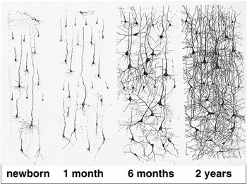
Golgi-stained sections showing the growth of pyramidal neuron soma and dendrites in the middle frontal gyrus. The normal newborn has sparse neural circuitry; then, with increasing age, there is a tremendous increase in the complexity of dendritic arborizations. In this frontal cortical area, the dendrite arbors for layer 3 pyramidal neurons, which are only 3% of mature size in the newborn, are still only about 50% by 2 years of age and do not reach 100% until the end of childhood (see text). From The Human Brain (3rd ed), by J. Nolte, 1993, St. Louis, MO: Mosby Year Book/Elsevier. Copyright 1993 by Mosby Year Book/Elsevier. Reprinted with permission from Elsevier.
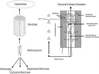
The minicolumn is a fundamental unit of information processing. (a) It is a roughly columnar vertical assembly of pyramidal neurons and interneurons, their interconnections, and input and output axons that extend from layer 6 up to the cortical surface. Each is 30–60 μm in diameter with perhaps 80–100 pyramidal neurons per minicolumn. (b) A schematic closeup of cortical layer 3 within a minicolumn. The shaded area represents the neuropil space that surrounds the vertical assembly of neurons. This area contains mostly synapses, dendrites, and unmyelinated axons. The unshaded section is where the majority of the cell soma reside and it contains myelinated axons. From “Morphological Differences Between Minicolumns in Human and Nonhuman Primate Cortex,” by D. P. Buxhoeveden, A. E. Switala, E. Roy, M. Litaker, and M. F. Casanova, 2001, American Journal of Physical Anthropology, 115. Copyright 2001 by American Journal of Physical Anthropology. Adapted with permission from Wiley–Liss, a subsidiary of John Wiley & Sons, Inc.
 You have
Access
- 150
- Cited by