Cardiovascular complication is one of the most common complications of diabetes mellitus with subsequent long-term morbidity and mortality. Cardiovascular complications include pre-mature atherosclerosis, coronary heart disease, and dilated cardiomyopathy. Reference Secrest, Becker, Kelsey, Laporte and Orchard1,Reference de Ferranti, de Boer and Fonseca2
Diabetic cardiomyopathy involves a wide range of spectrum ranging from pre-clinical left ventricular diastolic dysfunction, pre-mature atherosclerosis to overt left ventricular dysfunction and heart failure. Reference Salem, El Behery, Adly, Khalila and El Hadidib3–Reference Brunvand, Fugelseth, Stensaeth, Dahl-Jorgensen and Margeirsdottir5 Three-dimensional speckle-tracking echocardiography is a sensitive echocardiographic mode that can detect early myocardial dysfunction in children with type 1 diabetes mellitus. Reference El Razaky, El Amrousy, Elrifaey and Elgendy6 Moreover, several studies reported carotid intima–media thickness as an early marker of pre-mature atherosclerosis in diabetic children. Reference Rodriguez, Gómez-Díaz and Haj7
Early treatment of pre-clinical diabetic cardiovascular complications using cardioprotective drugs is a better practice than waiting for overt manifestation to occur. Very few randomised controlled studies have been performed in children with type 1 diabetes mellitus to assess the role of modification of risk factors and cardioprotective drugs to prevent the progression of cardiovascular complications. Reference Gourgari, Dabelea and Rother8
Several suggested mechanisms of diabetic cardiovascular complications include hyperglycaemia, insulin resistance, chronic inflammation, hyperlipidemia, microvascular disease, renin–angiotensin system activation, and excessive oxidative stress. Reference Chalakova, Yotov and Tzotchev9,Reference Schofield, Ho and Soran10 Among the various mechanisms, the role of oxidative stress has been strongly suggested in many studies, and therefore beneficial effects of antioxidants such as L-carnitine in the management of diabetic complications have been extensively investigated and reported. Reference Fu, Huang and Chen11
Angiotensin-converting enzyme inhibitors were found to reduce the incidence of vascular complications of diabetes mellitus as it prevents the increase in angiotensin II levels in cardiac muscles thus protecting against the oxidative stress, increasing bradykinin levels and cardiac dysfunction and hence reducing the afterload, and improving diastolic function. Reference Hornung, Reed, Mouat, Jefferies, Gunn and Hofman12–Reference Singh, Le, Khode, Baker and Kumar14 Statins are used to treat high lipid profile in type 1 diabetes mellitus, besides their anti-inflammatory and antioxidant effects that help to decrease cardiovascular complications. Reference Min, Shin and Lee15–Reference Blanco-Colio, Tuñón, Martín-Ventura and Egido17 In a large database, angiotensin-converting enzyme inhibitors and statins are the most commonly used cardiovascular medications among youth with type 1 diabetes mellitus. Reference Ahmadizar, Fazeli Farsani, Souverein, van der Vorst, de Boer and Maitland-van der Zee18
We aimed to assess the efficacy of captopril, simvastatin, and L-carnitine as cardioprotective drugs in children with type 1 diabetes mellitus and to evaluate the presence of any asymptomatic cardiovascular insult using different echocardiographic parameters, lipid profile, and carotid intima–media thickness.
Methods
Study population and design
This randomised controlled trial was conducted at the Endocrinology and Cardiology Units, Tanta University Hospital, on 100 children with type 1 diabetes mellitus for more than 3 years who were recruited from pre-screened 356 children with type 1 diabetes mellitus during the period from September 2018 to June 2020. Fifty healthy children of matched age and sex served as a control group. An informed consent was obtained from the parents of all patients of the study before enrolment. Local ethics committee of Faculty of Medicine, Tanta University, approved the study. The clinical trial was registered at www.cinicaltrial.gov (NCT03660293).
Inclusion criteria
Children and adolescents below 18 years of age with type 1 diabetes mellitus for more than 3 years.
Exclusion criteria
Children with CHD or acquired heart disease, other systemic diseases, children with diagnosed diabetic cardiomyopathy, children with non-type 1 diabetes mellitus, children with hypertension, children with renal or liver disease, dysmorphic children, and children with other endocrine disorders such as thyroid or adrenal disease.
Intervention
Children meeting the inclusion criteria were randomised into one of the four groups:
No-treatment group included 25 children with type 1 diabetes mellitus who received no cardioprotective drug besides their usual insulin treatment.
Simvastatin group included 25 children with type 1 diabetes mellitus who received simvastatin (10–20 mg/day) for 4 months; 10 mg for those weighing less than 30 kg and 20 mg for those weighing more than 30 kg. We used Zocor tablet (simvastatin), Merc & Co. Inc., USA.
Captopril group included 25 children with type 1 diabetes mellitus who received angiotensin-converting enzyme inhibitors in the form of captopril (0.2 mg/kg/day) for 4 months. We used Capoten tablet (captopril), SmithKline Beecham, Egypt.
L-carnitine group included 25 children with type 1 diabetes mellitus who received L-carnitine (50 mg/kg/day) for 4 months. We used Carnitol (L-carnitine tartrate), Global Pharmaceutical Industries, Egypt.
Randomisation and blinding
Random allocation software was used for generation of random sequence. Allocation concealment was performed by sequentially numbered sealed opaque envelopes. After signing the written consent, the sealed opaque envelope was opened and the child was enrolled into the respective group. All treating staff and outcome assessors were blinded to the treatment groups.
Measures
All children in the study were subjected to full history taking such as age, sex, age at onset and duration of diabetes mellitus, first presentation of diabetes mellitus, regimen, type and dose (unit/kg/day) of insulin used, and history of chronic diabetic complications. Moreover, anthropometric measurements such as weight, height, and body mass index were measured.
Laboratory investigations in the form of total cholesterol, triglycerides, high-density lipoprotein, low-density lipoprotein, and glycosylated haemoglobin were performed. High-sensitive serum troponin I was also measured by enzyme-linked immunosorbent assay technique.
Carotid intima–media thickness was measured using high-resolution B mode scan with the commercially available ultrasound transducer and equipment (Vivid 7, GE Healthcare, Horten, Norway) with a linear wide band of 5–12 MHz transducer. Before, ultrasound examination patients were rested quietly in a temperature-controlled dark room for 10–15 minutes. Patients were in supine position, the head slightly extended and rotated to the other side during the ultrasound examination.
Echocardiographic examination was performed using a commercially available ultrasound transducer and equipment (Vivid 7 or Vivid 9, GE Healthcare, Horten, Norway) with a 3.5-MHz, S7 and V3 matrix real-time three-dimensional probes. Digital loops were stored on the hard disc of the echocardiography machine and were transferred to a workstation (Echo PAC PC, 112 and 113; GE, Horten, Norway) for offline analysis.
Twelve lead surface electrocardiography (ECG) was performed using a three-channel α 1000 apparatus. QT interval was measured by manual method using handheld calipers accompanied by the use of magnification which were corrected by age (QTc). QT dispersion (QTd) is calculated as the difference between the maximum QT duration and minimum QT duration in different leads of 12-lead ECG tracing.
Echocardiography was performed to assess cardiac function using different echocardiographic modalities. Conventional echocardiography such as M mode was used to asses left ventricular ejection fraction where ejection fraction % = left ventricular end-diastolic volume-left ventricular end-systolic volume/left ventricular end-diastolic volume. Tissue Doppler imaging was used to evaluate systolic and diastolic mitral annulus velocities and left ventricular myocardial performance index, where left ventricular myocardial performance index was calculated as Tei index = a′ − b’/b’ where a’ is the time interval from the end of A'-wave to the onset of E'-wave and b’ is the time from the onset to the end of the S’-wave.
Three-dimensional speckle-tracking echocardiography was used to quantify the global radial, global area, global circumferential, and global longitudinal strain. Longitudinal strain was obtained from apical 2-, 3-, and 4-chamber views. Radial and circumferential strain were obtained from short-axis parasternal view. Then, the three-dimensional full volume of the left ventricle was obtained during breath hold to assess left ventricular ejection fraction. The three-dimensional strain tracking is performed starting from a region of interest defined at end systole. The three-dimensional strain of the region of interest is automatically generated in the end-systolic frame and is built up from an endocardial and an epicardial mesh. The user can correct the region of interest manually for better image.
Data collection and follow-up
All laboratory and echocardiographic measurements are performed before and 16 weeks after the treatment for all included patients. While in healthy control group, all measurements were performed once at the start of the study. We followed the participants by weekly phone call and face-to-face meeting every 2 months to provide the study materials and to take the used packages of drugs to ensure compliance to treatment. In addition, the parents were instructed to record any side effects during the treatment period.
Endpoints
The primary outcome was to assess the effect of captopril, simvastatin, and L-carnitine on the three-dimensional global longitudinal strain in children with type 1 diabetes mellitus. The secondary outcomes were to assess the effect of the three drugs on other echocardiographic parameters, lipid profile, serum troponin I, and carotid intima–media thickness. Side effects of the drugs were also assessed.
Statistical analysis
Sample size of 22 patients in each group was required to achieve the power of 90% with alpha = 0.05 to detect a difference of 3 in the mean of three-dimensional global longitudinal strain. Data were analysed using Statistical Package for Social Sciences software program V.21 (SPSS Inc. Chicago, IL, USA). Qualitative variables were presented as number and percentages and were compared by Chi-square test. Quantitative variables were presented as means ± standard deviation if normally distributed. Normal distribution of the data was checked by Shapiro–Wilk test. One-way analysis of variance was used to compare normally distributed quantitative variables between groups. Paired t-test was used to compare quantitative variables within the same group before and after the treatment. Post hoc test was used to detect significance between groups. To assess intra-observer variability, the same observer (O.E) performed the echocardiographic examination at an interval of 1 week to avoid recall bias. To assess inter-observer variability, the echocardiographic examinations were performed by a second observer (D.E) who was blinded to the results of the first observer. p-Value <0·05 was considered significant.
Results
Patients characteristics
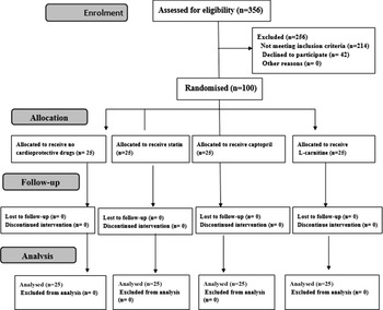
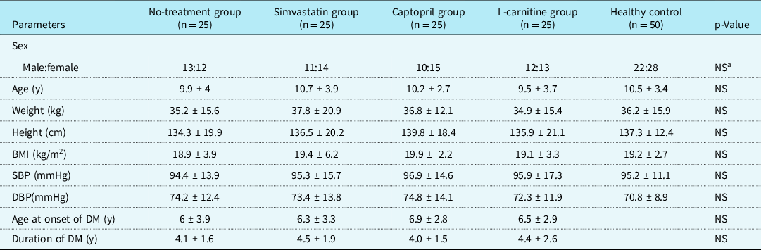
We examined 356 children with type 1 diabetes mellitus for eligibility criteria and only 100 children were eligible and included in the study (Fig 1). The characteristics of patients at baseline were comparable in the four groups (p > 0.05) (Table 1).
The randomised trial flow diagram, including enrolment, intervention allocation, and analysis.

Demographic and clinical data in the different studied groups

BMI = body mass index; DBP = diastolic blood pressure; DM = diabetes mellitus; SBP = systolic blood pressure.
a Chi-square test, NS: non-significant.
Echocardiographic and electrocardiographic data in the included children before the treatment
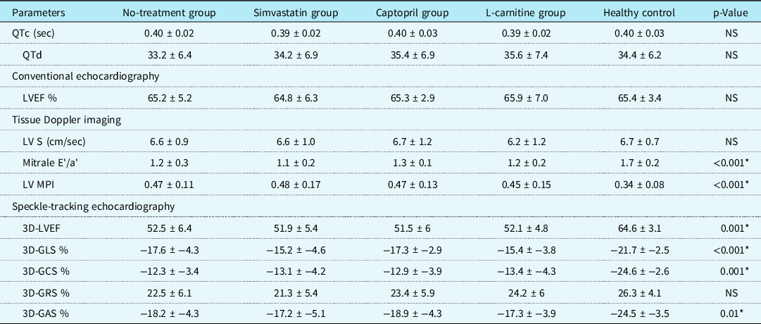
As regards echocardiographic and electrocardiographic examination before the treatment, there was no significant difference between the studied groups as regards QTc, QTd, and left ventricular systolic mitral annulus velocity measured by tissue Doppler imaging. However, there was a statistical significant decrease in mitral e'/a’ measured by tissue Doppler imaging in the four patient groups compared to the healthy control group. Moreover, there was a statistical significant increase in left ventricular myocardial performance imaging measured by tissue Doppler imaging in the four patient groups compared to the healthy control group. Furthermore, there was a statistically significant decrease in three-dimensional left ventricular ejection fraction %, three-dimensional global longitudinal strain, three-dimensional global circumferential strain, and three-dimensional global area strain in the four patients group compared to the healthy control group. However, there was no significant difference between the studied groups as regards three-dimensional global radial strain (p > 0.05) (Table 2).
Comparison between the different studied groups according to different echocardiographic and electrocardiographic parameters before the treatment

QTc = corrected QT; QTd = QT dispersion; LV S = left ventricular systolic mitral annulus velocity; mitral e'/a' = early diastolic mitral annulus velocity/atrial (late) mitral annulus velocity; LV MPI = left ventricular myocardial performance index; 3D-LVEF = three-dimensional left ventricular ejection fraction; GLS = global longitudinal strain; GCS = global circumferential strain; GRS = global radial strain; GAS = global radial strain.
* Means significant, NS: non-significant.
Effect of different treatment on echocardiographic and electrocardiographic data in the included children
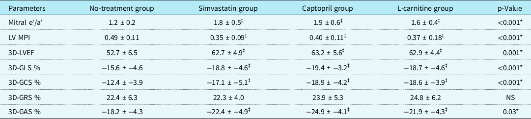
After the treatment, there was a significant improvement of left ventricular myocardial performance index, mitral e'/a’ ratio, three-dimensional left ventricular ejection fraction %, and three-dimensional global area strain in the simvastatin, captopril, and L-carnitine groups compared to no-treatment group. However, there was no significant difference between the three treatment groups as regards the improvement in the previous echocardiographic parameters which normalised in the three treatment groups. Moreover, there was a significant increase in the both three-dimensional global longitudinal strain % and three-dimensional global circumferential strain % in the simvastatin, captopril, and L-carnitine groups compared to no-treatment group. The improvement of three-dimensional global longitudinal strain was more in the captopril group than that of the simvastatin and L-carnitine groups and was comparable to the healthy control levels. In spite of the improvement in three-dimensional global circumferential strain, none of the patient groups reached the normal levels of the healthy control groups (Table 3).
Comparison between the different studied groups according to different echocardiographic parameters after the treatment

mitral e'/a' = early diastolic mitral annulus velocity/atrial (late) mitral annulus velocity; LV MPI = left ventricular myocardial performance index; 3D-LVEF = three-dimensional left ventricular ejection fraction; GLS = global longitudinal strain; GCS = global circumferential strain; GRS = global radial strain; GAS = global radial strain.
* Means significant, NS: non-significant.
‡ Significant compared to no-treatment group.
Different laboratory data in the included children before the treatment
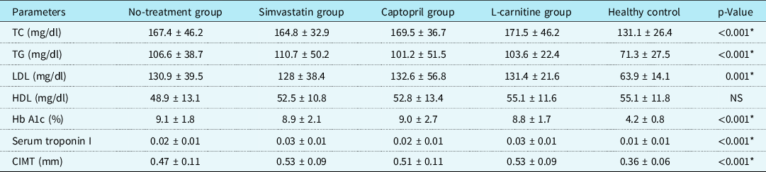
Before the treatment, there was the statistically significant higher levels of serum cholesterol, triglycerides, low-density lipoprotein, glycosylated haemoglobin, serum troponin I, and carotid intima–media thickness in the four patient groups compared to the healthy control group. However, there was no statistical significant difference between the patient groups and healthy control as regards serum high-density lipoprotein. There was no statistical significant difference between the four patient groups as regards serum cholesterol, triglycerides, low-density lipoprotein, glycosylated haemoglobin, serum troponin I, or carotid intima–media thickness (p > 0.05) (Table 4).
Comparison between the different studied groups according to the lipid profile and carotid intima–media thickness before the treatment

TC = total cholesterol; TG = triglycerides; LDL = low-density lipoproteins; HDL = high-density lipoproteins; CIMT = carotid intima–media thickness.
* Means significant, NS: non-significant.
Effect of different treatment on laboratory data in the included children
After the treatment, serum cholesterol and low-density lipoprotein levels significantly decreased in both simvastatin and L-carnitine groups but failed to decrease in both captopril group and no-treatment groups. Serum cholesterol and low-density lipoprotein levels in both simvastatin and L-carnitine groups were comparable to those of healthy control group and were also comparable to each other (Table 5).
Comparison between the different studied groups according to the lipid profile and carotid intima–media thickness (CIMT) after the treatment

CIMT = carotid intima–media thickness; HDL = high-density lipoproteins; LDL = low-density lipoproteins; TC = total cholesterol; TG = triglycerides.
* Means significant, NS: non-significant.
‡ Significant compared to no-treatment group.
Moreover, serum triglycerides significantly decreased in simvastatin group only compared to no-treatment group, captopril group, and L-carnitine group (p = 0.002, p = 0.01, and p = 0.06), respectively. Serum triglycerides level in simvastatin group was comparable to that of healthy control group. However, high-density lipoprotein levels significantly increased in both simvastatin and L-carnitine groups compared to captopril group and no-treatment groups. High-density lipoprotein levels in both simvastatin and L-carnitine groups were comparable to each other (Table 5). Furthermore, serum troponin I level significantly decreased in the simvastatin group, captopril group, and L-carnitine group after the treatment and was comparable to normal values. Interestingly, carotid intima–media thickness failed to show any significant change after the treatment in all treatment groups (Table 5).
Side effects of different treatment in different patient groups
There were no recorded serious side effects of the three drugs except for postural hypotension and dry cough in three patients receiving captopril that did not require cessation of the drug and headache in two patients receiving simvastatin that disappear with continuous use.
There was an excellent inter-observer and intra-observer reliability for all three-dimensional strain and three-dimensional left ventricular ejection fraction %. Intraclass correlation coefficient (ICC) of intra-observer reliability were 0.989 for three-dimensional left ventricular ejection fraction %, 0.913 for three-dimensional global longitudinal strain, 0.903 for three-dimensional global circumferential strain, 0.930 for three-dimensional global area strain, and 0.904 for three-dimensional global radial strain. Moreover, ICC of inter-observer reliability were 0.910 for three-dimensional left ventricular ejection fraction %, 0.856 for three-dimensional global longitudinal strain, 0.872 for three-dimensional global circumferential strain, 0.879 for three-dimensional global area strain, and 0.851 for three-dimensional global radial strain.
Discussion
For the best of our knowledge, the present study was the first to assess the efficacy of simvastatin, captopril, and L-carnitine as cardioprotective drugs in children with type 1 diabetes mellitus. In this study, we found that the three drugs are effective in varying degree to revert various cardiovascular dysfunctions caused by diabetes mellitus and could be used as cardioprotective drugs in these patients.
The mechanisms that lead to the development of the diabetic cardiomyopathy are poorly understood, although they appear to be a direct result of cellular damage caused by hyperglycaemia, and its consequences as hyperglycaemia lead to an increase in cardiac oxidative stress, upregulation of renin–angiotensin system, inflammation, myocardial fibrosis, and changes in calcium handling and mitochondrial function. In addition, increased formation of advanced glycation end products which are particularly prone to cross-linking with extracellular proteins such as collagen and elastin altering their structural properties and impairing cardiac relaxation leads to irreversible changes in myocardial structure. These changes may result in ventricular stiffness and consequently interfere with diastolic and systolic function. Reference Bugger and Abel19 Therefore, any drug that can eliminate one or more of these mechanisms can exert a cardioprotective effect in diabetic patients.
In our study, children with type 1 diabetes mellitus who received captopril showed a significant improvement in left ventricular myocardial performance index, mitral e'/a', three-dimensional global longitudinal strain, three-dimensional global circumferential strain, three-dimensional global area strain, and serum troponin I reflecting improvement of cardiac function and eliminating ongoing cardiac injury. However, captopril showed no significant effect on the lipid profile or carotid intima–media thickness that was similar to the results of Marcovecchio et al Reference Marcovecchio, Chiesa and Bond20 who studied the effect of captopril in adolescent with type 1 diabetes mellitus and reported no significant effect of captopril on lipid profile or carotid intima–media thickness.
Angiotensin-converting enzyme inhibitors such as captopril can exert its cardioprotective effect in diabetic patients regardless of its antihypertensive effect by several mechanisms. Angiotensin-converting enzyme inhibitors decrease local vascular inflammation through decreasing metalloprotenianse and plasminogen activator inhibitor 1 with subsequent decreased atherosclerosis. Furthermore, it decreases reactive oxygen species, increases nitric oxide level, decreases production of inflammatory cytokines, and increases vascular compliance. Reference McFarlane, Kumar and Sowers21 Moreover, angiotensin-converting enzyme inhibitors improve insulin sensitivity and increase glucose uptake by the skeletal muscle in diabetic patients. Reference Kudoh and Matsuki22
Children with type 1 diabetes mellitus who received simvastatin showed a significant improvement of their left ventricular myocardial performance index, mitral e'/a', three-dimensional global longitudinal strain, three-dimensional global circumferential strain, three-dimensional global area strain, and serum troponin I. To our knowledge, our study is the first clinical trial evaluating the effect of simvastatin on the cardiac function of diabetic children, so no available previous data to compare our results with. Moreover, patients treated with simvastatin showed a significant decrease in serum cholesterol and low-density lipoprotein, but a significant increase in high-density lipoprotein. However, no significant effect on triglycerides or carotid intima–media thickness was reported. In agreement with our results, Marcovecchio et al Reference Marcovecchio, Chiesa and Bond20 reported a significant decrease in lipid profile in adolescent with type 1 diabetes mellitus who received statin therapy with no effect on carotid intima–media thickness on their study. This can be explained by the fact that changes in carotid intima–media thickness need longer time of treatment to develop. Similarly, Canas et al Reference Canas, Ross and Taboada23 reported a significant decrease in the serum cholesterol and low-density lipoprotein after simvastatin therapy for 6 months in children with type 1 diabetes mellitus.
Statins are therapeutic drugs used to treat hyperlipidemia and are also known to reverse the progression of carotid intima–media thickness in diabetics with prolonged use. Reference Fang, Han, Gong, Chen and Fan24 Simvastatin has a lipid-lowering effect through inhibiting hepatic cholesterol synthesis, decreasing the secretion of apo-B lipoprotein, increasing receptor-mediated clearance of low-density lipoprotein, decreasing direct low-density lipoprotein production, and increasing low-density lipoprotein and very low-density lipoprotein clearance. Reference Ginsberg25 Independent of their lipid-lowering effects, statins have pleiotropic effects in the form of antioxidant and anti-inflammatory actions. Reference Shishehbor, Brennan and Aviles26–Reference Jain and Ridker28 Simvastatin may exert its cardioprotective effects through several mechanisms including lipid-lowering effect, antioxidant effect, anti-inflammatory effect, and upregulation of nitric oxide production. Reference Yamagishi, Matsui and Nakamura29,Reference Taqueti and Ridker30
Interestingly, children with type 1 diabetes mellitus who received L-carnitine had significantly improved cardiac function parameters such as left ventricular myocardial performance index, mitral e'/a', three-dimensional global longitudinal strain, three-dimensional global circumferential strain, and three-dimensional global area strain. They showed a significant decrease in serum cholesterol, triglycerides, low-density lipoprotein, and serum troponin I, but a significant increase in high-density lipoprotein. However, it showed no significant effect on carotid intima–media thickness. Our results were in agreement with the results of Santo et al Reference Santo, Sergio and Luigi31 who reported a significant decrease in cholesterol and low-density lipoprotein in diabetic patient who received L-carnitine treatment for 16 weeks.
Carnitine plays an important role in regulating carbohydrate metabolism by increasing peripheral glucose utilisation and in upregulation of free fatty acids within cardiac myocytes. Reference Ringseis, Keller and Eder32,Reference Malaguarnera, Vacante, Avitabile, Malaguarnera, Cammalleri and Motta33 Furthermore, L-carnitine has anti-inflammatory and antioxidant effects that play an important role in the prevention of diabetic cardiomyopathy. Reference Calo, Pagnin and Davis34,Reference Duranay, Akay, Yilmaz, Senes, Tekeli and Yücel35 Additionally, it has been reported that L-carnitine can improve diastolic dysfunction of the heart. Reference Serati, Motamedi, Emami, Varedi and Movahed36
Long-chain fatty acids are the main energy substrates for the myocardium. However, they cannot freely diffuse through the mitochondrial inner membrane. Carnitine plays an essential role in the transfer of long-chain fatty acids into the mitochondria for subsequent β-oxidation. So, carnitine deficiency, which is common in diabetes mellitus Reference Mamoulakis, Galanakis, Dionyssopoulou, Evangeliou and Sbyrakis37 , can lead to accumulation of long-chain fatty acid within the heart muscles with subsequent development of cardiomyopathy. Reference Calo, Pagnin and Davis34,Reference Serati, Motamedi, Emami, Varedi and Movahed36 Moreover, Malone et al Reference Malone, Malone and Morrison38 reported that carnitine levels in diabetic patients were below their normal range and were associated with cardiomyopathy and muscle weakness that improved with carnitine supplementation.
Based on the previous results of our study, we observed that simvastatin, captopril, and L-carnitine can reverse the harmful effect of diabetes mellitus on the heart of the children with type 1 diabetes mellitus and may be used as cardioprotective drugs in these patients. However, only simvastatin and L-carnitine can improve the lipid profile in these children. Lack of effect of the three drugs on carotid intima–media thickness was due to short duration of treatment. So, longer duration of treatment was recommended to study the effects of the three drugs on carotid intima–media thickness.
Limitation of the study: first, it is the relatively small sample size of the study. Second, it is a single-centre study with short duration of treatment. So, multi-centre studies with large sample size and longer duration of treatment and follow-up are recommended to validate our results.
Conclusion
Captopril, simvastatin, and L-carnitine have a significant beneficial effect on cardiac function in children with type 1 diabetes mellitus. However, only simvastatin and L-carnitine have beneficial effect on lipid profile. The drugs were safe and well tolerated. Long-term studies would provide a better insight on the impact of these drugs on the heart of children with type 1 diabetes mellitus.
Acknowledgments
Authors wish to thank all the patients who participated in the study.
Financial support
This research received no specific grant from any funding agency, commercial or not-for-profit sectors.
Conflict of interest
The authors have no conflicts of interest to disclose.
Ethical statement
The authors assert that all procedures contributing to this work comply with the ethical standards of the relevant national guidelines on human experimentation and with the Helsinki Declaration of 1975, as revised in 2008, and have been approved by the institutional committees of Faculty of Medicine, Tanta University.