I. INTRODUCTION
Recently, synthesis and surface-modification of upconversion nanocrystals (UCNs) have attracted considerable attention because of their unique optical properties and promising applications in biological fields.Reference Wang and Liu1, Reference Vetrone and Capobianco2 Lanthanide-doped near-infrared (NIR)–to–visible UCNs, which can convert NIR excitation to visible light, are of particular interest.Reference Wang, Tan, Zhang, Fan and Wang3 Since most biological species absorb minimally in the NIR window, their use in fluorescent imaging minimizes the induced autofluorescence and photodamage, as well as enables high penetration depth of light in tissues.Reference Chatterjee, Rufalhah and Zhang4–Reference Idris, Li, Ye, Sim, Mahendran, Ho and Zhang6 UCNs exhibit superior photostability, as the lanthanide ions responsible for their fluorescence emission are doped within the nanocrystal core such that they are well shielded from the surroundings.Reference Shen, Sun and Yan7 Moreover, taking advantage of sharp emissions and long lifetime, the UCNs can also serve as efficient electron donors and improve the efficiency and sensitivity in fluorescence resonance energy transfer (FRET) detection, a popular technology widely used in biological detection.Reference Wang, Yan, Hao, Wang, Zeng, Bao, Wang, Peng and Li8, Reference Chen, Chen, Hu, Yu, Li, Zhang, Zhou, Yi and Huang9 Among various host materials for lanthanide-doped UCNs, NaGdF4 nanocrystals are attractive because of the low photo energy associated with high upconversion efficiency.Reference Liu, Wang, Zhang and Chen10, Reference Ptacek, Schafer, Kompe and Haase11 At the same time, Gd3+ ions possess a large number of unpaired electrons, and thus NaGdF4 nanocrystals can also serve as solid-state contrast agents in magnetic resonance imaging (MRI).Reference Zhou, Sun, Du, Xiong, Hu and Li12, Reference Park, Kim, Lee, Jeon, Na, Yu, Kim, Leei, Cho, Baik, Kim, Park, Park, Kim, Lee, Yoon, Song, Moon, Suh and Hyeon13 Therefore, synthesis of high-quality and biocompatible NaGdF4 nanoparticles may support the development of multimodal bioimaging nanoplatform for diagnosis, detection, and therapy in future bionanotechnology.
Many successful attempts have been made to synthesize NaGdF4 nanocrystals in the past years, but unfortunately high-quality and monodisperse nanocrystals are generally prepared from nonpolar solvents and bear a hydrophobic surface.Reference Liu, Wang, Zhang and Chen10–Reference Park, Kim, Lee, Jeon, Na, Yu, Kim, Leei, Cho, Baik, Kim, Park, Park, Kim, Lee, Yoon, Song, Moon, Suh and Hyeon13 To harness these hydrophobic nanocrystals to biological fields, surface modification is demanded to make them water dispersible, biocompatible, and capable of conjugating to various biomolecules. Zhou et al. have used an organic route to convert hydrophobic NaGdF4 nanocrystals to its hydrophilic analogs by directly oxidizing the surfactants.Reference Zhou, Sun, Du, Xiong, Hu and Li12 While this route was novel and straightforward, the organic reaction was time consuming and the reaction yield was unsatisfactory. Poly(ethylene glycol) phospholipids were also used to encapsulate the hydrophobic NaGdF4 nanocrystals through van der Waals interaction, but the bond between nanocrystals and phospholipids were weak, and thus these UCNs might be unable to sustain complex chemical/biological processes.Reference Park, Kim, Lee, Jeon, Na, Yu, Kim, Leei, Cho, Baik, Kim, Park, Park, Kim, Lee, Yoon, Song, Moon, Suh and Hyeon13 Naccache et al. used a ligand-exchange method to modify hydrophobic NaGdF4 nanocrystals and made them water dispersible.Reference Naccache, Vetrone, Mahalingam, Cuccia and Capobianco14 However, notable fluorescent quench was induced because of some unexpected surface defects during the ligand-exchange process. As such, it is still very important to develop facile and general methods to surface modify the NaGdF4 nanocrystals, not only making them water dispersible, biocompatible, and easily conjugated but also maintaining their strong upconversion photoluminescence.
Owing to the excellent chemical and thermal stability coupled with novel electronic and optical properties, carbon nanomaterials have been intensively explored for biosensors, bioimaging probes, and therapy nanomedicines in recent years.Reference Yang, Ratinac, Ringer, Thordarson, Gooding and Braet15 If a carbon layer could cover luminescent nanocrystals, such a layer might be able to serve as a robust shell for the purpose of maintaining both chemical and photostability of the luminescent nanocrystals. Conventional carbon nanomaterials prepared by thermal methods such as chemical vapor deposition (CVD) generally exhibit a highly hydrophobic surface. However, carbon materials synthesized by polymerization of carbohydrates under hydrothermal conditions display a hydrophilic surface consisting of cross-linked biocompatible molecules.Reference Titirici and Antonietti16, Reference Hu, Wang, Wu, Yu, Antonietti and Titirici17 This kind of carbonaceous shell is water dispersible, biocompatible, and easily conjugated to biomolecules. Producing a carbon shell on hydrophilic nanocrystals has been realized through hydrothermal process on a few nanoparticles.Reference Sun and Li18, Reference Wan, Min and Yu19 However, how to coat a carbon shell on a hydrophobic nanocrystal remains challenging, because these nanocrystals can hardly be dispersed in water, which is normally used in a hydrothermal route.
In this work, we demonstrate a microemulsion route to coat a thin carbon shell on hydrophobic nanocrystals and use upconversion NaGdF4:Yb,Er nanocrystals as an example. The prepared NaGdF4:Yb,Er@Carbon nanoparticles are uniform in size and can be well dispersed in water. These nanoparticles, covered with a thin carbon shell, exhibit good cell biocompatibility and strong fluorescence and can be used as efficient cell imaging probes. Such biocompatible UCNs might serve as a multimodal bioimaging nanoplatform for the purpose of diagnosis, detection, and therapy in future bionanotechnology.
II. EXPERIMENTAL
A. Materials
YCl3, YbCl3, and ErCl3 were prepared by dissolving the corresponding lanthanide oxides [Gd2O3, Yb2O3, and Er2O3 (purity of 99.99%), purchased from Shanghai Yuelong Nonferrous Metals Limited, China] in dilute HCl and followed evaporation. Octandecene (ODE) and oleic acid (OA, purchased from Sigma-Aldrich, China) and other reagents/solvents (obtained from Shanghai Chemical Reagent Co, Ltd.) were used without further purification.
B. Synthesis of NaGdF4:Yb,Er nanoparticles
Monodisperse NaGdF4:20%Yb,2%Er nanocrystals were synthesized according to a protocol we previously developed with slight modification.Reference Li and Zhang20 In brief, 0.78 mmol GdCl3, 0.2 mmol YbCl3, and 0.02 mmol ErCl3 powders were dissolved in a mixed solution of 2 mL OA and 20 mL ODE at 160 °C. At room temperature, a methanol solution containing 4 mmol NH4F and 10 mmol NaOH was then added into the solution and reacted for 30 min. Subsequently, the methanol was evaporated off and the solution was degassed for 10 min before slowly heating to 260 °C. After heating at 260 °C for 40 min, the solution was cooled to room temperature naturally. NaGdF4:Yb,Er nanocrystals were separated by adding acetone and centrifuging at 5000 rpm for 10 min. The nanocrystals were then washed with ethanol/water at least three times and finally dispersed in cyclohexane to form a transparent dispersion.
C. Synthesis of NaGdF4:Yb,Er@Carbon nanoparticles in microemulsion
The water-in-oil microemulsion was formed by sonicating a mixed solution of 8 mL cyclohexane, 0.25 mL CO-520, and 50 μL aqueous glucose (1 M). After a clear solution was formed, 2 mL NaGdF4:Yb,Er nanoparticles (0.01 M) was added into the microemulsion and stirred for 10 min. The solution was then transferred into a 20 mL sealed autoclave and slowly heated to 120 °C at 3 °C/min. After 2 h at 120 °C, the autoclave was cooled to room temperature naturally. The products were separated from the solution by adding acetone, and then washed with three cycles of ethanol and water each.
D. Characterization
Transmission electron microscopy (TEM) images were recorded on a JEOL 2010F TEM (JEOL Ltd., Japan). X-ray powder diffraction (XRD) was carried out on a Japan Rigaku D/max rA x-ray diffractometer equipped with a Cu Kα radiation (Rigaku Ltd., Japan). Fourier transform infrared spectroscopy (FTIR) was performed on an attenuated total reflection FTIR (Varian Inc., Polo Alto, CA). Upconversion photoluminescence spectra were acquired on a F-4500 fluorescence spectrometer equipped with a commercial 980 nm NIR laser (Hitachi Ltd., Japan). Biocompatibility of the nanoparticles was assessed using murine bladder cancer cell lines (MB49). The cells were seeded on a 96-well plate and then incubated with 200 μL culture medium that contained different concentrations of the nanoparticles for 2 days. Cell viability was examined using an MTS solution and quantified by measuring the absorbance at 490 nm using a standard microplate reader (Bio-Tek Instruments, Winooski, VT). Cell-imaging experiments were carried out under a confocal laser scanning microscope (Nikon C1 Confocal) equipped with a continuous-wave 980 nm NIR laser excitation source (Nikon Instruments Inc., Japan). The cells were initially cultured with the nanoparticles for around 3 h and washed with phosphate-buffered saline (PBS) twice before imaging test.
III. RESULTS AND DISCUSSION
A. Surface-modification approach and possible formation mechanism
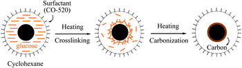
A schematic of the surface-modification approach is shown in Fig. 1. To form a thermodynamically stable microemulsion, cyclohexane/CO-520/aqueous glucose with a certain ratio was used, acting as oil phase, surfactant (CO-520), and water phase, respectively. This kind of microemulsion has proved to be a powerful tool to encapsulate hydrophobic nanoparticles inside the water pool and had been widely used to coat a silica layer on nanocrystals using ammonia and TEOS as reagents.Reference Yi, Selvan, Lee, Papaefthymiou, Kundaliya and Ying21, Reference Li, Zhang and Jiang22 In this work, however, we filled the water pools of the microemulsion with glucose solution, followed by elevating the temperature to induce hydrothermal carbonization of the glucose molecules. Since NaGdF4:Yb,Er nanocrystals were inside the water pools, carbonization of glucose proceeds around the nanocrystals, using the surface of the nanocrystals as seeding substrates. Therefore, a carbon layer was formed around these NaGdF4:Yb,Er nanocrystals as a result of the hydrothermal carbonization of glucose molecules at elevated temperature. The microemulsion was thought to provide a microscale site for each single nanocrystal and confine them in the pools during carbonization of glucose molecules.
Schematic of the formation process of a carbon shell on NaGdF4:Yb,Er nanocrystals in a water-in-oil microemulsion.

B. Structural and morphological characterizations
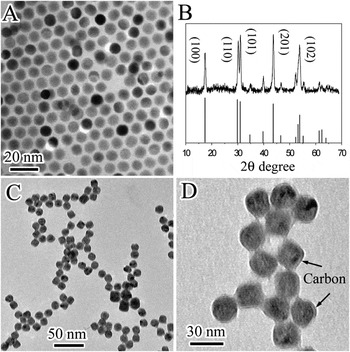
Morphology of the prepared NaGdF4:Yb,Er nanocrystals are shown in Fig. 2(a). The nanocrystals are uniform in size and self-assembled on the copper grid because of their hydrophobic surface. The XRD patterns of these nanocrystals are displayed in Fig. 2(b). All the diffraction peaks are indexed to hexagonal-phase NaGdF4 crystals (JCPDS Card No. 27-0699), confirming high-phase purity and good crystallinity of these nanocrystals. After hydrothermal carbonization of the glucose molecules on these nanocrystals in microemulsion at 120 °C for 2 h, an obvious layer emerged on the nanocrystals as shown in Figs. 2(c) and 2(d), suggesting that a carbon shell of around 2–5 nm was formed on the NaGdF4:Yb,Er nanocrystals. The size and morphology of the core NaGdF4:Yb,Er nanocrystals were maintained after hydrothermal treatment, revealing that the hydrothermal process did not affect the core nanocrystals.
(a) TEM image and (b) XRD patterns of the NaGdF4:Yb,Er nanocrystals. (c) and (d) are TEM images of the as-prepared NaGdF4:Yb,Er@Carbon nanoparticles at different magnifications.

C. Influence of concentration and temperature
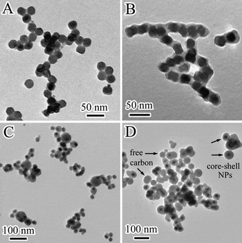
The concentration of glucose in the microemulsion and reaction temperature played important roles in determining the final morphology of the core–shell nanoparticles. With lower glucose concentration inside the water pool (such as 0.5 M), no obvious carbon shell was found on the nanocrystals [Fig. 3(a)], although most of the products could still be dispersed in water solution, revealing that their surface property was modified. This was probably because only partial glucose molecules cross-linked on the nanocrystals surface and were not well carbonized. On the other hand, a thicker layer of carbon around 3 to 8 nm emerged if the glucose concentration increased to 2 M [Fig. 3(b)]. These NaGdF4:Yb,Er@Carbon nanoparticles had a tendency to bond through their surface carbon shells, which might be from subsequent cross-linking and carbonization of surface glucose between adjacent nanoparticles. The above results indicated that it was possible to control the thickness of the carbon shell on a nanocrystal by adjusting the glucose concentration, although some nanoparticles bond at high concentration. When the reaction temperature was increased to 140 °C, it was found that some big carbon nanoparticles were also formed along with the core–shell nanoparticles. This result suggested that high reaction temperature would induce homogenous nucleation of carbon in addition to heterogeneous nucleation of carbon on the nanocrystals surface. This hypothesis was confirmed when the reaction temperature was increased to 160 °C. In this case, more free carbon spheres were produced as a result of the increased homogeneous nucleation and growth of carbon seeds at elevated temperature.
TEM images of NaGdF4:Yb,Er@Carbon nanoparticles prepared under different experimental conditions. (a) Using 0.5 M glucose at 120 °C. (b) Using 2 M glucose at 120 °C. (c) 1 M glucose at 140 °C. (d) 1 M glucose at 160 °C.

D. Surface groups and luminescent properties of nanoparticles
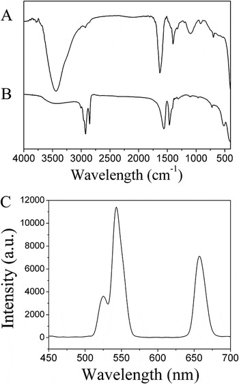
FTIR spectra of the as-prepared NaGdF4:Yb,Er@Carbon and NaGdF4:Yb,Er nanocrystals before modification are shown in Figs. 4(a) and 4(b), respectively. In Fig. 4(b), two peaks at 2924 and 2858 cm−1 are characteristic of –CH2 stretching, and the other two peaks at 1565 and 1463 cm−1 were attributed to –COOH stretching. These peaks indicate that oleic acid was attached on the NaGdF4:Yb,Er nanocrystals during nanocrystal synthesis. After formation of a carbon shell on these nanocrystals, a strong peak at 3438 cm−1 (–OH stretching), 1632 cm−1 (C = O stretching) and two peaks at 1402 and 1093 cm−1 (C–OH stretching) appeared [Fig. 4(a)], showing that –OH, C–OH, and –COOH groups were produced on the nanoparticles surface. The FTIR spectrum of NaGdF4:Yb,Er@Carbon nanoparticles was similar to that of the carbon spheres prepared from pure glucose solution, which confirmed the formation of a carbonaceous shell on the NaGdF4:Yb,Er nanocrystals.Reference Sun and Li23 Note that the –OH and –COOH groups on the nanoparticle surface not only confer hydrophilicity of these nanoparticles, but also provide many favorable sites for grafting various biomolecules for targeting applications.
FTIR spectra of the as-prepared (a) NaGdF4:Yb,Er@Carbon nanoparticles and (b) NaGdF4:Yb,Er nanocrystals before modification that covered with a oleic acid surface. (c) Upconversion photoluminescent spectrum of the NaGdF4:Yb,Er@Carbon nanoparticles.

The upconversion spectrum of NaGdF4:Yb,Er@Carbon nanoparticles is shown in Fig. 4(c). The NaGdF4 nanocrystals codoped with Yb and Er ions displayed two green peaks (525 and 543 nm) and a red peak (655 nm) under the excitation of a commercial continuous-wave (CW) diode NIR laser, which were ascribed to the energy transitions from 2H11/2, 4S3/2, and 4F9/2 to 4I15/2 of Er3+ ions, respectively.Reference Liu, Wang, Zhang and Chen10 The upconversion luminescence of these nanoparticles was strong enough to be observed by the naked eye under the NIR excitation. For comparison, we also tried to modify the NaGdF4:Yb,Er nanocrystals with a 10 nm SiO2 layer, whose photoluminescence was obviously weaker than the prepared NaGdF4:Yb,Er@Carbon nanoparticles under the same concentration (based on NaGdF4:Yb,Er nanocrystals). This result might be attributed to the thinner layer of carbon (2–5 nm) having lower scattering of the incident and emission light than the thick silica layer (10 nm).
E. Biocompatibility and imaging capability of nanoparticles
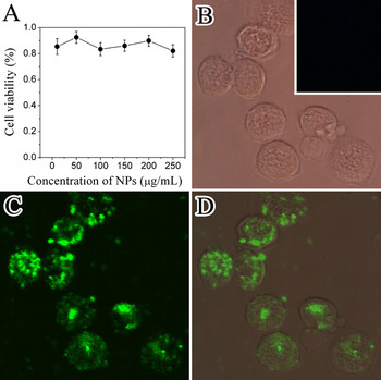
To evaluate the biocompatibility of NaGdF4:Yb,Er@Carbon nanoparticles, MB49 cells were grown with the cell culture medium containing nanoparticles, and then cell viability was determined using the MTS assay. Figure 5(a) depicts the viability of cells grown for 2 days with different concentrations of nanoparticles. This result shows that the nanoparticles were biocompatible to the MB49 cells in the range of 10 to 250 μg/mL. Even at the high concentration, which was far above the conventional concentration used in cell-labeling experiments, cell viability above 80% still could be achieved.
(a) Viabilities of cells after being cultured with different concentrations of NaGdF4:Yb,Er@Carbon nanoparticles for 2 days. (b) Bright-field image, (c) fluorescence image, and (d) their combined image of MB49 cells labeled with NaGdF4:Yb,Er@Carbon nanoparticles. Inset in (b) is the control imaging of cells under NIR imaging without incubation of nanoparticles.

The use of the NaGdF4:Yb,Er@Carbon nanoparticles for cell-imaging applications was also studied. Before the imaging test, the MB49 cells were cultured with the nanoparticles for 3 h and unbounded nanoparticles were washed off with PBS water. Under the bright-field mode of the microscope equipped with a Xe lamp, no fluorescence from these cells could be observed [Fig. 5(b)]. However, bright blue emissions were clearly presented when the microscope switched to the fluorescence mode with a NIR laser as light resource [Fig. 5(c)]. If we combine the bright-field image and the fluorescence image, it is easy to see that these two images match well, suggesting that the NaGdF4:Yb,Er@Carbon nanoparticles have been well internalized by the MB49 cells, and the location of these cells could be quickly distinguished by the nanoparticles. In a control experiment, no fluorescence was observed under the NIR imaging if the cells did not incubate with the NaGdF4:Yb,Er@Carbon nanoparticles [inset of Fig. 5(b)], confirming that the fluorescence comes from the nanoparticles. The aforementioned results indicate that the prepared NaGdF4:Yb,Er@Carbon nanoparticles possess a promising potential to serve as a powerful cell-imaging tool for various bioimaging applications. Since the emissions of lanthanide-doped nanocrystals can be easily tuned by the doping ions, it is expected that multiplexing bioimaging can be achieved by combining use of different lanthanide-doped NaGdF4@Carbon nanoparticles and suitable filters under the NIR excitation.
IV. CONCLUSION
In summary, we developed a water-in-oil microemulsion route to prepare core–shell structured NaGdF4:Yb,Er@Carbon nanoparticles. By encapsulating the hydrophobic NaGdF4:Yb,Er nanocrystals inside the water pools of a microemulsion, a thin layer of carbon could be produced from hydrothermal carbonization of glucose molecules. Structural and surface characterizations showed that these nanoparticles were uniform in size and had functional groups for bioconjugation. Owing to the thin carbon shells, these nanoparticles displayed good water dispersibility, good biocompatibility, and satisfactory upconversion fluorescence. Their potential for use as efficient cell-imaging probes was also demonstrated. This type of nanoparticle might serve as multimodal bioimaging nanoplatforms for the purpose of diagnosis, detection, and therapy in future bionanotechnology.
ACKNOWLEDGMENTS
The authors acknowledge financial support from National Nature Science Foundation of China (20901068) and Zhejiang Qianjiang Talent Project. The authors also thank Dr. Huichen Guo at National University of Singapore for her help in the cell-imaging experiments.