Introduction
Acute retinal ischemia due to arterial or venous occlusion is a major cause of blindness and visual impairment (Hayreh, Reference Hayreh2005). Retinal neurons are particularly susceptible to ischemic injury because they rely on mitochondrial oxidative phosphorylation to meet their high energy demands. During ischemia, a lack of oxygen and glucose decreases the rate of oxidative phosphorylation resulting in decreased ATP generation, depletion of phosphocreatine (PCr), subsequent disruption of ion homeostasis, neurotransmitter dysregulation, and increased formation of reactive oxygen species (ROS) and reactive nitrogen species (RNS) (Sun et al., Reference Sun, Bui, Vingrys and Kalloniatis2007a,Reference Sun, Vingrys and Kalloniatisb). During reperfusion, there may be a transient normalization of ATP and PCr concentrations and partial recovery of retinal function (Sun et al., Reference Sun, Bui, Vingrys and Kalloniatis2007a). However, escalating mitochondrial dysfunction caused by oxidative and nitrosative stress, coupled with high energy demands for the restitution of cellular homeostasis, leads to secondary energy deficiency and further retinal injury. The subsequent elevation of intracellular Ca2+ coupled with ATP depletion triggers mitochondrial permeability transition (MPT) leading to apoptosis and/or necrosis (Johnson et al., Reference Johnson, Perkins, Giddabasappa, Chaney, Xiao, White, Brown, Waggoner, Ellisman and Fox2007; Kakkar & Singh, Reference Kakkar and Singh2007; Juhaszova et al., Reference Juhaszova, Wang, Zorov, Nuss, Gleichmann, Mattson and Sollott2008). Retinal ischemia/reperfusion frequently results in the loss of amacrine and ganglion cells in the inner retina due to their expression of ionotropic glutamate receptors and consequent vulnerability to glutamate excitotoxicity (Lipton, Reference Lipton1999; Sun et al., Reference Sun, Bui, Vingrys and Kalloniatis2007a; Osborne, Reference Osborne2008). In contrast, photoreceptors often survive acute retinal ischemia/reperfusion, although post ischemic electroretinographic (ERG) studies in rats indicate that many surviving photoreceptors are dysfunctional (Sun et al., Reference Sun, Bui, Vingrys and Kalloniatis2007a,Reference Sun, Vingrys and Kalloniatisb). This is not surprising given the high energy demands of photoreceptors to maintain plasma membrane ion gradients, phototransduction, and the high turnover of outer segments (Johnson et al., Reference Johnson, Perkins, Giddabasappa, Chaney, Xiao, White, Brown, Waggoner, Ellisman and Fox2007; Okawa et al., Reference Okawa, Sampath, Laughlin and Fain2008; Fulton et al., Reference Fulton, Akula, Mocko, Hansen, Benador, Beck, Fahl, Seeliger, Moskowitz and Harris2009; Warrant, Reference Warrant2009).
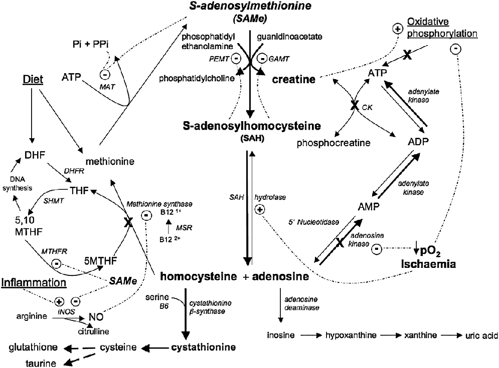
Reducing energy deficiency after retinal ischemia/reperfusion is a rational neuroprotective strategy that targets a central cause of neuronal loss and dysfunction (Osborne, Reference Osborne2008; Schober et al., Reference Schober, Chidlow, Wood and Casson2008). A promising candidate for bioenergetic neuroprotection in the retina is S-adenosyl-l-methionine (SAMe), a reactive form of methionine that is synthesized from methionine and ATP by methionine adenosyltransferase (MAT) predominantly in the liver (MAT I and III) in and central nervous system (MAT II) (Schubert et al., Reference Schubert, Blumenthal and Cheng2003) (Fig. 1). SAMe acts as a methyl donor in more than 60 methyltransferase reactions, as well as a source of aminoisopropyl groups for polyamine synthesis, and 5′-deoxyadenosyl radicals and sulfur atoms for lipoic acid synthesis (Baric, Reference Baric2009). However, in humans, the majority of SAMe is used for phosphatidylcholine (PC) synthesis by phosphatidylethanolamine N-methyltransferase (PEMT), creatine synthesis by guanidinoacetate N-methyltransferase (GAMT), and sarcosine synthesis by glycine N-methytransferase (Mudd et al., Reference Mudd, Brosnan, Brosnan, Jacobs, Stabler, Allen, Vance and Wagner2007; Grillo and Colombatto, Reference Grillo and Colombatto2008).
A simplified scheme of one carbon metabolism in the retina showing the postulated effects of ischemia (X, flux through pathway that is downregulated by ischemia) and the upregulation of pathways by post ischemic administration of SAMe (bold arrows). AMP, adenosine monophosphate; ADP, adenosine diphosphate; ATP, adenosine triphosphate; B6, vitamin B6; B12, vitamin B12; CK, creatine kinase; DHF, dihydrofolate; DHFR, dihydrofolate reductase; GAMT, guanidinoacetate N-methyltransferase; iNOS, inducible nitric oxide synthase; MAT, methionine adenosyltransferase; MSR, methionine synthase reductase; 5MTHF, 5-methyltetrahydrofolate; 5,10 MTHF, 5,10 methylenetetrahydrofolate; MTHFR, 5,10 methylenetetrahydrofolate reductase; NO, nitric oxide; PEMT, phosphatidylethanolamine N-methyltransferase; Pi, inorganic phosphate; PPi, pyrophosphate; SAMe, S-adenosyl-l-methionine; SAH, S-adenosylhomocysteine; SHMT, serine hydroxymethyltransferase; THF, tetrahydrofolate; ---O, enzyme activity or expression is upregulated (+) or downregulated (−).

A number of animal and human studies suggest that SAMe may be neuroprotective in the retina. Kikuchi et al. (Reference Kikuchi, Kashii, Honda, Tamura, Kaneda and Akaike1997) found that administration of SAMe (1 μ m) to rat retinal cell cultures before exposure to high glutamate concentrations decreased neuronal cell death. Furthermore, in vivo studies in human (Silveri et al., Reference Silveri, Parow, Villafuerte, Damico, Goren, Stoll, Cohen and Renshaw2003) and rat (Young & Shalchi, Reference Young and Shalchi2005) demonstrated that SAMe crosses the blood-brain barrier and increases cerebral PCr and ATP concentrations, respectively (Sato et al., Reference Sato, Hariyama and Moriguchi1988; Silveri et al., Reference Silveri, Parow, Villafuerte, Damico, Goren, Stoll, Cohen and Renshaw2003; Glick, Reference Glick2006; de Brouwer et al., Reference de Brouwer, Williams, Duley, van Kuilenburg, Nabuurs, Egmont-Petersen, Lugtenberg, Zoetekouw, Banning, Roeffen, Hamel, Weaving, Ouvrier, Donald, Wevers, Christodoulou and van Bokhoven2007). Moreover, intraperitoneal administration of SAMe during or after transient ischemia in the rat brain has been shown to be neuroprotective and to ameliorate post ischemic energy deficiency (Sato et al., Reference Sato, Nakano, Horita, Takahashi and Moriguchi1987a,Reference Sato, Tobita, Ohtomo, Izumiyama and Kogureb, Reference Sato, Hariyama and Moriguchi1988; Kozuka & Iwata, Reference Kozuka and Iwata1989).
We postulated that administration of SAMe to adult rats following acute retinal ischemia would reduce retinal injury and improve photoreceptor function. SAMe acts as a methyl donor in a number of transmethylation reactions that are vital for photoreceptor function, including the synthesis of creatine. Creatine and creatine kinase (CK) constitute an energy buffering system that modulates the rate of oxidative phosphorylation and facilitates the temporal and spatial distribution of ATP in neurons (Monge et al., Reference Baric2008). Photoreceptors contain cytosolic, brain type (BB-CK), and ubiquitous mitochondrial (μMiCK) CK isoenzymes, as well as high concentrations of phosphocreatine (PCr) and creatine (Wallimann et al., Reference Wallimann, Wegmann, Moser, Huber and Eppenberger1986; Wegmann et al., Reference Wegmann, Huber, Zanolla, Eppenberger and Wallimann1991).
Post ischemic mitochondrial dysfunction and concomitant inhibition of CK isoenzymes caused by ROS and RNS could impair photoreceptor function by limiting the supply of ATP (Stachowiak et al., Reference Stachowiak, Dolder, Wallimann and Richter1998; Schlattner et al., Reference Schlattner, Tokarska-Schlattner and Wallimann2006). Increasing retinal creatine after ischemia may afford neuroprotection because creatine not only directly scavenges ROS and RNS but also binds to CK isoenzymes and protects their active sites from free radical attack and subsequent inhibition (Lawler et al., Reference Lawler, Barnes, Wu, Song and Demaree2002; Sestili et al., Reference Sestili, Martinelli, Bravi, Piccoli, Curci, Battistelli, Falcieri, Agostini, Gioacchini and Stocchi2006). However, it is likely that intracellular creatine concentrations in retinal neurons decrease after ischemia/reperfusion due to a number of factors. The first is down-regulation of the activity and/or expression of the creatine transporter, SLC6-A8. Creatine is not synthesized in retinal neurons but is transported into the retina and neurons by the creatine transporter (CRT-1) (Nakashima et al., Reference Nakashima, Tomi, Katayama, Tachikawa, Watanabe, Terasaki and Hosoya2004; Acosta et al., Reference Acosta, Kalloniatis and Christie2005). Creatine transport occurs against the creatine concentration gradient and is driven by Na+ and Cl− ion gradients across the plasma membrane. Disruption of these ion gradients during ischemia/reperfusion may impair creatine transport into neurons, as well as creatine transport into the retina from the inner vascular and choroidal circulation (Perez-Sala et al., Reference Perez-Sala, Tan, Canada and Rando1991, Reference Perez-Sala, Gilbert, Tan and Rando1992; Wyss & Kaddurah-Daouk, Reference Wyss and Kaddurah-Daouk2000). CRT-1 expression may also be altered after ischemia (Strutz-Seebohm et al., Reference Strutz-Seebohm, Shojaiefard, Christie, Tavare, Seebohm and Lang2007).
The second factor that could adversely affect retinal creatine concentrations after ischemia is inhibition of creatine synthesis in Müller cells. Müller cells express GAMT that catalyses the transfer of a methyl group from SAMe to guanidinoacetate to yield S-adenosylhomocysteine (SAH) and creatine, respectively (Nakashima et al., Reference Nakashima, Tomi, Tachikawa, Watanabe, Terasaki and Hosoya2005) (Fig. 1). Methyltransferases such as GAMT are sensitive to product inhibition by SAH, which is normally maintained at low concentrations by SAH hydrolase (Kloor et al., Reference Kloor, Delabar, Muhlbauer, Luippold and Osswald2002). However, hydrolysis of SAH is reversible, and its equilibrium favors the synthesis of SAH from adenosine and homocysteine. Under normoxic conditions, hydrolysis of SAH prevails because adenosine and homocysteine are maintained at low concentrations. However, during ischemia/reperfusion, low oxygen tension inhibits adenosine kinase resulting in the elevation of intracellular adenosine and inhibition of SAH hydrolysis (Kloor & Osswald, Reference Kloor and Osswald2004; Hermes et al., Reference Hermes, Osswald and Kloor2007). A concomitant increase in homocysteine concentrations due to inhibition of methionine synthase by increased levels of nitric oxide and hydrogen peroxide would favor the synthesis of SAH (Persa et al., Reference Persa, Pierce, Ma, Kabil and Lou2004). Low methionine and ATP concentrations during ischemia/reperfusion may also decrease the rate of SAMe synthesis by MAT (Fig. 1). Thus, ischemia/reperfusion could decrease the methylation potential (SAMe:SAH ratio) and the rate of creatine synthesis (Finkelstein, Reference Finkelstein2007; Kharbanda, Reference Kharbanda2007).
A decrease in the methylation potential may not only inhibit creatine synthesis but also decrease the activity of other SAMe-dependent methylases that are important for photoreceptor function. For example, efficient phototransduction in the outer segment is dependent on methylation of the Tγ subunit of transducin catalyzed by isoprenylcysteine carboxylmethyltransferase (Perez-Sala et al., Reference Perez-Sala, Tan, Canada and Rando1991, Reference Perez-Sala, Gilbert, Tan and Rando1992). PEMT in photoreceptors also utilizes SAMe to synthesize PC, which is an essential component of disc membranes in the outer segments (Roque & Giusto, Reference Roque and Giusto1995; Lamb & Pugh, Reference Lamb and Pugh2004). Trovarelli et al. (Reference Trovarelli, De Medio, Porcellati, Stramentinoli and Porcellati1983) demonstrated that depletion of PC in the brain after cerebral ischemia could be reversed by post ischemic adminstration of SAMe, suggesting that PEMT activity was compromised by decreased SAMe concentrations.
Maintenance of retinal SAMe concentrations after ischemia is also important for ensuring adequate glutathione and taurine synthesis (Persa et al., Reference Persa, Pierce, Ma, Kabil and Lou2004; Chen et al., Reference Chen, Zhang, Wang, Liu, Mi, Xu, Chen and Zeng2009). Both these antioxidants can be synthesized in the retina from cysteine, which is derived from homocysteine via the transsulfuration pathway. SAMe increases the flux of homocysteine down the transsulfuration pathway by allosteric activation of cystathionine β-synthase (Persa et al., Reference Chan, Tchantchou, Rogers and Shea2006). Winkler (Reference Winkler2008) has proposed that the negligible concentrations of glutathione in outer photoreceptor segments may provide insufficient protection from free radical injury, therefore necessitating their constant renewal. Post ischemic SAMe administration may not only help to maintain lipid synthesis for outer segment renewal but also has the potential to reduce oxidative stress and lipid peroxidation in photoreceptors by increasing retinal glutathione and taurine synthesis (De La Cruz et al., Reference De La Cruz, Villalobos, Cuerda, Guerrero, Gonzalez-Correa and Sanchez De La Cuesta2002).
Despite the central role played by SAMe-dependent methyltransferases in photoreceptor function, few studies have considered the effect of ischemia/reperfusion on methyltransferase activity and its ramifications for the pathogenesis of ischemic retinal diseases. We postulated that administration of SAMe to adult rats after acute retinal ischemia would reduce retinal injury and improve photoreceptor function. We also assessed whether CRT-1 expression in the retina is decreased after acute ischemia because any increase in retinal creatine afforded by SAMe administration is unlikely to be neuroprotective if creatine cannot be transported into retinal neurons.
Materials and methods
Animals
Forty-eight adult female Dark Agouti rats (150–180 g), obtained from the Animal Resources Centre (Perth, Australia), were housed in a temperature-controlled animal house and maintained in a 12-h light/12-h dark cycle (light on at 6.00 am). Standard rat chow and water were available ad libitum. The rats were divided into four treatment groups: control (no ischemia, vehicle only, n = 12), SAMe (no ischemia + SAMe supplementation, n = 12), ischemia (ischemia + vehicle, n = 12), and ischemia + SAMe (n = 12). All experiments were conducted in accordance with the principles regarding the care and use of animals adopted by the American Physiological Society and the Society for Neuroscience, the Australian Code of Practice for the Care and Use of Animals for Scientific Purposes, and with approval of the University of Queensland Animal Ethics Committee.
Anesthesia
Rats were anesthetized with an intraperitoneal injection of 75 mg/kg ketamine, 15 mg/kg xylazine, and 2 mg/kg acepromazine. The animals were placed on a homeothermic electric heating mat to maintain body temperature at 37°C at all times under anesthesia.
Induction of acute retinal ischemia
Unilateral retinal ischemia was induced as previously described (Holcombe et al., Reference Holcombe, Lengefeld, Gole and Barnett2008). Briefly, the rat’s head was immobilized by resting the front teeth over a horizontal stabilizing bar and securing the skull with adjustable rods inserted in the bony external ear canal. The anterior chamber of the left eye was cannulated with a 30-G needle attached to a reservoir containing 0.9% saline. A micromanipulator was used to insert the needle vertically into the anterior chamber at the 12 o’clock position. Ischemia was obtained by raising the reservoir to elevate intraocular pressure to 110 mm Hg for 60 min. Corneal hydration was maintained with carmellose sodium (10 mg/ml, Celluvisc; Allergan Irvine, CA). Intraocular pressure was confirmed with a TonoLab rodent tonometer (Colonial Medical Supply Inc., Franconia, NH) factory calibrated for rats.
Post ischemic SAMe supplementation
Immediately after ischemia, 2 μl SAMe (S-(5′-adenosyl)-l-methionine p-toluenesulfonate salt; Sigma-Aldrich, Sydney, Australia), dissolved in isotonic phosphate-buffered saline, pH 7.4, was injected intravitreally with a Hamilton syringe to give a final vitreous concentration of approximately 100 μ m. During the reperfusion phase, daily oral SAMe was administered by oral gavage of 0.33 MoodLift tablet (Naturopathica, Sydney, Australia) dissolved in distilled water containing SAMe (69 mg), folic acid (32 μg), vitamin B6 (3 mg), and vitamin B12 (17 μg) for 5 or 10 days after the intraocular injection (n = 6 for each time point). Folic acid and vitamins B6 and B12 were concurrently administered to help maintain low concentrations of homocysteine and S-adenosylhomocysteine (Finkelstein, Reference Finkelstein2007; Mudd et al., Reference Mudd, Brosnan, Brosnan, Jacobs, Stabler, Allen, Vance and Wagner2007). A similar protocol was followed for non-ischemic animals to assess the effects of SAMe supplementation on the non-ischemic retina.
Electroretinography
Full-field (Ganzfeld) ERGs were recorded preischemia and at 5 and 10 days of reperfusion following the acute ischemic insult. Animals were dark adapted overnight, and all procedures were executed under dim red light (λ max = 650 nm). The pupils were dilated with 0.5% tropicamide drops (Mydriacyl; Alcon Laboratories, Fort Worth, TX), and the corneas were kept moist with carmellose sodium (10 mg/ml, Celluvisc, Allergan). Responses were amplified, recorded and averaged with a bioamplifier/analogue to digital converter (Powerlab/4ST; ADInstruments, Colorado Springs, CO), band-pass filtered between 0.3 and 1000 Hz, and digitized at 4 kHz. A single- or twin-flash paradigm was used to elicit mixed (rod and cone) or isolated cone responses, respectively (Nixon et al., Reference Nixon, Bui, Armitage and Vingrys2001). Flash stimuli for mixed responses were provided by a photographic flash unit (Mecablitz 60CT4, Metz, Zirndorf, Germany), over a stimulus intensity range of −4.0 to 3.0 log cd·s·m−2. Interstimulus interval was increased from 2 s for the lowest intensities to 5 min for the highest intensities to allow complete recovery of the b-wave between stimuli. Isolated cone responses were obtained over the same intensity range following a rod-saturating stimulus of 2.1 log cd·s·m−2 given 400 ms before the test stimulus (Friedburg et al., Reference Friedburg, Thomas and Lamb2001; Nixon et al., Reference Nixon, Bui, Armitage and Vingrys2001; Peachey & Ball, Reference Peachey and Ball2003). This short interval after a rod-saturating flash does not allow recovery of rod function, thereby revealing cone-only responses (Birch et al., Reference Birch, Hood, Nusinowitz and Pepperberg1995). Isolated rod responses were obtained by digital subtraction of the cone response from the mixed response at each stimulus intensity (Sun et al., Reference Sun, Bui, Vingrys and Kalloniatis2007a).
The photoreceptoral response (PIII)
The photoreceptoral response (PIII) was modeled from the leading edge of the ERG a-wave using the equation proposed by Hood and Birch (Reference Hood and Birch1990, Reference Hood and Birch1993).

This model determines the PIII for a given stimulus intensity (i, log cd·s·m−2) at time t (s) after the flash by modeling three parameters: a saturated PIII amplitude (RmP3, μV), a sensitivity parameter (S, cd−1·m2·s−3), and a fixed time delay (t d, s) that accounts for delays in the phototransduction cascade and the recording system. PIII modeling was achieved by floating RmP3 and S with a t d fixed to the average of control data (t d=3.3 s) using the Solver function in Excel to minimize the sum of squares for collected data between the t d and the a-wave minimum or to a maximum of 18 ms against the model.
The postreceptoral response (PII)
Hood and Birch (Reference Hood and Birch1992) proposed that the b-wave of the electroretinogram underestimates the postreceptoral response. While the fast component of the PIII waveform decays shortly after the onset of the PII, another receptorally derived component maintains a relatively constant negative influence during the duration of the PII (Green & Kapousta-Bruneau, Reference Green and Kapousta-Bruneau1999; Kapousta-Bruneau, Reference Kapousta-Bruneau1999; Kang Derwent & Linsenmeier, Reference Kang Derwent and Linsenmeier2001). Consequently, to estimate bipolar response, the modeled PIII was subtracted from the raw waveform. To avoid interference of oscillatory potentials (OPs), the PII was low-pass filtered with a sliding 7 ms average. This successfully attenuated the oscillatory potential responses, prevented their intrusion into the PII, and had minimal effect on the PII amplitude.
The oscillatory potentials
The amplitudes of the inner retinal–derived OPs, which are superimposed on the rising phase of the b-wave, were calculated as previously described (Bui et al., Reference Bui, Armitage and Vingrys2002; Sun et al., Reference Sun, Bui, Vingrys and Kalloniatis2007a). Briefly, following digital subtraction of the modeled PIII and PII from the raw waveform, the signal was filtered between 70 and 280 Hz and modeled with a Gabor function, which is the product of a Gaussian envelope and a sine wave. The maximum amplitude of the Gaussian envelope was analyzed to determine OP amplitude. This method of OP analysis has been shown to be a simple quantitative model that is able to describe differences in OPs in normal and abnormal states (Bui et al., Reference Bui, Armitage and Vingrys2002, Reference Bui, Kalloniatis and Vingrys2004; Phipps et al., Reference Phipps, Fletcher and Vingrys2004).
Statistical analysis
Following confirmation of normal distribution, a one-way analysis of variance (ANOVA) with a Bonferroni post test was used to compare all experimental groups for each ERG parameter (RmP3, log S, rod PII, cone PII, and OPs). P < 0.05 was considered significant for all tests.
Immunohistochemistry and histology
The distribution and expression of creatine transporter (CRT-1) in the control and post ischemic rat retina were assessed by fluorescence immunohistochemistry using standard techniques. Retinas were isolated from control and experimental rats after 5 or 10 days of reperfusion and immersion fixed in 4% paraformaldehyde for 1 h. Following cryoprotection, 10-μm frozen sections were incubated overnight at 4°C with a polyclonal rabbit anti-CRT-1 antibody, 1:300 dilution, previously characterized as specific for CRT-1 by western blot (Dodd & Christie, Reference Dodd and Christie2001) (kindly donated by David Christie, University of Auckland, New Zealand). Primary antibody binding was detected using a biotinylated anti-rabbit antibody (Amersham, Castle Hill, Australia) diluted 1:500, followed by a streptavidin-Cy2 fluoroprobe (Amersham) diluted 1:300. Semithin, resin-embedded, sections of retina obtained approximately 2 mm superior to the optic disc were also stained with toluidine blue to assess gross morphological changes in response to ischemia and SAMe supplementation. Digital images were obtained with a Spot RT digital camera mounted on a Zeiss Axioscope microscope (Zeiss, Oberkochen, Germany). Images were imported into Adobe Photoshop CS3 for minor editing of contrast and sharpness.
Results
Histology
Retinal histology was examined after 5 or 10 days of daily SAMe administration obtained from control or post ischemic eyes.
Neither 5 days (Fig. 2C) nor 10 days (Fig. 2D) of SAMe supplementation affected the normal morphological appearance of the retina, which was indistinguishable from that obtained from control, non-SAMe-treated animals (Fig. 2A and 2B). However, an acute ischemic insult followed by reperfusion-induced morphological changes as previously described (Barnett & Grozdanic, Reference Barnett and Grozdanic2004). After 5 days, disruption of the laminar structure was evident, as were vacuoles in the plexiform layers, swelling of the inner plexiform layer, and cellular disruption in the outer nuclear and ganglion cell layers (Fig. 2E). Following 10 days of reperfusion (Fig. 2F), the histological damage was more pronounced with severe disruption to the laminar organization of the inner and outer retina. Furthermore, infiltration of macrophages was also apparent. SAMe supplementation for 5 days after the ischemic insult (Fig. 2G) partially ameliorated the post ischemic damage when compared to ischemic retinas obtained from animals without SAMe (Fig. 2E) and examined at the same time point. However, some retinal damage and disruption remained. Similarly, retinal morphology was better preserved in SAMe-treated animals 10 days after the ischemic insult (Fig. 2H) than in non-SAMe-treated animals (Fig. 2F). Although evidence of cell death and dropout was present, and there was disruption to the outer nuclear layer morphology, post ischemic SAMe administration clearly reduced the severity of the ischemic injury to the retina.
Semithin (0.5 μm) sections of rat retina stained with toluidine blue showing the effects of ischemia/reperfusion and SAMe supplementation on retinal histology. (A and B) Control and non-ischemic retinas. (C) No apparent effect of SAMe supplementation alone for 5 days or (D) 10 days. (E) 5 days of reperfusion following an acute ischemic insult led to disruption of retinal morphology, including swelling of the inner and outer plexiform layers. (F) Severe disruption of the inner and outer retina, plus infiltration of macrophages 10 days after an acute ischemic insult. (G) SAMe supplementation appeared to reduce the severity of the ischemic injury at 5 days and (H) 10 days. ONL, outer nuclear layer; INL, inner nuclear layer; IPL, inner plexiform layer; GCL, ganglion cell layer. Scale bar = 40 μm.

Electroretinography: Effect of ischemia/reperfusion
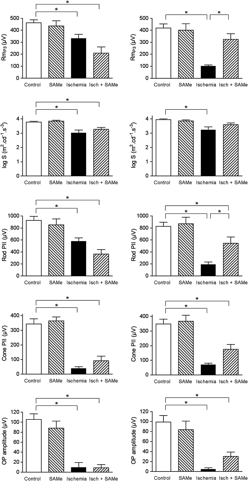
Fig. 3 shows the effects of an acute ischemic insult followed by 5 days (left column) or 10 days of reperfusion (right column) on isolated parameters of the ERG.
The effect of ischemia/reperfusion and SAMe supplementation on isolated and modeled parameters of the full-field electroretinogram (ERG). Left column: 5 days posttreatment. Right column: 10 days posttreatment. SAMe supplementation alone did not affect any parameter of the ERG. An acute ischemic insult followed by 5 days reperfusion caused a significant decrease in the maximum rod photoreceptoral response (RmP3), the gain parameter (S), the rod-driven bipolar response (rod PII), the cone-driven bipolar response (cone PII), and the OPs (OP amplitude). These responses were also significantly suppressed after 10 days. SAMe supplementation for 5 days after the ischemic insult did not significantly improve any ERG parameter. However, SAMe supplementation for 10 days postinsult resulted in a significant improvement in the RmP3 and rod PII responses. *P < 0.05, one-way ANOVA followed by Bonferroni’s multiple comparison test.

Five days after the ischemic insult, there was a small but significant (P < 0.05) decrease in photoreceptoral RmP3 from 462 ± 25 to 332 ± 35 μV. Post ischemic RmP3 dropped to 101 ± 10 μV after 10 days of reperfusion. A small but significant reduction in the photoreceptor sensitivity parameter (log S) was apparent at 5 days (from 3.78 ± 0.01 to 3.02 ± 0.19 m2·cd−1·s−3) and 10 days (from 3.95 ± 0.05 to 3.23 ± 0.22 m2·cd−1·s−3). Inner retinal responses were also suppressed by the acute ischemic insult. The rod bipolar cell PII response, which was moderately suppressed at 5 days (from 927 ± 70 to 579 ± 56 μV, P < 0.05), was greatly reduced 10 days after the insult (192 ± 39 μV). The cone PII was affected to a much greater degree than the rod PII response at 5 days. The ischemic insult induced a significant reduction in the cone bipolar cell response from 342 ± 36 to 40 ± 11 μV after 5 days and from 348 ± 34 to 70 ± 10 μV after 10 days of reperfusion. Ischemia and reperfusion caused an almost total loss of OPs. At 5 days, the OP amplitude was reduced from 106 ± 11 to 10 ± 10 μV, and at 10 days, the OP amplitude fell to only 5 ± 3 μV (P < 0.05).
Electroretinography: Effect of SAMe
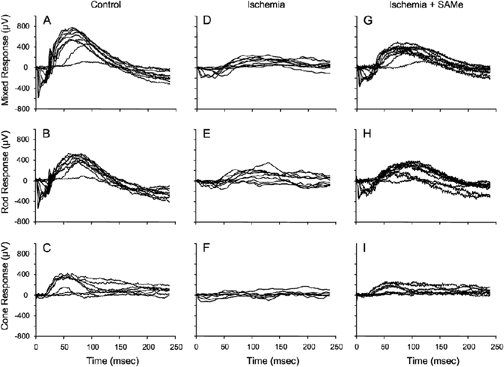
SAMe administration for 5 or 10 days did not affect the ERGs recorded from non-ischemic retinas. No significant difference in any of the analyzed parameters (RmP3, S, rod PII, cone PII, and OPs) was observed between SAMe-treated and non-SAMe-treated control retinas (Fig. 3). Similarly, SAMe supplementation did not alter any ERG parameter recorded 5 days after the acute ischemic insult. The apparent differences in RmP3, rod PII, and cone PII between the ischemia without SAMe group and ischemia with SAMe supplementation group at 5 days (Fig. 3 left column) were not statistically significant. However, SAMe supplementation for 10 days after the ischemic insult significantly restored photoreceptor function (Figs. 3 and 4).
Full-field electroretinogram (ERG) responses recorded over the stimulus range of −4 to +3 cd·s·m−2 from control eyes (A, B, and C), 10 days after an acute ischemic insult (D, E, and F), and 10 days postinsult with SAMe supplementation (G, H, and I). Rod responses (B, E, and H) were isolated from the mixed (rod and cone) responses (A, D, and G) by subtraction of the isolated cone responses (C, F, and I). The suppression of the ERG by ischemia was partially ameliorated by SAMe supplementation.

RmP3 recovered to 325 ± 46 μV in SAMe-treated rats compared to 101 ± 10 μV in non-SAMe-treated animals (P < 0.05). The restoration of RmP3 by SAMe was of such magnitude that SAMe-treated ischemic RmP3 values were no longer significantly lower than non-ischemic RmP3 values (P > 0.05). SAMe treatment for 10 days also significantly improved the post ischemic rod PII response, from 192 ± 39 to 546 ± 105 μV (P < 0.05). The apparent improvement in cone PII responses and OPs with SAMe supplementation (Fig. 3, right column) was not statistically significant (P > 0.05).
Creatine transporter localization
In the control non-ischemic rat retina, the localization and expression of CRT-1 were as described previously (Acosta et al., Reference Acosta, Kalloniatis and Christie2005).
Intense CRT-1 labeling of photoreceptor inner segments and perikarya in the outer retina was apparent (Fig. 5A). Ganglion cell bodies were strongly labeled, as were their dendrites ramifying through the inner plexiform layer and axon bundles in the nerve fiber layer. Weaker labeling of cells in the inner nuclear layer, including amacrine cells, was evident. CRT-1 immunoreactivity was not observed in Müller cells or astrocytes. Five days after the acute ischemic insult, there was a general decrease in the intensity of CRT-1 labeling throughout the retina (Fig. 5B). However, the localization of the transporter did not appear to be altered. The strongest labeling remained in the photoreceptor inner segments, although the intensity of immunofluorescence was greatly reduced when compared to the control retina. Very weak labeling was evident in the inner nuclear and ganglion cell layers. When examined 10 days after the ischemic insult, a patchy recovery in CRT-1 immunolabeling was evident (Fig. 5C). Intense CRT-1 labeling was observed in areas across the outer retina, with photoreceptor labeling in these patches comparable to that of control retinas. The labeling of ganglion cell dendrites and axons was also more pronounced in retinas observed after 10 days of reperfusion than in the 5-day recovery group. Weak labeling in the inner nuclear layer was comparable to that seen in control tissue. Fig. 5C shows immunofluorescence in blood vessels in the ganglion cell layer. This localization was also seen in control retinas.
Fluorescence micrographs of creatine transporter immunolabeling in the adult rat retina. (A) In the control retina, creatine transporter was localized to photoreceptor (PR) cell bodies, inner segments (IS), and ganglion cells (GC) and their dendrites in the inner plexiform layer. Weak immunolabeling was also evident in amacrine cells (Am). (B) 5 days after an acute ischemic insult, creatine transporter immunoreactivity was reduced throughout the retina. Weak labeling remained in the outer retina. (C) 10 days after the ischemic insult, intense creatine transporter immunoreactivity was again evident in the PRs. Ganglion cell dendrites and blood vessels were also stained in the inner retina. ONL, outer nuclear layer; INL, inner nuclear layer; IPL, inner plexiform layer; GCL, ganglion cell layer. Scale bar = 40 μm.

Discussion
Five days after an acute ischemic insult, retinal histology showed a disruption of photoreceptor perikarya; an apparent reduction in the length of photoreceptor inner and outer segments; loss of cells in the inner nuclear layer (INL); and swelling in the inner plexiform layer (IPL), outer plexiform layer (OPL), and outer nuclear layer (ONL)(Fig 2E). Infiltration of macrophages was also apparent and is consistent with post ischemic vascular edema and breakdown of the blood-retinal barrier. These morphological changes were more pronounced 10 days after ischemia, indicating progressive retinal injury. The ERG responses mirrored these histological changes. The photoreceptor PIII response amplitude (RmP3) and sensitivity parameter (log S) were significantly reduced 5 days after ischemia. There was a further marked reduction in RmP3 by day 10, most likely caused by a loss of photoreceptors, a greater number of photoreceptors with shortened or disorganized outer segments (Fig. 2F), and possibly a decrease in the number of cyclic guanosine monophosphate (cGMP) channels gated off in response to phototransduction in the outer segment (Nusinowitz et al., Reference Nusinowitz, Nguyen, Radu, Kashani, Farber and Danciger2003). In contrast, log S did not change markedly between day 5 and day 10, consistent with a depressed but stable rate of phototransduction. This suggests that methyltransferases involved in the methylation of transducin and other G proteins in rod outer segments (Perez-Sala et al., Reference Perez-Sala, Tan, Canada and Rando1991) may be inhibited after ischemia, but their inhibition does not play a major role in escalating photoreceptor dysfunction.
Post ischemic administration of SAMe for 5 or 10 days reduced the loss of photoreceptors and helped to maintain their structural integrity. Retinal edema and macrophage infiltration was also markedly reduced and the laminar structure of the retina better preserved. The protection of the outer photoreceptor structure afforded by SAMe treatment may be partly due to the maintenance of PC synthesis by PEMT localized in disc membranes. There is a high turnover of disc membranes in rod outer segments, and PEMT is essential for supplying PC rich in long-chain polyunsaturated fatty acids for new membrane synthesis (Roque & Giusto, Reference Roque and Giusto1995; Castagnet et al., Reference Castagnet, Roque, Pasquare and Giusto1998). The lipid milieu of the disc and plasma membranes in rod outer segments also modulates rhodopsin’s behavior and the rate of phototransduction (Sangiovanni & Chew, Reference Sangiovanni and Chew2005; Wensel, Reference Wensel2008). PEMT is the only biochemical pathway for PC synthesis in outer photoreceptor segments, but it is sensitive to inhibition by SAH (Roque & Giusto, Reference Roque and Giusto1995; Tan & Guiloff, Reference Tan and Guiloff1998; Kharbanda, Reference Kharbanda2007; Mudd et al., Reference Mudd, Brosnan, Brosnan, Jacobs, Stabler, Allen, Vance and Wagner2007) (Fig. 1). The post ischemic elevation of SAH could downregulate flux through the PEMT pathway, significantly altering the lipid milieu of disc membranes and reducing the rate of disc renewal (Tan & Guiloff, Reference Tan and Guiloff1998; Danishpajooh et al., Reference Danishpajooh, Gudi, Chen, Kharitonov, Sharma and Boss2001).
Decreased recycling of phospholipids for disc renewal may have also contributed to photoreceptor dysfunction following ischemia. Phospholipids derived from the breakdown of disc membranes in the retinal pigmented epithelium (RPE) are normally transported to the photoreceptor inner segments where they are catabolized and reutilized for synthesis of new disc membranes. However, high concentrations of polyunsaturated fatty acids and photosensitizing compounds in photoreceptor disc membranes, coupled with negligible glutathione concentrations and elevated ROS and RNS after ischemia, could increase the rate of phospholipid oxidation in disc membranes (Sangiovanni & Chew, Reference Sangiovanni and Chew2005; Winkler, Reference Winkler2008). It is likely that photoreceptor discs containing oxidized phopholipids are not completely digested in RPE phagosomes, resulting in the deposition of oxidized PC in intracellular lipofuscin and in debris exocytosed from the RPE (Suzuki et al., Reference Suzuki, Kamei, Itabe, Yoneda, Bando, Kume and Tano2007). This would not only reduce the amount of phospholipid available for recycling into new discs but may also lead to excessive lipofuscin deposition in RPE, RPE dysfunction, and subsequent photoreceptor cell death (Suzuki et al., Reference Suzuki, Kamei, Itabe, Yoneda, Bando, Kume and Tano2007). Thus, aberrant phospholipid metabolism may be an important etiological factor in photoreceptor loss, decreased outer segment length, and changes in membrane lipid composition underlying the decrease in the sensitivity and amplitude of the ERG a-wave observed after acute ischemia.
Aberrant phospholipid metabolism and excessive lipofuscin deposition in RPE also occur in response to chronic oxidative stress, an etiological factor in age-related atrophic macular degeneration characterized by RPE and photoreceptor loss. Elevated homocysteine is a risk factor for arterial disease, retinal artery and vein occlusions, and macular degeneration (Wright et al., Reference Wright, Martin and Dodson2008). The importance of maintaining low homocysteine concentrations for retinal health has recently been demonstrated by a large human trial in which long-term administration of folate, vitamin B12, and pyridoxine decreased the incidence of macular degeneration in women with existing or increased risk of cardiovascular disease (Christen et al., Reference Christen, Glynn, Chew, Albert and Manson2009). In our experiments, SAMe was administered together with folic acid, cyanocobalamin, and pyridoxine to optimize the remethylation and transsulfuration of homocysteine generated by PEMT and other methyltransferases. The fact that SAMe treatment had no effect on photoreceptor function or morphology in the absence of ischemia and significantly reduced morphological injury and loss of photoreceptors after acute ischemic injury suggests that co-administration of these vitamins prevented deleterious elevation of homocysteine in the retina.
Although post ischemic SAMe administration for 5 days improved retinal morphology, photoreceptor function was still significantly depressed compared to non-ischemic controls. There was also no improvement in RmP3 and log S values compared to the ischemic non-SAMe-treated group. However, treatment with SAMe for 10 days significantly increased RmP3 values compared to the ischemic non-SAMe-treated group with RmP3 and log S increasing to levels approaching non-ischemic control values. This dramatic restoration of photoreceptor function coincided with an apparent increase in CRT-1 expression 10 days after ischemia (Fig. 5). The expression of CRT-1 in control non-ischemic retinas was highest in photoreceptor inner segments, in photoreceptor perikarya and cone pedicles, and in ganglion cells. Immunolabeling for CRT-1 in these areas was clearly reduced 5 days after ischemia but showed partial recovery on day 10. This recovery in CRT-1 expression and presumably creatine transporter activity may underpin the simultaneous improvement in photoreceptor function. Further studies are required to examine the temporal changes of CRT-1 expression in perivascular astrocytes, inner retinal blood vessels, and RPE in order to determine how creatine uptake from the inner vascular and choroidal circulation is affected by ischemia. This will be important for determining if and when creatine supplementation should be administered after acute retinal ischemia (Osborne, Reference Osborne2008). However, one of the presumed advantages of SAMe administration over creatine administration is that SAMe increases creatine synthesis directly in the retina as well as in other organs, such as the liver and kidney. Accordingly, the recovery of neuronal CRT-1 expression allows SAMe-facilitated creatine synthesized in Müller cells to be transported into the neurons. This elevation of neuronal creatine by SAMe would occur despite any possible post ischemic disruption of blood-retinal creatine transport, which could prevent exogenously administered creatine from reaching the retina.
The simultaneous increase in RmP3 and CRT-1 expression in SAMe-treated rats underlines the importance of the creatine kinase buffering system in photoreceptor function. Retinal dysfunction is more likely to occur in the dark when retinal energy demand by inner segment plasma membrane Na+–K+ ATPases is at its highest (Okawa et al., Reference Okawa, Sampath, Laughlin and Fain2008). The supply of ATP to these ion pumps is facilitated by close coupling of BB-CK to Na+–K+ ATPases and the close proximity of inner segment mitochondria (Hemmer et al., Reference Hemmer, Riesinger, Wallimann, Eppenberger and Quest1993). Disruption of energy supply to inner segment Na+–K+ ATPases caused by mitochondrial dysfunction, inhibition of CK isoenzymes, and/or decreased intracellular creatine concentrations may lead to increased intracellular Na+ concentrations in the outer segment discs. Consequent down-regulation of outer segment Na+/Ca2+–K+ exchanger activity would lead to hyperelevation of cytosolic Ca2+ concentrations during the dark and prolonged elevation of Ca2+ concentrations following exposure to light. Subsequent inhibition of guanylyl cyclase activity would lead to slower or inadequate restoration of cGMP levels after phototransduction, thereby increasing the number of cGMP-gated channels in the closed configuration, decreasing the circulating dark current, and reducing glutamate release from photoreceptors. This impairment of photoreceptor function may be advantageous for the long-term survival of photoreceptors because it would limit the flux of Ca2+ through voltage gated channels into rod synaptic terminals in the dark. Consequently, the energy required for Ca2+ efflux by plasma membrane Ca2+ ATPases in photoreceptor terminals would be reduced, thus decreasing the likelihood of energy failure, Ca2+ overload, and MPT in rod spherule mitochondria. Post ischemic mitochondrial dysfunction and energy deficiency in photoreceptor synaptic terminals could also decrease glutamate uptake into synaptic vesicles and the rate of vesicle priming. Secondary energy deficiency in the photoreceptors may therefore decrease glutamate release by photoreceptors in the dark, resulting in increased post ischemic cation channel permeability in rod ON bipolar cells, as reported by Sun et al. (Reference Sun, Bui, Vingrys and Kalloniatis2007a,Reference Sun, Vingrys and Kalloniatisb).
The percentage decrease in the amplitude of the rod ON bipolar cell response (PII) was similar to that of the photoreceptors (RmP3) at both 5 and 10 days after the ischemic insult (Fig. 3). Photoreceptor loss and dysfunction are therefore likely to be the major cause of the depressed post ischemic rod bipolar PII response. The much larger decrease in the cone ON bipolar response 5 days after ischemia may have been due to greater cone loss or dysfunction as a result of their higher energy requirements for phototransduction and signaling compared to that of rods (Okawa et al., Reference Okawa, Sampath, Laughlin and Fain2008; Warrant, Reference Warrant2009). Although ischemic cone PII and amacrine OP amplitudes were less than 15% of non-ischemic control values 5 days after ischemia, both responses improved markedly after 10 days of SAMe treatment (Figs. 3 and 4). This demonstrates that many of these cells were still viable and that improved energy metabolism due to SAMe treatment and recovery of CRT-1 expression in photoreceptors may have contributed to their functional recovery.
The significant recovery of RmP3 and rod PII values coincident with increased CRT-I expression demonstrates that the CK buffering system is a key regulator of photoreceptor energy homeostasis and viability. Disruption of plasma membrane ion gradients during ischemia/reperfusion may impair creatine transport into neurons and/or allow diffusion of creatine out of neurons down the concentration gradient into the extracellular fluid. Creatine also acts as an intracellular osmolyte and may be released from neurons in response to cytotoxic edema that occurs as a result of anoxic depolarization of neurons and subsequent energy failure (Rumpel et al., Reference Rumpel, Lim, Chang, Chan, Ho, Wong and Tan2003; Moxon-Lester et al., Reference Moxon-Lester, Sinclair, Burke, Cowin, Rose and Colditz2007). Intracellular creatine and PCr also undergo constant irreversible nonenzymatic conversion to creatinine, which diffuses freely out of cells into the bloodstream and is excreted in the urine (Wyss & Kaddurah-Daouk, Reference Wyss and Kaddurah-Daouk2000). All these factors may contribute to the depletion of intracellular creatine in retinal neurons during ischemia/reperfusion. In vivo magnetic resonance spectroscopy studies of the brain have shown decreased creatine concentrations in the ischemic penumbra following stroke (Rumpel et al., Reference Rumpel, Lim, Chang, Chan, Ho, Wong and Tan2003) and in the neonatal brain after perinatal hypoxia–ischemia (Cheong et al., Reference Cheong, Cady, Penrice, Wyatt, Cox and Robertson2006). The recovery of CRT-1 expression in retinal neurons 10 days after ischemia may be triggered by increased expression of serum and glucocorticoid-inducible kinase-1 (SGK-1). Increased transcription of SGK-1 occurs in response to post ischemic osmotic stress, oxidative stress, increased cytosolic Ca2+, and nitric oxide (Nishida et al., Reference Nishida, Nagata, Takahashi, Sugahara-Kobayashi, Murata and Asai2004; Lang et al., Reference Lang, Bohmer, Palmada, Seebohm, Strutz-Seebohm and Vallon2006). SGK-1 activates Na+–K+ ATPases and increases the trafficking of CRT-1 to the plasma membrane, both of which may increase neuronal CRT-1 activity (Strutz-Seebohm et al., Reference Strutz-Seebohm, Shojaiefard, Christie, Tavare, Seebohm and Lang2007).
Restitution of CRT-1 activity and neuronal creatine concentrations may play a vital role in protecting neurons from ROS and RNS. Ischemia/reperfusion increases the expression of inducible nitric oxide synthase (iNOS) in the retina resulting in the elevation of nitric oxide free radicals (Hangai et al., Reference Hangai, Yoshimura, Hiroi, Mandai and Honda1996; Aslan et al., Reference Aslan, Cort and Yucel2008), inhibition of multiple metabolic pathways, including creatine kinase and methionine synthase, and subsequent tissue damage. Accordingly, inhibiting iNOS expression and activity or protecting proteins from modification by ROS and RNS can reduce retinal injury (Sennlaub et al., Reference Sennlaub, Courtois and Goureau2002). Interestingly, SAMe downregulates iNOS expression in the liver and may similarly decrease iNOS expression in glial cells following retinal ischemia and inflammation (Majano et al., Reference Majano, Garcia-Monzon, Garcia-Trevijano, Corrales, Camara, Ortiz, Mato, Avila and Moreno-Otero2001; Saha & Pahan, Reference Saha and Pahan2006).
SAMe may also indirectly protect various proteins from modification by ROS and RNS by stimulating creatine synthesis in Müller cells. The first step in creatine synthesis requires arginine and glycine. Depletion of arginine by creatine synthesis could limit substrate availability for iNOS and also inhibit the translation of iNOS mRNA (Lee et al., Reference Lee, Ryu, Ferrante, Morris and Ratan2003). Increasing creatine synthesis would therefore decrease NO production in Müller cells while increasing neuronal creatine concentrations. Direct scavenging of free radicals by creatine would help protect vulnerable proteins from free radical modification and maintain cellular energy homeostasis (Lawler et al., Reference Lawler, Barnes, Wu, Song and Demaree2002; Sestili et al., Reference Sestili, Martinelli, Bravi, Piccoli, Curci, Battistelli, Falcieri, Agostini, Gioacchini and Stocchi2006). Creatine binding to μMiCK prevents the oxidation of sulfhydryl groups at the active site, thus maintaining the octameric conformation and activity of μMiCK. The conversion of octameric μMiCK to the dimeric form has been implicated in MPT, although the role of CK in the mitochondrial transition pore has recently been questioned (Juhaszova et al., Reference Juhaszova, Wang, Zorov, Nuss, Gleichmann, Mattson and Sollott2008). The binding of creatine and phosphocreatine similarly protects the active site of BB-CK, which is particularly vulnerable to oxidative modification in the outer photoreceptor segments due to very low glutathione concentrations. Glutathione plays an important role in maintaining the energy buffering capacity in neurons because it is capable of reducing S-S bonds in oxidatively modified CK (Winkler, Reference Winkler2008).
The depletion of neuronal glutathione leads to the accumulation of mitochondrially derived ROS and RNS and eventual cell death (Tan et al., Reference Tan, Schubert and Maher2001; Maher & Hanneken, Reference Maher and Hanneken2005). Creatine synthesis in Müller cells may be a key driver of glutathione synthesis because it generates homocysteine that can be either methylated to form methionine or further metabolized to form cysteine. Treatment with SAMe not only facilitates creatine synthesis but also increases the formation of cysteine (Prudova et al., Reference Prudova, Bauman, Braun, Vitvitsky, Lu and Banerjee2006) (Fig. 1). Cysteine combines with glutamate to form γ-glutamylcysteine, a precursor of glutathione. Glutamate transport into Müller cells by GLutamate ASpartate Transporter (GLAST) continues after ischemia (Barnett et al., Reference Barnett, Pow and Bull2001; Barnett & Grozdanic, Reference Barnett and Grozdanic2004) and is essential for glutathione synthesis and the prevention of glutamate excitotoxicity (Harada et al., Reference Harada, Harada, Nakamura, Quah, Okumura, Namekata, Saeki, Aihara, Yoshida, Mitani and Tanaka2007). However, the uptake of cystine via the cystine–glutamate antiporter requires the efflux of glutamate, thereby decreasing glutathione synthesis and exacerbating glutamate excitotoxicity (Pow, Reference Pow2001). Increased intracellular generation of cysteine due to SAMe treatment may be beneficial because glutathione synthesis may be sustained without the need for cystine transport into Müller cells via the antiporter. The last step in glutathione synthesis is the addition of glycine to γ-glutamylcysteine, catalyzed by glutathione synthetase. This enzyme is highly expressed in Müller cells and RPE and is essential for maintaining photoreceptor function and viability (Burstedt et al., Reference Burstedt, Ristoff, Larsson and Wachtmeister2009). Pretreatment of rats with SAMe decreases lipid peroxidation and increases total glutathione production in rat brain slices exposed to oxygen–glucose deprivation (De La Cruz et al., Reference De La Cruz, Villalobos, Cuerda, Guerrero, Gonzalez-Correa and Sanchez De La Cuesta2002). Further studies are needed to verify whether SAMe similarly increases glutathione synthesis in the retina, particularly after ischemic injury.
SAMe may also afford neuroprotection in the retina by supplying exogenous adenosine, which is generated by creatine synthesis in Müller cells. During ischemia, inhibition of adenosine kinase increases intracellular adenosine, which is then further catabolized leading to depletion of the adenine nucleotide pool (Fig. 1). Restitution of normal O2 tension reverses adenosine kinase inhibition allowing adenosine to be salvaged to AMP, which can be further phosphorylated to form ADP and ATP. Increasing intracellular adenosine concentrations by SAMe administration after ischemia may therefore help to allay secondary energy deficiency as demonstrated in previous studies (Sato et al., Reference Sato, Tobita, Ohtomo, Izumiyama and Kogure1987b; Silveri et al., Reference Silveri, Parow, Villafuerte, Damico, Goren, Stoll, Cohen and Renshaw2003; Young & Shalchi, Reference Young and Shalchi2005). SAMe treatment may also increase extracellular adenosine, which is thought to play a major role in alleviating retinal and Müller cell edema so clearly evident in the untreated retinas at 5 and 10 days postischemia (Fig. 2). Müller cells play a pivotal role in clearing water generated by retinal metabolism into inner retinal vasculature (Reichenbach et al., Reference Reichenbach, Wurm, Pannicke, Iandiev, Wiedemann and Bringmann2007; Goodyear et al., Reference Goodyear, Crewther and Junghans2009). Osmotic regulation by Müller cells is markedly improved by activation of adenosine A1 receptors that open K+ and Cl− ion channels in the Müller cell plasma membrane, allowing the extrusion of ions and water from Müller cells into the bloodstream (Reichenbach et al., Reference Reichenbach, Wurm, Pannicke, Iandiev, Wiedemann and Bringmann2007). We postulate that the dramatic improvement in retinal edema we observed after treatment with SAMe may be due to the elevation and subsequent breakdown of intracellular AMP secondary to increased creatine synthesis in Müller cells (Nagase et al., Reference Nagase, Tomi, Tachikawa and Hosoya2006). Triamcinolone acetonide, an anti-inflammatory corticosteroid used to treat macula edema, also reduces Müller cell cytotoxic edema by stimulating adenosine release (Uckermann et al., Reference Uckermann, Kutzera, Wolf, Pannicke, Reichenbach, Wiedemann, Wolf and Bringmann2005).
Given these promising findings, the potential of SAMe as a neuroprotectant in ischemic retinal diseases warrants further investigation, especially as SAMe is already in human use for other indications, such as depression and Alzheimer’s disease (Bottiglieri, Reference Bottiglieri2002; Goren et al., Reference Goren, Stoll, Damico, Sarmiento and Cohen2004; Chan et al., Reference Chan, Tchantchou, Rogers and Shea2009). SAMe is generally a safe drug except when administered to patients with a history of (hypo)mania (Sarris et al., Reference Sarris, Schoendorfer and Kavanagh2009).
Conclusions
In conclusion, postinsult treatment with SAMe together with vitamins B6, B12, and folate reduced neuronal loss and ameliorated retinal degeneration 10 days after acute retinal ischemia. Photoreceptor and rod bipolar cell function was significantly improved by SAMe treatment. The recovery in retinal function coincided with increased expression of CRT-1 in the photoreceptors, amacrine, and ganglion cells, indicating the vital importance of creatine in maintaining neuronal energy homeostasis. We postulate that SAMe may act as a neuroprotectant by increasing phosphatidylcholine synthesis in photoreceptors and creatine synthesis in Müller cells, thereby maintaining photoreceptor structure and function. Further metabolism of adenosine and homocysteine generated by SAMe-dependent creatine synthesis in Müller cells may play an important role in reducing retinal edema and oxidative injury. This study demonstrates that recovery of retinal function after ischemia may be dependent on restitution of energy homeostasis afforded by SAMe treatment and subsequent recovery of the creatine kinase energy buffering capacity in photoreceptors.
Acknowledgments
We thank David Christie, University of Auckland, for the kind donation of the CRT-1 antibody. This work was supported by an Australian National Health & Medical Research Council (NHMRC) grant to N.L.B. L.M.-L. was supported by the Lions Medical Research Foundation.