Introduction
Cryopreservation of human ovarian tissue has successfully resulted in 13 live births (Donnez et al., Reference Donnez, Silber, Andersen, Demeestere, Piver, Merow, Pellicer and Dolmans2011). However, there is a paucity of efficient cryopreservation protocols for primordial follicles enclosed in the ovarian tissue from non-human primates (NHP), in special New World primates. Due to species-specific differences, cryopreservation protocols developed using mice as the model cannot be applied directly in primates (Santos et al., Reference Santos, Amorim, Cecconi, Fassbender, Imhof, Lornage, Paris, Schoenfeldt and Martinez-Madrid2010). As well, protocols developed using human ovarian tissue should be tested in NHP species before application. Therefore, tissue collection from NHP and experimentation is crucial. Ovarian tissue can be collected from animals found dead at zoos or from live animals by laparoscopy. The first option is a simple procedure, but does not depend solely on an efficient network between zoo and research institutions, but on the healthy and reproductive condition of the females. For example, most of the females that die naturally are senile, which means that they have ovaries with a depleted population of primordial follicles. Laparoscopy, although more traumatic, allows a controlled collection of ovarian biopsies from healthy animals within their normal reproductive lifespan. Furthermore, ovarian biopsy may be performed by laparoscopy simultaneously with antral follicles puncture, under ultrasound monitoring (Domingues et al., Reference Domingues, Caldas-Bussiere, Petretski, Ohashi, Lima, Santos, Cordeiro and Gomes de Castro2010). An efficient method to collect ovarian biopsies from Cebus apella (capuchin monkeys), has not been described as yet. Therefore, we have performed ovarian tissue biopsies by modifying a method developed for gingival tissue, the so-called trap door (Edel, Reference Edel1974). Success of the technique was assessed by ultrasound monitoring, as well as morphological and quantitative analysis of primordial follicles.
Materials and methods
Animals
Our study was approved by the local institutional ethics committee (no. 029/2009/CEPAN/IEC/SVS/MS) and the Brazilian Institute for Wildlife and Environment (IBAMA). Five healthy and sexually mature C. apella females (15 to 20 years old, weight range: 1.9 to 2.8 kg) were selected for our study. Their daily diet consisted of fresh fruits and commercial pellet chow (FOXY Junior Supreme, 28% crude protein; Provimi, São José dos Pinhais, Paraná, Brazil). Milk, vitamins, minerals, and eggs were supplied once a week. Tap water was available ad libitum. The females were kept indoors under a natural photoperiod and housed individually in cages (80 × 90 × 80 cm) at the National Primate Center, Ananindeua, Pará, Brazil. All females presented regular reproductive history.
Ultrasound procedures
All procedures were performed under general anaesthesia. Each animal was anesthetized with ketamine hydrochloride (10 mg/kg; IM; Vetanarcol, Köning S.A., Avellaneda, Argentina) and xylazine hydrochloride (1 mg/kg; IM; Kensol, Köning S.A.). Before and after surgeries, the females were submitted to two-dimensional (2D) ultrasound and triplex Doppler examination to evaluate uterus and ovaries dimensions, as well as colour and spectral Doppler of uterine and uterus–ovary–ligament (UOL) arteries (Domingues et al., Reference Domingues, Caldas-Bussiere, Martins and Carvalho2007). To this end, the females were placed in dorsal recumbence. Trans-abdominal 2D ultrasound was used to visualize the uterus, ovaries and ovarian structures. A real-time B-mode diagnostic ultrasound with a multi-frequency (5–8 MHz) microconvex transducer (US/conducted with an HDI 4000 PHILIPS scanner – Philips Medical Systems, Bothell, W.A., USA) was used.
Uterine length was calculated as the distance from the fundus of the uterus to the cervix (long axis of the uterus). The uterine height was perpendicular to this axis (dorsum ventral diameter), measured on the long axis view from anterior to posterior walls. The width (transverse diameter) was measured in the axial view. Similarly, the length, height, and width of both ovaries were measured. All measurements were made by obtaining the maximal size in the appropriate dimension, freezing the image, and using internal callipers. Uterine and ovarian volumes were calculated by the ultrasound system using a formula for an ellipsoid [π/6 (length versus width versus height)].
Uterine and UOL arteries were visualized using colour Doppler ultrasound. The right and left uterine arteries and the UOL arteries were evaluated on the long axis and the axial view, respectively. Blood flow velocity waveforms were obtained by positioning a sample volume to a coloured spot and activating the spectral Doppler; resistance index (RI) and pulsatility index (PI) were also determined. In brief, the RI was calculated as the difference between the maximal systolic and end diastolic blood flow, divided by the value of maximal systolic flow. The RI value was calculated as the difference between the peak systolic shifted frequency and the diastolic frequency, divided by the time-averaged maximum frequency over the entire cardiac cycle.
Biopsy procedures
Ovarian biopsies were collected by exploratory laparoscopy. For this, a ventral midline skin incision was made to reach the ovaries, as described by Domingues et al. (Reference Domingues, Caldas-Bussiere, Martins and Carvalho2007). To obtain the ovarian tissue samples, we have modified a technique, the so-called trap door, previously described to harvest gingival biopsies (Edel, Reference Edel1974). Briefly, we have performed a 1 mm thick circular incision in the surface of the ovarian tissue and an ovarian cortical envelope flap was raised by means of a No. 15 blade (Paramount Surgimed LTDA -New Delhi, Delhi, India). The tissue was then removed, kept in pre-warmed (37°C) 0.9% NaCl solution and subjected to fragmentation of 1 mm × 5 mm × 8 mm ovarian tissue pieces. Suture was unnecessary and bleeding was repressed by gently pressing the sampled area with cold (15°C) 0.9% saline-soaked gauze.
Microscopy procedures
On completion of the procedure, ovarian fragments were subjected to microscopic examination of stromal cells and primordial follicles. Ovarian tissue biopsies were fixed in 2% paraformaldehyde and 2.5% glutaraldehyde in 0.1M sodium cacodylate buffer (pH 7.2) for 3 h. Semi-thin sections of 1 μm thickness were stained with toluidine blue. Primordial follicles were defined as follicles with an oocyte surrounded either by one flattened and/or cuboidal layer of granulosa cells. To avoid counting more than once follicle, primordial follicles were counted only in the sections where their oocyte nucleus was observed. Follicular quality was evaluated based on the morphological integrity of the oocyte, the granulosa cells and the basement membrane as described previously (Santos et al., Reference Santos, Rodrigues, Costa, Silva, Matos, Lucci, Báo, Van den Hurk and Figueiredo2006). Briefly, primordial follicles were classified as morphologically normal when they contained an intact oocyte and intact granulosa cells, or degenerated when their oocyte nucleus has become pycnotic, ooplasm was shrunken or when granulosa cells have detached from the basement membrane.
Results
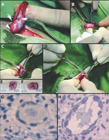
All the procedures were performed within 68 ± 7 min. Biopsy harvesting was uneventful and ovarian biopsy specimens were collected successfully in all animals. Bleeding was minimal and it was sufficiently controlled by slightly pressing the tissue and using cold 0.9% saline solution-soaked gauze. After recovering eight biopsy specimens of 1.0 mm × 5 mm × 8 mm, most of the ovarian surface remained intact. There were no postoperative complications and all animals were recovered within 24 h. The complete procedure is detailed in Figure 1 (A–F).
Ovarian biopsy from a female capuchin monkey (Cebus apella). (A) The macroscopic normal appearance of one ovary presenting functional corpora lutea (see arrows). (B) 1 mm thickness incision in the ovarian cortex. (C) Preparation of the ovarian cortical envelope flap. (D) Biopsy recovery. (E, F) ovarian biopsies 1 mm × 5 mm × 8 mm. (G,H) Light microscopic appearance of morphologically normal (G) and degenerated (H) primordial follicles after toluidine blue staining (×400). A colour version of this figure is available online.

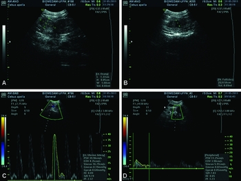
Ultrasound analysis confirmed that ovaries and uterine structures were normal, without adherences or fibrosis after surgery (Figure 2A). Furthermore, it was possible to visualize growing antral follicles 5 months after surgery, confirming ovarian functionality (Figure 2B). Ovarian length, height, width, and volume were 1.22 ± 0.08, 0.83 ± 0.05, 0.91 ± 0.11, and 520 ± 10 mm3. There was no difference between RI and PI values of the uterine arteries (RI: 0.93 ± 0.08; PI: 2.83 ± 0.8) and uterine-ovarian ligament (RI: 0.65 ± 0.005; PI: 1.39 ± 0.9) before and after surgery. Wave flow of blood vessels presented normal systolic and diastolic components (Figure 2C, D). Sections of the ovarian biopsies stained with toluidine blue showed that normal (Figure 1G) and degenerated follicles (Figure 1H) were found. The follicular density in the biopsies was of 41 ± 4.25 primordial follicles/mm2 and > 75 % of them was morphologically normal.
Ultrasonography of the reproductive tract from a female capuchin monkey (Cebus apella). (A) Longitudinal view of the ovary. (B) Longitudinal view of an antral follicle 5 months after surgery. (C, D) Wave form for uterine arteries (C) and uterus–ovary–ligament (UOL) arteries (D). A colour version of this figure is available online.

Discussion
We have adapted an efficient biopsy method to recover ovarian tissue from capuchin monkeys. The trap door method (Edel, Reference Edel1974) allowed the recovery of primordial follicles and resulted in minimal bleeding. This finding can be explained as the specimens were only 1 mm thick, and by the hypothermia caused by pressing the sampled tissue with cold saline-soaked gauze. Cold compression is an effective way to reduce bleeding by vasoconstriction. Moreover, cold also reduces edema and decreases the production of inflammatory agents (Taskin et al., Reference Taskin, Yigit, Bilici, Kuvat, Sisman and Celebi2011). Probably, this procedure also avoids adherences after surgery, which was confirmed by ultrasound analysis. We measured the ovarian dimensions using ultrasound evaluation. Meirow et al. (Reference Meirow, Fasouliotis, Nugent, Schenker, Gosden and Rutherford1999) estimated that the surface area of a human ovary was 540 mm2. In the present study, the volume of the ovary of capuchin monkeys was approximately 500 mm3. Our measurements were all similar to those previously reported by Domingues et al. (Reference Domingues, Caldas-Bussiere, Martins and Carvalho2007), when measuring ovarian dimensions (length, height, width and volume) from healthy mature capuchin monkeys. Taking these findings together, we have observed that follicular density was within the normal parameters (Domingues et al., Reference Domingues, Diniz, Furtado, Ohashi, Rondina and Silva2004); more than 75% of the primordial follicles were healthy, confirming the success of our procedure.
Cryopreservation of ovarian tissue is already a reality in large mammals (Santos et al., Reference Santos, Amorim, Cecconi, Fassbender, Imhof, Lornage, Paris, Schoenfeldt and Martinez-Madrid2010; Figueiredo et al., Reference Figueiredo, Rodrigues, Silva and Santos2011), which is a precedent for the successful preservation and complete development of primordial follicles from NHP found dead or being part of an assisted reproductive programs. Therefore, the next steps to be conducted are the cryopreservation and the development of in vitro culture systems of primordial follicles enclosed in ovarian fragments. We conclude that laparoscopic collection of ovarian tissue using the modified trap door method together with tissue compression with cold saline-soaked gauze is an efficient procedure to harvest healthy primordial follicles. The use of Cebus apella as a model for other species will help many researchers preserve female gametes from endangered threatened NHP species.
Acknowledgements
This study was supported by the project No. 483439/2009-6 from CNPq, Brazil. The authors thank the CENP for the logistical support.