Introduction
The spine is a frequent site of osseous metastases. Common cancers that metastasise to the spine include breast, lung and prostate cancer.Reference Constans, de Divitiis, Donzelli, Spaziante, Meder and Haye 1 Metastases to the cervical spine are less common and comprise 10 per cent of all spinal metastases, compared with 70 per cent that involve the thoracic spine and 20 per cent the lumbar spine.Reference Aydinli, Ozturk, Bayram, Sarihan, Evrensel and Yilmaz 2
Cervical spine metastases usually present with severe neck pain.Reference Jenis, Dunn and An 3 However, they can lead to epidural spinal cord compression, an oncologic emergency with the possibility of paralysis, sensory loss, and urinary incontinence.Reference Byrne 4
Cervical spine metastases are not commonly seen in the ENT clinic and therefore run the risk of being overlooked. We report three cases of unusual first presentation to the ENT surgeon of cervical spine metastases from undiagnosed primary tumours.
Case reports
Case report one: presentation with stridor
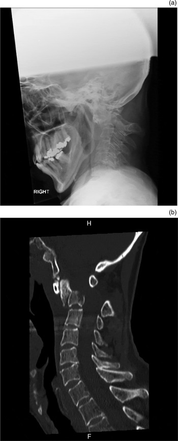
An 80-year-old woman presented to the emergency department with inspiratory stridor. The patient had considerable cervical kyphosis and urgent anaesthetic and senior ENT assistance were required to secure the airway. Flexible laryngoscopy revealed a narrowed supraglottic space but mobile vocal folds. On further questioning, the patient confirmed that she had been having severe neck pains for a week and was undergoing investigations for a pancreatic lump. An urgent lateral cervical spine X-ray revealed a fracture through the C1 odontoid peg with gross anterior displacement in relation to the body of C2 (Figure 1a). The patient was transferred to intensive care and the cervical spine immobilised using a halo device. This intervention stabilised the breathing. A computed tomography (CT) scan confirmed the metastatic pathological fracture of C1 (Figure 1b). Unfortunately, the cervical spine dislocated again a few days later leaving the patient with paraplegia. The patient subsequently died.
(a) Sagittal plain X-ray showing atlas subluxation in an 80-year-old woman who presented with stridor. (b) Sagittal computed tomography scan of this patient confirming the presence of a pathological fracture at C1. H = head; F = feet

Case report two: presentation with dysphagia
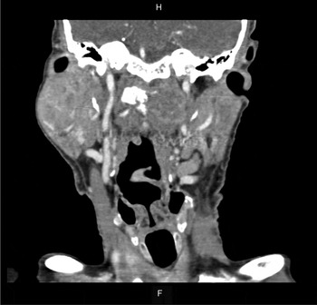
An 81-year-old man presented with an 8-month history of neck pain and increasing dysphagia with weight loss. A flexible laryngoscopy revealed a suspicious lesion in the left pyriform fossa. Rigid endoscopy and CT scan of the neck and thorax were arranged. Aside from candidiasis, the endoscopy did not reveal any significant pathology. The CT scan of the neck showed a large soft tissue mass on the left side of the C1 vertebra. This had caused significant bony erosion that extended to the left occiput and C2 vertebral body (Figure 2). The CT scan of the chest showed a spiculated mass in the central left lower lobe. The patient was placed in a hard neck collar and commenced on radiotherapy. Unfortunately, after seven fractions of radiotherapy he developed massive haematemesis and passed away. A post-mortem examination confirmed three lesions in the lung with metastases in the neck and kidneys. Pathology confirmed squamous cell carcinoma consistent with cancer originating in the lung.
Coronal computed tomography scan of an 81-year-old man who presented with dysphagia, which shows a large soft tissue mass on the left side of the C1 vertebra causing significant bony erosion that extended to the left occiput and left side of the C2 vertebral body. (Note the incidental pleomorphic adenoma of the right parotid gland.) H = head; F = feet

Case report three: presentation with dysphonia
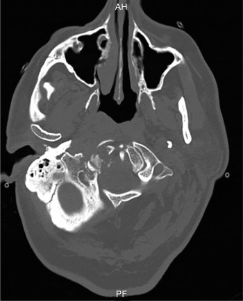
A 68-year-old man with a high body mass index (BMI) was admitted to the medical unit with severe pain in the neck and back causing immobility. The back pain was of long-standing duration, but no neurological deficit or new cause for the neck pain was identified. On the third day of admission the patient developed difficulty with speech. The ENT examination revealed an isolated right-sided hypoglossal weakness. A CT scan of the head revealed a destructive lesion of the right occipital condyle involving the margins of the hypoglossal canal (Figure 3). Subsequent whole-body CT scanning showed widespread lytic lesions of the spine and iliac crests. Biopsy from the right iliac crest revealed moderately differentiated adenocarcinoma with a possible gastrointestinal primary tumour. In view of the widespread disease, palliative therapy was commenced, however, the patient died shortly afterwards.
Transverse computed tomography scan of a 68-year-old man who presented with dysphonia, showing a destructive lesion of the right occipital condyle involving the margins of the hypoglossal canal. AH = anterior; PF = posterior

Discussion
Patients with conditions related to the cervical spine present frequently to the ENT surgeon. Typical presentations include neck pain due to spondylotic changes, vertigo related to the cervical spine and referred pain to the ear. Although neck pain is the classical symptom of cervical spondylosis, it is also a presenting feature of cervical spine metastasis. As pain related to the spine is very common in the general population (with a reported lifetime prevalence of 84 per cent), many cases of vertebral metastasis have a delayed diagnosis when the presenting symptom is a new-onset back or neck pain.Reference Sciubba and Gokaslan 5 Also, as is evident from our case reports, cervical spine metastases can be a source of severe neck pain even in elderly patients without cancer-related history. Indeed, pain due to metastases in the spine may be the first sign of malignancy in up to 10 per cent of cancer patients.Reference Livingston and Perrin 6 Atypical presentations of metastases to the cervical spine should always be considered in the elderly.
Stridor due to cervical dislocation from metastases (as in case report one) is a very rare presentation indeed. A literature search revealed scattered reports of stridor related to cervical spine pathology, all of which were of a non-malignant nature.Reference Caminos, Cenoz, Louise, Otano, Esain and Perez de Ciriza 7 In one report, stridor was the result of bilateral abductor cord palsy associated with degenerative (non-malignant) subluxation of the odontoid peg.Reference Brooker 8 In another case, hypertrophic anterior cervical osteophytes resulted in airway compromise following a diagnostic biopsy.Reference Lin, Quesnel, Holman, Curry and Rho 9 In our experience, stridor due to cervical spine dislocation is extremely problematic. Attempts to intubate or perform a tracheostomy, especially in an emergency, could result in irreversible spinal cord damage associated with neck mobilisation. Our first patient had her cervical spine stabilised following admission but went on to develop paraplegia a few days later because of re-dislocation of the cervical spine.
The predominant symptom of our second patient was dysphagia, which is another rare presentation of cervical spine metastases. The literature reports have associated dysphagia with diffuse hyperostosis of the cervical spine (Forestier disease) and anteriorly placed cervical osteophytes.Reference Lin, Quesnel, Holman, Curry and Rho 9 , Reference Verlaan, Boswijck, de Ru, Dhert and Oner 10 To the best of our knowledge, this is the first report of cervical spine metastases in a case presenting predominantly with dysphagia.
Our third patient had isolated hypoglossal weakness, which is another rare condition. This was the only objective physical finding, and the investigative CT scan confirmed the diagnosis. Skull base metastases account for a significant proportion of isolated hypoglossal weakness.Reference Combarros, Alvarez de Arcaya and Berciano 11
Owing to their specific circumstances, CT scans of the neck were the primary method of investigation for all of our patients. (For patient one, a quick investigation was required in an emergency situation, patient two was initially suspected to have a hypopharyngeal tumour and patient three had a high BMI which precluded a magnetic resonance imaging (MRI) scan.) However, the radiological investigation of choice when cervical metastases are suspected is MRI; this method is very sensitive in detecting cord and nerve compression and changes in the bone marrow.Reference Lecouvet, Geukens, Stainier, Jamar, Jamart and d'Othee 12 , Reference Sarpel, Sarpel, Yu, Hyder, Kaufman and Hindo 13 Plain radiographs of the neck can reveal gross abnormalities, but are not sensitive enough to allow the identification of osteolysis due to metastases.Reference Aydinli, Ozturk, Bayram, Sarihan, Evrensel and Yilmaz 2 In our experience, CT is a good modality of investigation if MRI is not immediately available, or in an emergency situation. The entire spine should be imaged in order to investigate for metastases elsewhere. A pathological diagnosis can be obtained using CT-guided needle biopsy, depending on accessibility and the size of the lesion. Open biopsy is a good option when CT-guided needle biopsy fails to be diagnostic.Reference Datir, Pechon and Saifuddin 14 Metastases at other sites may be more amenable to biopsies than the cervical spine itself, as seen with our third patient.
-
• Patients with cervical spine metastases commonly present with neck pain but symptoms may be more unusual
-
• Stridor, dysphagia and hypoglossal weakness are rare symptoms of cervical spine metastases
-
• High suspicion of cervical spine metastases should be maintained in older patients with neck symptoms
The prognosis of patients with spinal metastases is guarded, and patients with non-cervical spinal disease are reported to survive longer than those with cervical spine disease.Reference Aizenberg, Fox, Suki, McCuthcheon, Rao and Rhines 15 Treatment options available for metastatic spine disease include radiation therapy, surgery and chemotherapy. Several factors will determine what the best treatment modality is. These include tumour radiosensitivity, spinal stability, neurological symptoms, pain and the patient's overall prognosis. Radiotherapy is the first-line choice for most patients with metastatic spinal disease. This treatment can provide effective (local) pain control in 50–80 per cent of patients,Reference Lutz, Berk, Chang, Chow, Hahn and Hoskin 16 and may prevent the onset of neurological complications.Reference Bartels, van der Linden and van der Graaf 17 Surgical treatment is indicated in patients with spinal cord compression or spinal instability in order to stabilise the spine. Recent surgical advances have significantly improved surgical options for patients with cervical spine metastases.Reference Harel and Angelov 18
Conclusion
Patients with cervical spine metastases will occasionally present directly to the ENT surgeon. A high index of suspicion should be adopted in an older patient with severe neck pain and/or additional symptoms, and radiological investigations should be requested accordingly.