INTRODUCTION
Distortion of host sex ratios by parasitic microorganisms is widespread in arthropods. In general, sex ratio distorting microbes are vertically transmitted through the female line only, so inducing the host to reallocate resources from male to female offspring benefits the parasite (Hurst, Reference Hurst1993). At a population level, these parasitic manipulations of host sex ratios can cause dramatic effects, including transitions from male to female heterogamety (Rigaud and Juchault, Reference Rigaud and Juchault1993) and reversal of sex roles as males become scarce (Jiggins et al. Reference Jiggins, Hurst and Majerus2000). In most arthropod hosts, only bacterial sex ratio distorters have been discovered to date and sex ratio distortion usually occurs through the early death of infected male offspring (Hurst and Jiggins, Reference Hurst and Jiggins2000). In contrast, parasites of Crustacea appear to be capable of subverting their hosts’ mechanisms of sex determination, causing infected individuals to develop as females, regardless of any genetic or environmental predisposition (Bulnheim, Reference Bulnheim1978; Ginsburger-Vogel, Reference Ginsburger-Vogel1985; Bouchon et al. Reference Bouchon, Rigaud and Juchault1998). This highly efficient feminizing form of sex ratio distortion is associated with infection by bacteria (Valette et al. Reference Valette, Essono, Le Clec'h, Johnson, Bech and Grandjean2013) and also by protists, including paramyxids (Ginsburger-Vogel and Desportes, Reference Ginsburger-Vogel and Desportes1979b ) and several species of microsporidia (Ironside et al. Reference Ironside, Smith, Hatcher, Sharpe, Rollinson and Dunn2003). Paramyxids and microsporidia can exhibit high levels of co-infection (Short et al. Reference Short, Guler, Yang, Kille and Ford2012), possibly due to hyperparasitism of paramyxids by microsporidia (Stentiford et al. Reference Stentiford, Ramilo, Abollo, Kerr, Bateman, Feist, Bass and Villalba2017). This has led to controversy concerning whether the ability to feminize has evolved independently in paramyxids and microsporidia or whether one group of parasites merely hitchhikes on the feminizing ability of the other (Short et al. Reference Short, Guler, Yang, Kille and Ford2012; Ironside and Alexander, Reference Ironside and Alexander2015).
Sex ratio distortion by paramyxid parasites was first described in populations of the talitrid amphipod host Orchestia gammarellus and Orchestia aestuarensis in the Penze estuary (near Roscoff, France), which exhibited female-biased sex ratios and high proportions of intersex individuals (Ginsburger-Vogel, Reference Ginsburger-Vogel1973; Ginsburger-Vogel, Reference Ginsburger-Vogel1991). Breeding experiments demonstrated that these populations contained thelygenic females, which produced mostly female and intersex offspring (Ginsburger-Vogel, Reference Ginsburger-Vogel1973). The thelygenic trait was temperature sensitive, occurring only in females maintained below a critical temperature in the laboratory (Ginsburger-Vogel, Reference Ginsburger-Vogel1975). The feminizing factor could also be transmitted experimentally by injecting tissue from intersex males or thelygenous females into normal males (which became intersex) and amphigenous females (which became thelygenous) (Ginsburger-Vogel and Carrelecuyer, Reference Ginsburger-Vogel and Carrelecuyer1976). Observation of paramyxid-like ‘micro-cells’ in thelygenous females (Ginsburger-Vogel and Desportes, Reference Ginsburger-Vogel and Desportes1979a ) led eventually to the description of a new paramyxid genus and species Paramarteilia orchestiae (Ginsburger-Vogel and Desportes, Reference Ginsburger-Vogel and Desportes1979b ). The parasite occurred only in intersex males and thelygenous females and exhibited similar temperature sensitivity to the thelygenic trait (Ginsburger-Vogel and Desportes, Reference Ginsburger-Vogel and Desportes1979a ). A recent paramyxid molecular phylogeny (Ward et al. Reference Ward, Bennett, Bateman, Stentiford, Kerr, Feist, Williams, Berney and Bass2016) includes a DNA sequence from a paramyxid parasite of O. gammarellus, sampled from populations at Weymouth and the Gann estuary (Dale), both in the UK. Light and ultrastructural observations of this parasite were concordant with the original description of P. orchestiae (Ginsburger-Vogel and Desportes, Reference Ginsburger-Vogel and Desportes1979b ) and parasites were discovered in the oocytes of female hosts, indicating the potential for vertical transmission.
In the gammarid amphipods Gammarus duebeni and Corophium curvispinum, breeding experiments have associated thelygeny and intersexuality with infection by three microsporidian species; Dictyocoela duebenum and Nosema granulosis in G. duebeni (Ironside et al. Reference Ironside, Smith, Hatcher, Sharpe, Rollinson and Dunn2003; Kelly et al. Reference Kelly, Hatcher and Dunn2004) and an undescribed microsporidian species in C. curvispinum (Mautner et al. Reference Mautner, Cook, Forbes, McCurdy and Dunn2007). Populations the gammarid Echinogammarus marinus also exhibit intersexuality associated with co-infection of a microsporidian closely related to D. duebenum and a paramyxid closely related to P. orchestiae (Short et al. Reference Short, Guler, Yang, Kille and Ford2012). In this case, the paramyxid is associated with intersexuality even in the absence of the microsporidian, and so it was concluded that the paramyxid is the cause of feminization. However, in G. duebeni the microsporidia N. granulosis and D. duebenum are associated with feminization in the absence of co-infecting paramyxids (Ironside and Alexander, Reference Ironside and Alexander2015). The available evidence from gammarid host therefore supports multiple origins of the ability to feminize amphipod hosts, in microsporidia and paramyxids.
Microsporidian parasites closely related to the feminizing parasite D. duebenum have been discovered in Orchestia populations. In a UK population of Orchestia cavimana, a microsporidian described as Dictyocoela cavimanum occurred at higher prevalence in female hosts than males, suggesting that it could be a feminizer (Terry et al. Reference Terry, Smith, Sharpe, Rigaud, Littlewood, Ironside, Rollinson, Bouchon, MacNeil, Dick and Dunn2004), but the population was not screened for co-infecting paramyxids. In French populations of Orchestia mediterranea and Orchestia aestuarensis, intersex individuals were co-infected with the paramyxid P. orchestiae and an unidentified microsporidian (Ginsburger-Vogel, Reference Ginsburger-Vogel1991). The microsporidian was also found in normal males while P. orchestiae was not (Ginsburger-Vogel, Reference Ginsburger-Vogel1991; Reference Ginsburger-Vogel1992), suggesting that the microsporidian did not cause feminization in the absence of the paramyxid. However, none of these earlier studies is able to demonstrate unambiguously whether feminization of Orchestia species is caused by a paramyxid or a microsporidian. The aims of this study were therefore to identify the paramyxid and microsporidian parasites infecting known thelygenic populations of O. gammarellus and O. aestuarensis using molecular methods and to use molecular screening techniques to establish whether feminization was caused directly by paramyxid parasites or whether the phenomenon could be attributed to co-infection with feminizing microsporidia.
MATERIALS AND METHODS
Collection and maintenance of Orchestia spp.
Individuals of the genus Orchestia were collected from two of the original locations on the Penzé estuary in France at which thelygenic individuals were discovered by Ginsburger-Vogel (Reference Ginsburger-Vogel1973). These were under a railway bridge (latitude 48·629 016, longitude −3·952 417) and under the D58 road bridge (Pont de la Cord – latitude 48·644 557, longitude −3·951 988), the locations of Ginsburger-Vogel (Reference Ginsburger-Vogel1973) station 1 and station 2, respectively. Additional samples were collected from similar habitat on the Gann estuary in the UK (latitude 51·722 914, longitude −5·165 377). Orchestia were collected by hand from beneath stones above the strand line and transferred to a container of soil for transport. In the laboratory, each individual was transferred to a plastic Petri dish lined with filter-paper, which had been soaked in sea water and maintained at a temperature of 17 °C. In order to maintain humidity, the filter paper was regularly replenished with fresh water. Dried sections of Laminaria digitata frond were soaked in sea-water and provided as food.
Identification of host sex and species
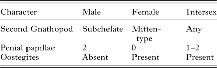
The Orchestia were anaesthetized in carbonated water and categorized according to their sex on the basis of primary and secondary sexual characters (Ginsburger-Vogel, Reference Ginsburger-Vogel1975; McCurdy et al. Reference McCurdy, Forbes, Mautner, Cook, Logan and Lancaster2004) (Table 1). Females with eggs or young in their brood pouches were maintained until their next moult, when their young were released. The females were then separated from their young and stored in ethanol. Males were immediately stored in ethanol.
Primary and secondary sexual characters used to categorize Orchestia according to sex

At all three locations sampled, an assemblage of Orchestia species occurred, containing at least two of the species O. gammarellus, O. mediterranea and O. aestuarensis. Females of these species cannot be discriminated reliably using existing dichotomous keys (e.g. Wildish, Reference Wildish1987; Lincoln, Reference Lincoln1979) and so a DNA barcoding methodology was used. Orchestia specimens were dissected under a microscope using watchmaker's forceps to remove the cuticle. The forceps were flamed between dissections to avoid cross-contamination. Genomic DNA was extracted using a QIAGEN Dneasy blood and tissue kit, following the manufacturer's instructions. The Orchestia mitochondrial cytochrome oxidase subunit 1(CO1) gene was amplified by PCR, using the primers OGCO1f (forward) (5′-CTGGAAATTTAATTGGTGAC-3′) and OGCO1r (reverse) (5′-GGAGAGAAGAAGTAGAAT-3′). These primers were designed on the basis of published O. gammarellus CO1 sequences available in Genbank (Accession numbers: EU276377, EU276358, EU276191, EU276207, EU276208). 35 PCR cycles were used, with an annealing temperature of 50 °C for one minute and an extension time of 1 min. PCR products were Sanger-sequenced using an Applied Biosystems, ABI3730xl Genetic Analyser. Phylogenetic analysis of the COI sequences was performed according to the maximum-likelihood criterion, with 2000 bootstrap replicates. The substitution model of best fit was selected according to the corrected Akaike information criterion (AICc, Hurvich and Tsai, Reference Hurvich and Tsai1989).
Screening for parasite infection
Microsporidian infection was detected through the amplification of the microsporidian small subunit ribosomal RNA gene (SSUrRNA), using the general microsporidian primers V1 (forward) (5′-CACCAGGTTCTGCCTGAC-3′) and 1492r (reverse) (5′-GGTTACCTTGTTACGACTT-3′), which amplify a DNA fragment of approximately 1·2 kbp (Weiss et al. Reference Weiss, Zhu, Cali, Tanowitz and Wittner1994). Paramyxid infection was detected through the amplification of the paramyxid SSUrRNA gene, using the primers ParaJI_F (forward) (5′-GGACCATTGCTGAGACTAAA-3′) and ParaJI_R1 (reverse) (5′-GAGTTCAGAGAAACAGTTG-3′) which amplify a 980 bp fragment (Ironside and Alexander, Reference Ironside and Alexander2015). In both cases, 35 PCR cycles were used, with an annealing temperature of 50 °C for one minute and an extension time of 1 min.
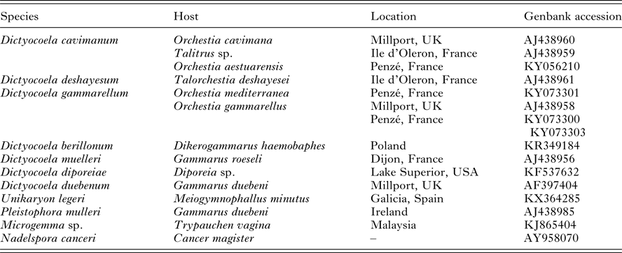
The PCR products were sequenced using an ABI3730xl Genetic Analyser (Applied Biosystems, Foster City, California). A preliminary BLASTn search of the NCBI nucleotide database indicated that the microsporidian sequences closely matched those of the genus Dictyocoela. The new sequences were therefore aligned using Clustal W (Thompson et al. Reference Thompson, Higgins and Gibson1994) with reference Dictyocoela sequences on the NCBI nucleotide database (Table 2). The new paramyxid sequences were aligned with a published sequence obtained from a putative feminizing parasite of E. marinus (Short et al. Reference Short, Guler, Yang, Kille and Ford2012) and other published paramyxid sequences showing greater than 90% coverage (Table 3). Phylogenetic analysis of microsporidian and paramyxid SSUrRNA sequenceswas performed according to the maximum-likelihood criterion, with 2000 bootstrap replicates. The substitution models of best fit were selected according to the corrected Akaike information criterion (Hurvich and Tsai, Reference Hurvich and Tsai1989).
Microsporidian small subunit ribosomal RNA sequences used in phylogenetic analysis

Paramyxid small subunit ribosomal RNA sequences used in phylogenetic analysis

95% confidence intervals for the prevalence of each parasite were calculated using the binomial distribution. A χ 2 test, based on a 2 × 2 contingency table, was used to test for excesses or deficits in the number of individuals co-infected with multiple parasites. Separate χ 2 contingency tests were used to test for differences in prevalence between the host sexes and host species at each location.
Sex ratios of offspring
Young Orchestia released from the brood pouches of females in the laboratory were separated into individual Petri dishes, lined with damp filter paper and provided with food as described above. At 30 weeks of age, the laboratory-reared Orchestia were anaesthetized and categorized by sex as described above. 95% binomial confidence intervals were calculated for the total number of offspring of each sex produced by infected and uninfected females. A χ 2 test was performed on a 2 × 3 table to test for differences between the expected and observed ratio of males, females and intersex individuals for both the uninfected and infected broods.
After being categorized by sex, the laboratory-reared Orchestia were screened for microsporidian and paramyxid parasites as described above. 2 × 2 Pearson's χ 2 contingency tables were used to test whether there were significant differences between the number of infected offspring produced by infected females and the number of infected offspring produced by uninfected females.
The initial brood size of each female was recorded and compared with the size of the brood at adulthood (30 weeks after release from the brood pouch). The proportion of surviving offspring was calculated for each brood and binomial confidence intervals were calculated for the proportion of surviving offspring in broods produced by infected and uninfected females. 2 × 2 Pearson's χ 2 contingency tables were used to test whether there were significant differences between the number of infected offspring produced by infected females and the number of infected offspring produced by uninfected females.
RESULTS
Host species and sex ratios in natural populations
DNA barcoding revealed that all three sites contained an assemblage of several Orchestia species. Samples from the two Penzé estuary sites both contained O. mediterranea and O. aestuarensis in addition to O. gammarellus, with the upstream site containing a higher proportion of O. aestuarensis. The Gann estuary site contained O. gammarellus and O. aestuarensis, with O. gammarellus predominating. A random sample of 56 adult O. gammarellus from the Gann Estuary contained 32 males and 24 females. Other samples of all three species were similarly male-biased at all sites.
Parasite phylogenetics
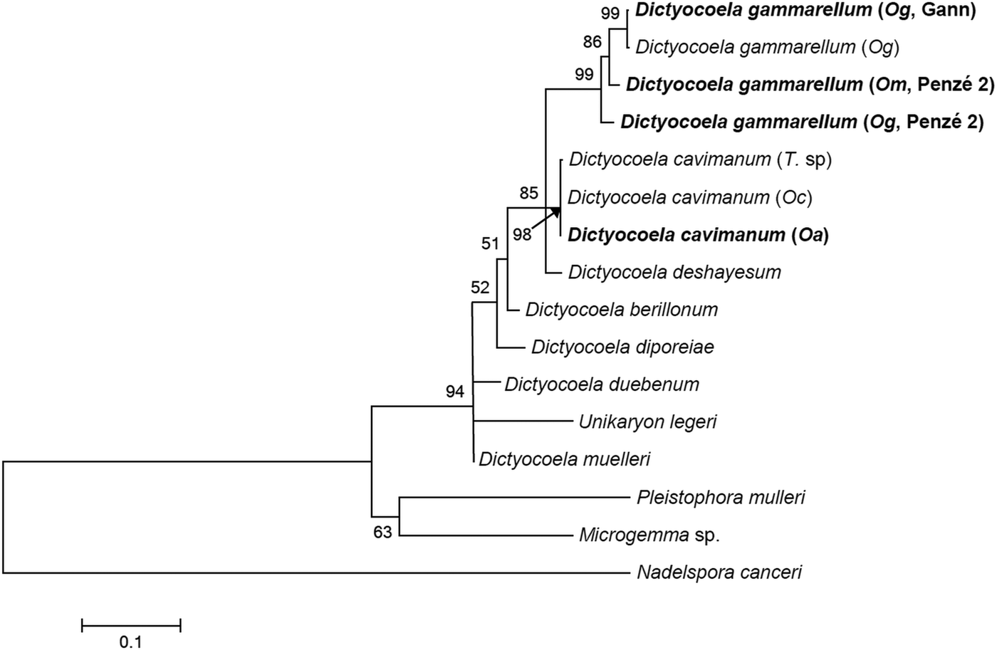
All 23 microsporidian SSU sequences amplified from the three Orchestia species fall within the genus Dictyocoela. The SSU of Dictyocoela contains a hypervariable region, and so the sequences exhibit considerable variability. However, all sequences fall into two well-supported clades, the first corresponding to D. cavimanum and the second to Dictyocoela gammarellum (Fig. 1). Both species have previously been reported from hosts of the genus Orchestia (Terry et al., Reference Terry, Smith, Sharpe, Rigaud, Littlewood, Ironside, Rollinson, Bouchon, MacNeil, Dick and Dunn2004).
Maximum-likelihood phylogenetic tree of Dictyocoela small subunit ribosomal RNA sequences. Constructed using a general time-reversible model of evolution with gamma-distributed rate variation among sites. Abbreviations in brackets indicate host species: Oc, Orchestia cavimanum; Oa, Orchestia aestuarensis; T. sp, Talitrus sp.; Og, Orchestia gammarellus; Om, Orchestia mediterranea. Sequences obtained in this study are indicated in bold.

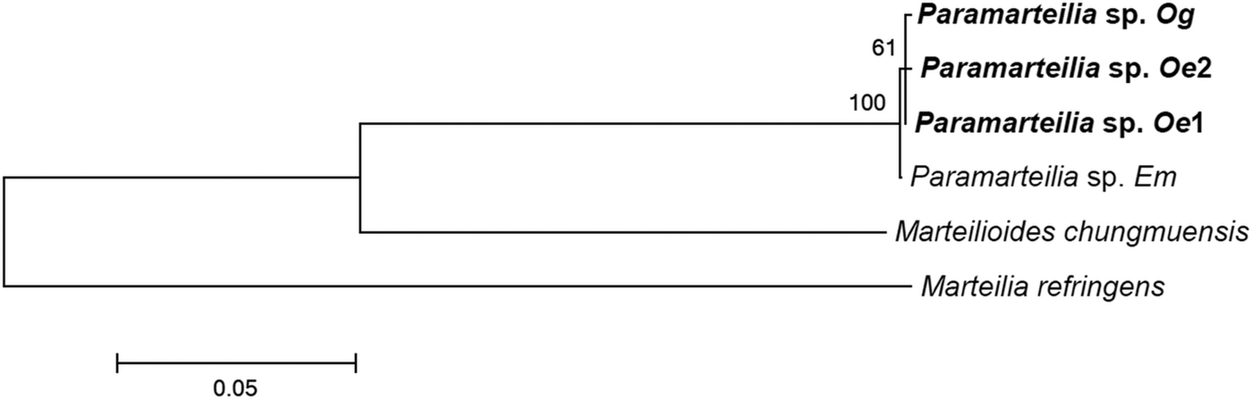
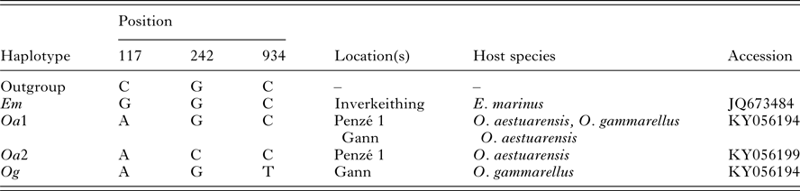
The 26 paramyxid sequences amplified from O. gammarellus and O. aestuarensis are all very similar, but not identical, to a sequence amplified from E. marinus (Short et al. Reference Short, Guler, Yang, Kille and Ford2012) (Fig. 2). All of the sequences amplified from Orchestia species belong to three haplotypes (Table 4). The first of these haplotypes (designated Oa1) differs from the E. marinus sequence by a transition from G to A at position 117. This haplotype was found in five O. aestuarensis females and one O. gammarellus female from the Penzé 1 site and also in the three O. aestuarensis females obtained from the Gann site. A single O. aestuarensis male from the Penzé 1 site yielded a second haplotype (designated Oa2), which contained the transition at position 117 and a transversion from G to C at position 242. A third haplotype (designated Og), containing the transition at position 117 and an additional transition from C to T at position 934, was found in nine O. gammarellus females from the Gann site. These sequences also closely match a short (348 bp) sequence amplified by Ward et al. (Reference Ward, Bennett, Bateman, Stentiford, Kerr, Feist, Williams, Berney and Bass2016) from O. gammarellus and confirmed by electron microscopy as matching the description of P. orchestiae. The three haplotypes identified here are therefore described as Paramarteilia sp. Oa1, Oa2 and Og.
Maximum-likelihood phylogenetic tree of Paramyxid small subunit ribosomal RNA sequences. Constructed using a Tamura–Nei model of evolution with gamma-distributed rate variation among sites. Sequences obtained in this study are indicated in bold.

Paramyxid small subunit ribosomal RNA haplotypes amplified from Orchestia hosts

Parasite prevalence
The Microsporidium D. gammarellum was detected at low prevalence in samples of O. gammarellus from the Penzé and Gann estuaries, and was also detected in samples of O. mediterranea from the Penzé estuary. Both males and females were infected. Dictyocoela cavimanum was detected in samples collected at all three sites and in all three host species. Although both male and female hosts were infected, prevalence in females was significantly higher than prevalence in males in O. aestuarensis (χ 2 = 4·339, d.f. = 1, P < 0·05). From samples of O. gammarellus and O. mediterranea, only a single female individual of each species was infected.
The paramyxid parasite was detected in samples of O. gammarellus and O. aestuarensis from the Penzé and Gann estuaries. Of the 26 infected individuals identified, only one was male, representing a significant female bias to infection in O. aestuarensis (χ 2 = 13·68, d.f. = 1, P < 0·001) and O. gammarellus (χ 2 = 14·90, d.f. = 1, P < 0·001).
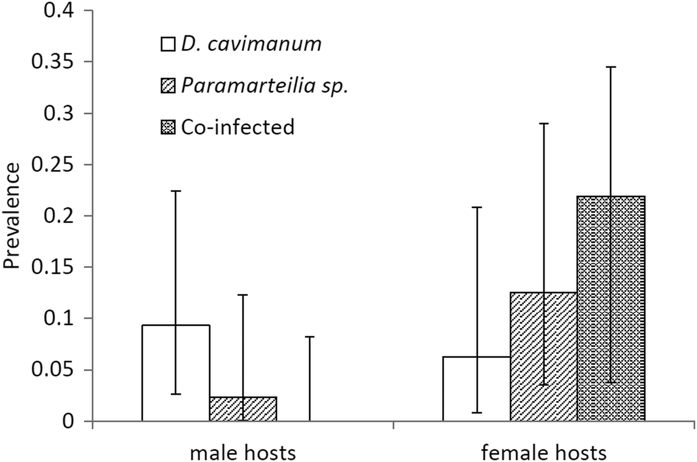
At the Penzé 1 and Gann sites, where Paramarteilia occurred, seven individuals (all female O. aestuarensis) were co-infected with D. cavimanum and Paramarteilia. Given the individual prevalences of the two parasites, the number of co-infected individuals is significantly higher than would be expected in O. aestuarensis at these two sites by chance (Fisher's exact P < 0·005). Once the co-infected individuals are excluded, the prevalence of D. cavimanum shows no significant sex bias (Fisher's exact P > 0·05) (Fig. 3).
Prevalence of single infections with the putative feminizing parasites Dictyocoela cavimanum and Paramarteilia sp. together with co-infections with both parasites in the host Orchestia aestuarensis. Error bars indicate 95% binomial confidence intervals for parasite prevalence.

Sex ratios of offspring
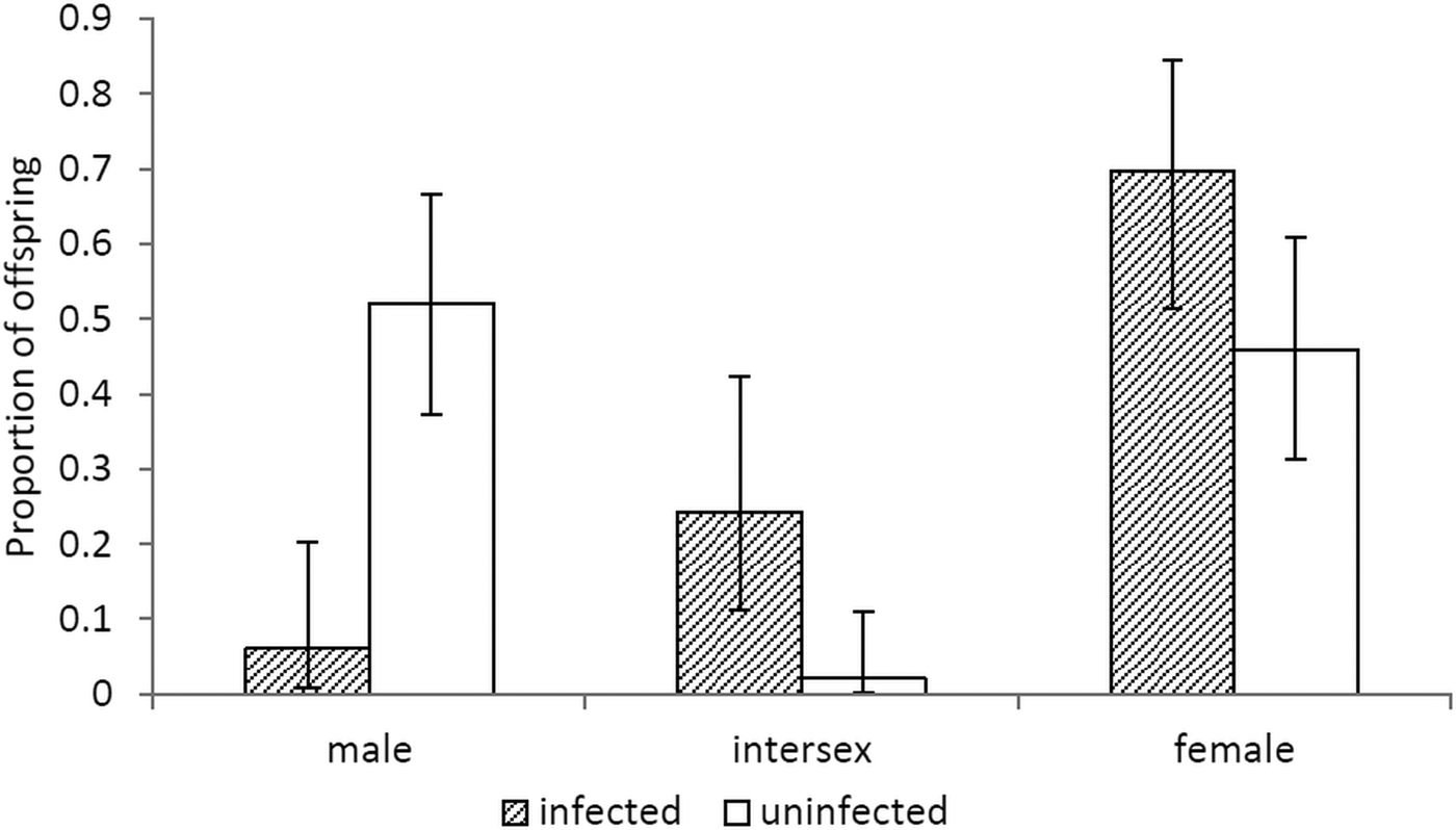
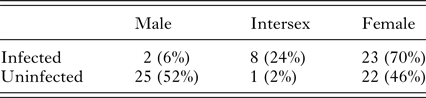
Twenty-one O. gammarellus females produced offspring under laboratory conditions, which survived to a point where sex could be determined. Eight of these females were infected with paramyxid parasites while none were infected with microsporidia. Sex ratio data for offspring produced by infected females were pooled and compared with data for offspring of uninfected females (Table 5, Fig. 4). A Pearson's χ 2 test indicated that there was a significant difference in the proportions of male, female and intersex individuals between the broods of infected and uninfected females (χ 2 = 22·038, d.f. = 2, P < 0·001), with the broods of infected females exhibiting a lower proportion of males and more intersex individuals than the broods of uninfected females.
Proportions of male, intersex and female offspring produced by Orchestia gammarellus females infected with the paramyxid Paramarteilia sp. and by uninfected females. Error bars indicate 95% binomial confidence intervals for sex ratio.

Numbers of male, intersex and female offspring produced by Orchestia gammarellus females infected with the paramyxid Paramarteilia sp. and by uninfected females

Survival of offspring
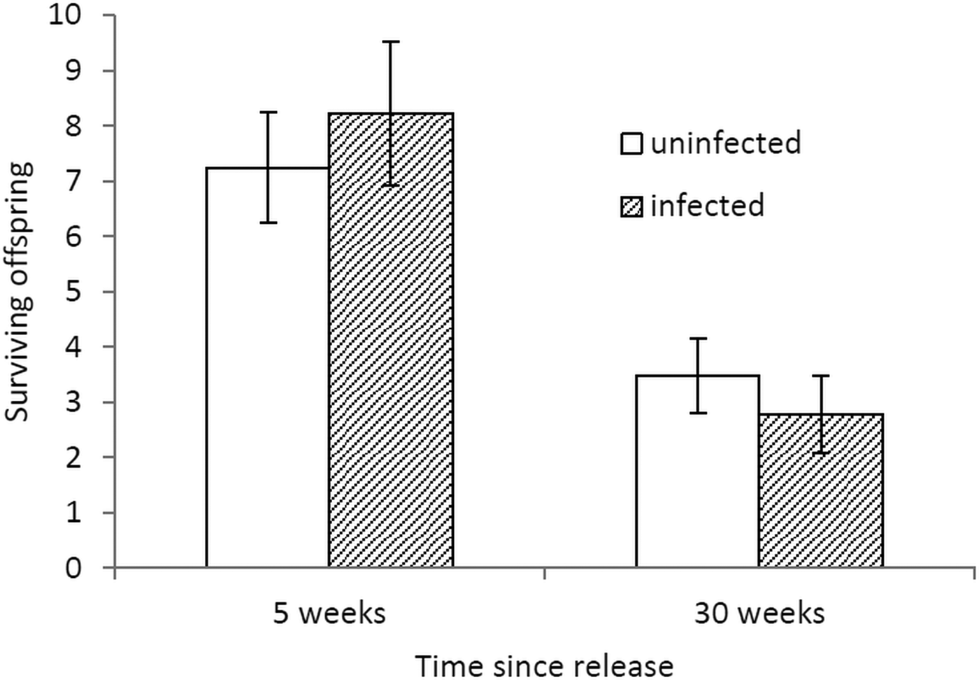
T-tests indicated no significant differences between female O. gammarellus infected with Paramarteilia sp. and uninfected females in the number of offspring surviving 5 weeks (t = 0·59, d.f. = 17, P > 0·05) or 30 weeks (t = 0·71, d.f. = 20, P > 0·05) after release from the brood pouch (Fig. 5).
Proportion of offspring produced by Orchestia gammarellus females infected with the paramyxid Paramarteilia sp. and by uninfected females surviving 5 weeks and 30 weeks after release from the brood pouch. Error bars indicate 95% binomial confidence intervals for survival.

Vertical transmission
Forty-three offspring of female O. gammarellus infected with Paramarteilia sp. were screened for infection by PCR. The PCR screen detected Paramarteilia sp. in 16 of these offspring, a transmission efficiency of 0·35. All of the offspring in which paramyxid infection was confirmed by PCR were female or intersex.
DISCUSSION
Paramarteilia orchestiae is currently the only paramyxid in which feminizing activity has been demonstrated experimentally (Ginsburger-Vogel and Desportes, Reference Ginsburger-Vogel and Desportes1979a ). This study provides the first molecular phylogenetic analysis to include DNA sequences from the type site (Penzé Estuary) and host species (O. gammarellus) of P. orchestiae. Although these sequences are highly similar to those obtained previously from E. marinus (Short et al. Reference Short, Guler, Yang, Kille and Ford2012), they show clear and consistent differences. Given the low levels of variation in the paramyxid SSU (Ward et al. Reference Ward, Bennett, Bateman, Stentiford, Kerr, Feist, Williams, Berney and Bass2016), even these small differences suggest that the E. marinus parasite belongs to a different species, host race or geographical variant to the parasite originally described as P. orchestiae (Ginsburger-Vogel and Desportes, Reference Ginsburger-Vogel and Desportes1979b ). Confirmation of the E. marinus parasite as P. orchestiae will require a complete description based on electron microscopy and, if possible, additional genetic evidence indicating a lack of reproductive isolation. As has been demonstrated in microsporidia (Ironside and Alexander, Reference Ironside and Alexander2015), even very closely related parasites may differ in their feminizing activity. Therefore, although there is circumstantial evidence for feminization by the E. marinus paramyxid, in the form of high levels of intersexuality in infected individuals (Short et al. Reference Short, Guler, Yang, Kille and Ford2012), this parasite should not be described as a feminizer until feminization has been demonstrated experimentally.
All paramyxid SSU sequences obtained from O. aestuarensis from the Gann Estuary display a haplotype identical to the most common haplotype found in O. aestuarensis and O. gammarellus at the Penzé Estuary (Haplotype Oa1). However, all sequences obtained from O. gammarellus from the Gann Estuary display a haplotype (Og), which is slightly different from any sequences obtained from the Penzé Estuary. The organism described by Ward et al. (Reference Ward, Bennett, Bateman, Stentiford, Kerr, Feist, Williams, Berney and Bass2016) as P. orchestiae is likely to correspond to haplotype Og, given that it was obtained from the Gann Estuary O. gammarellus population. Haplotype Og was not detected at the Penzé Estuary and is therefore unlikely to correspond to the exact species or strain described by Ginsburger-Vogel and Desportes (Reference Ginsburger-Vogel and Desportes1979a , Reference Ginsburger-Vogel and Desportes b ) as P. orchestiae. The clear association of haplotypes with host species at the Gann Estuary site suggests that the haplotypes Oa1 and Og may correspond to different host races, or even different species. The only male individual found to be infected with a paramyxid parasite (an O. aestuarensis male from the Penzé Estuary) exhibited another haplotype (Oa2), which was not detected in any other individual. This raises the possibility that non-feminizing species or strains of Paramarteilia may also occur in Orchestia species.
The experiments described in this study indicate that the parasite associated with haplotype Og feminizes O. gammarellus in the absence of co-infecting microsporidia, causing infected females to produce fewer male and more intersex offspring than uninfected females. The proportion of offspring surviving does not differ significantly between the broods of infected and uninfected females, indicating that the difference in sex ratio is not due to reduced survival of infected male offspring, as occurs in some cases of parasitic infection (Hurst et al. Reference Hurst, Jiggins, von der Schulenburg, Bertrand, West, Goriacheva, Zakharov, Werren, Stouthamer and Majerus1999). Although the vertical transmission efficiency of the paramyxid parasite is low compared with microsporidian feminizers such as N. granulosis (Terry et al. Reference Terry, Smith and Dunn1998), feminization of infected offspring appears to be efficient. No males infected with Paramarteilia sp (Og) were produced by infected females in the laboratory or discovered in any O. gammarellus population.
The microsporidia D. gammarellum and D. cavimanum also infect Orchestia communities of the Penzé and Gann Estuaries. Some members of the genus Dictyocoela cause feminization of amphipod hosts such as G. duebeni, while others have no apparent feminizing effects (Ironside and Alexander, Reference Ironside and Alexander2015). Previously, Terry et al. (Reference Terry, Smith, Sharpe, Rigaud, Littlewood, Ironside, Rollinson, Bouchon, MacNeil, Dick and Dunn2004) detected a female bias in the prevalence of D. cavimanum in an Orchestia host, which they interpreted as evidence that this parasite was a feminizer. In the current study, a similar female bias was noted in the prevalence of D. cavimanum in O. aestuarensis. However, it was also observed that Paramarteilia and D. cavimanum co-infected the same host far more frequently than would be expected by chance. Where D. cavimanum occurred in the absence of Paramarteilia, there was no sex bias to its prevalence, indicating that this microsporidian parasite is hitchhiking upon the feminizing ability of Paramarteilia rather than causing feminization itself. Similar associations between Paramarteilia and Dictyocoela infection have also been described in E. marinus (Short et al. Reference Short, Guler, Yang, Kille and Ford2012). The mechanism of co-infection remains unknown but hyperparasitism of the paramyxid by the microsporidian, as observed in Marteilia cochillia (Stentiford et al. Reference Stentiford, Ramilo, Abollo, Kerr, Bateman, Feist, Bass and Villalba2017), provides a possible mechanism.
Previous studies have demonstrated that microsporidia can feminize amphipod hosts in the absence of paramyxids (Ironside and Alexander, Reference Ironside and Alexander2015). By demonstrating unambiguously that paramyxids can also cause feminization in the absence of microsporidia, the current study strengthens the evidence that the ability to feminize amphipods has evolved on multiple occasions in very distantly related groups of parasites.
FINANCIAL SUPPORT
This research received no specific grant from any funding agency, commercial or not-for-profit sectors.