Implications
Digital dermatitis (DD) causes painful lesions on the skin above the heels of dairy cows. Prevalence appears to be influenced by many factors, including the amount of contact between the animals and slurry. In this study, in vitro treatment with slurry increased the permeability of dairy cow skin to methylene blue dye. This offers one mechanism by which slurry could affect the occurrence of DD by increasing skin permeability, which could facilitate pathogen entry. This finding adds to evidence of the role that slurry might play in the pathogenesis of DD, highlighting the importance of good slurry management in dairy cow housing.
Introduction
Digital dermatitis (DD) is a major problem for dairy industries in many countries. The disease is thought to be caused by spirochaete bacteria (Walker et al., Reference Walker, Read, Loretz and Nordhausen1995; Demirkan et al., Reference Demirkan, Carter, Murray, Blowey and Woodward1998; Evans et al., Reference Evans, Brown, Demirkan, Singh, Getty, Timofte, Vink, Murray, Blowey, Birtles, Hart and Carter2009) and leads to painful lesions, commonly at the skin-horn junction on the heels of dairy cattle (Read and Walker, Reference Read and Walker1998). There appears to be natural variation in susceptibility to DD, with some animals in a group infected repeatedly while others never develop the disease (Laven, Reference Laven1999). Indeed, estimates for heritability of DD range from 0.10 (based on 430 herds in The Netherlands, van der Waaij et al., Reference van der Waaij, Holzhauer, Ellen, Kamphuis and De Jong2005) to 0.40 (based on three dairy farms in California, USA, Oberbauer et al., Reference Oberbauer, Berry, Belanger, McGoldrick, Pinos-Rodriquez and Famula2012). There are numerous factors which could potentially affect an individual's susceptibility, including morphological and functional characteristics of the skin.
Bovine skin shows typical mammalian skin morphology, with a slightly different composition of epidermal lipids (Netzlaff et al., Reference Netzlaff, Schaefer, Lehr, Meiers, Stahl, Kietzmann and Niedorf2006) and a higher density of hair follicles (Pitman and Rostas, Reference Pitman and Rostas1982) than human skin. The mammalian skin acts as a protective barrier for the body by preventing the entry of pathogens and protecting against physical and chemical damage (Proksch et al., Reference Proksch, Brandner and Jensen2008). The most important layer of the skin in maintaining this barrier function is the outer layer, the stratum corneum (Madison, Reference Madison2003). How well the skin functions as a barrier varies between individuals and could be affected by a number of factors including the thickness of the stratum corneum (Holbrook and Odland, Reference Holbrook and Odland1974), the number of hair follicles in the skin (Wosicka and Cal, Reference Wosicka and Cal2010) and genetic mutations (Palmer et al., Reference Palmer, Irvine, Terron-Kwiatkowski, Zhao, Liao, Lee, Goudie, Sandilands, Campbell, Smith, O'Regan, Watson, Cecil, Bale, Compton, DiGiovanna, Fleckman, Lewis-Jones, Arseculeratne, Sergeant, Munro, El Houate, McElreavey, Halkjaer, Bisgaard, Mukhopadhyay and McLean2008). There are links between skin permeability and occurrence of bacterial skin disease, for example, humans who suffer from an inflammatory skin condition called atopic dermatitis, which is associated with an impaired skin barrier function (Di Nardo et al., Reference Di Nardo, Wertz, Giannetti and Seidenari1998), are more susceptible to some bacterial, viral and fungal infections of the skin (Jung and Stingl, Reference Jung and Stingl2008). Potential relationships between skin characteristics and susceptibility to DD have not yet been examined.
There is a growing body of evidence that the prevalence of DD is affected by the environment, and that contact with slurry or damp, muddy conditions can contribute to the development of infection (Blowey and Sharp, Reference Blowey and Sharp1988; Rodriguez-Lainz et al., Reference Rodriguez-Lainz, Hird, Carpenter and Read1996; Laven, Reference Laven1999). Slurry may increase disease prevalence by acting as a reservoir for infectious agents (Laven, Reference Laven1999), although the causative agents have not yet been found in slurry – a recent examination of dairy cow houses containing infected animals did not find the treponeme groups generally found in DD lesions in the slurry, even though they were found in rectal and oral tissues of some animals (Evans et al., Reference Evans, Timofte, Isherwood, Brown, Williams, Sherlock, Lehane, Murray, Birtles, Hart and Carter2012). An additional possibility is that slurry may damage the skin barrier and make it easier for bacteria to enter and cause infection, as the mixture of urine and faeces is thought to have keratolytic properties (Laven, Reference Laven2001).
The first aim of this study was to determine whether there was a difference between dairy cows with and without a history of DD in the thickness of the stratum corneum, the number of hair follicles per unit area or skin permeability. The second aim was to determine whether skin permeability was affected by contact with slurry.
Material and methods
Animals and skin sampling
The study involved 29 dairy cows from the Agri-Food and Biosciences Institute dairy herd. The animals were various breeds (Holstein–Friesian, Jersey × Holstein–Friesian, Montbeliarde × Holstein–Friesian, Norwegian Red and Norwegian Red × Holstein–Friesian), and parities (from 1 to 6). Animals had been monitored once weekly in the milking parlour for signs of DD infection in the rear feet throughout the preceding autumn and winter (from November 2009 until date of cull (see below)). The hind feet were washed using a medium-pressure hose and then illuminated using a torch and carefully examined for the presence of any lesions. Cows were classed as either ‘without a history of DD’ (DD−, n = 12, mean parity 3.6) if no DD had been recorded, or ‘with a history of DD’ if they had a history of DD on one or both rear feet (DD+, n = 17, mean parity 3.6). Animals were kept in cubicle housing with automatically scraped floors. Cubicles were fitted with rubber mats and bedded with sawdust thrice weekly. The cows passed through a 2.5% copper sulphate footbath for four consecutive milkings once per fortnight in order to prevent DD. Animals with painful active lesions were treated by stockpersons with a topical antibiotic aerosol spray containing 2.45% wt/wt chlortetracycline hydrochloride EP (CEVA Animal Health Ltd, Chesham, UK) as per standard practice in this herd. No animals had DD lesions at the time of culling and sample collection.
Animals were culled in five groups between 2 February 2010 and 26 April 2010 for herd management reasons. Both rear feet of each animal were collected and a rectangle of skin 4 cm high × 3 cm wide was cut from each heel, with the base of the rectangle ∼0.5 cm above the top of the interdigital cleft. Samples were transported to the laboratory in a cool box. A 1 × 3 cm portion was cut off from the proximal edge of the sample from the left heel of each animal and stored separately for examination of skin morphology. Samples were then patted dry and frozen at −20°C within 2 h and remained frozen for a mean of 112 days (minimum 85, maximum 127).
Preparation of samples for skin permeability testing
Samples were tested in batches of nine (due to the capacity of equipment) during a 10-week period. On the day before testing, required samples were allowed to defrost at ambient temperature, shaved and the subcutaneous fat removed using a scalpel. The next morning the samples were soaked in PBS Dulbecco A solution (pH 7.3; Oxoid Limited, Hampshire, UK) for 30 min. One sample from each animal was then tested immediately to determine the skin's permeability to methylene blue. The second sample was treated with slurry for 24 h before permeability testing.
Slurry treatment
The slurry was collected the day before use from a group of lactating animals, fed a diet containing ∼55% concentrate and 45% forage and kept in an automatically scraped cubicle house with a solid concrete floor. Slurry collected from this system had a mean pH of 7.25. After collection the slurry was refrigerated overnight and then allowed to warm to ambient temperature before application to the skin samples. One ml of slurry was added to the epidermal surface of each skin sample. The samples were kept in lidded petri dishes at ∼21°C for 24 h. The slurry was then washed off and the samples tested for permeability as described below.
Permeability testing
Each skin sample was set up in a Franz diffusion chamber with a 9 mm orifice (PermeGear Inc., Hellertown, PA, USA). The receptor compartment was filled with PBS Dulbecco A solution (pH 7.3) and a magnetic bar was added to stir the solution. The skin sample was stretched taut and attached to the donor chamber using cyanoacrylate glue. The donor chamber was then clamped on top of the receptor chamber with the dermal side of the skin in contact with the receptor solution.
Methylene blue solution (1 ml at 3 mg/ml) was added to the epidermal surface of the skin in the donor chamber. The top of the donor chamber was covered in laboratory film (Parafilm, Alcan Packaging Ltd, Neenah, WI, USA) to prevent evaporation. The receptor solution was continuously stirred and maintained at 37°C using a thermostatically controlled water bath (V9A Stirrer, PermeGear Inc., Hellertown, PA, USA). Forty eight hours after the application of methylene blue, 400 μl of receptor solution was withdrawn via a sampling port. This was filtered using a 0.2 μ, 4 mm acetate syringe filter (Chromacol Ltd, Welwyn Garden City, UK) and 200 μl of the filtrate was transferred to a blank microtitre plate. Samples of receptor solution were tested for absorption at 664 nm using a microplate spectrophotometer (PowerWave XS, BioTek, Winooski, VT, USA) blanked using PBS Dulbecco A solution. The absorbtion was used to calculate the concentration of dye in the receptor solution using the following equation:
 $$ {&{\rm{Concentration}}\ {\rm{of}}\ {\rm{dye}}\ {\rm{(}}\mu {\rm{g/ml)}} = \cr &\quad{\rm{(0}}{\rm{.0369}}\times {\rm{absorbance}}\ {\rm{value)}}\, + \,{\rm{0}}{\rm{.0538}}{\rm{.}}$$
This equation was developed by one of the authors (M. Garland) from a calibration curve constructed using the absorbance values of known concentrations of methylene blue dye in solution.
Control samples
The aim of the experiment was to compare paired samples from the same animal (one from each rear foot) with and without 24 h of slurry treatment, to account for the fact that there may be individual differences between cows in skin permeability. The fact that the slurry-treated samples were kept at 21°C for 24 h while covered with slurry, however, added an additional difference between the samples. Five control samples were therefore used to determine the effect of being left at room temperature for 24 h on skin permeability, so that this could be separated from the effect of slurry treatment. The control samples were obtained by cutting a sample that was to be covered in slurry in half and were therefore ‘internal controls’. It was not possible to supply this type of internal control for all samples as areas of some samples contained small imperfections or damage and would not yield two large enough samples of perfect quality when cut in two. One half of the sample was then treated with slurry as described above. The other half (the control sample) was not covered with slurry but was kept under the same conditions (∼21°C for 24 h). The control and slurry-treated samples for these animals were then tested to determine skin permeability in the same way as for the other samples. The absorbance values from the control samples were then used to correct the absorbance values of the slurry-treated samples as described in Data manipulation and statistical analysis.
Skin morphology
The samples were allowed to defrost at ambient temperature and shaved. Seven of the samples were unsuitable for use, which left 22 samples (12 DD+, 10 DD−). One vertical and one horizontal section slide were prepared from each sample. Samples were fixed by placing in 10% Neutral Buffered Formalin solution for 24 h, then embedded in paraffin (Leica ASP200 Tissue Processor, Leica Microsystems Ltd, Milton Keynes, UK), sectioned (3 μm thick) and stained with Haematoxylin and Eosin (Leica Autostainer, Leica Microsystems Ltd). Slides were examined under a binocular microscope (B3 Professional Series, Motic, Causeway Bay, Hong Kong). The stratum corneum thickness was measured from the vertical sections at five points using an eyepiece graticule and ×400 magnification. The mean of these values was used for analysis. The density of the hair follicles was determined by counting all of the primary hair follicles in the field of view at ×40 magnification in two non-overlapping areas of the horizontal sections and calculating the mean. The area of the field of view was calculated and then the mean values were adjusted to give the number of hairs that would be present in 1 cm2.
Data manipulation and statistical analysis
All data analysis was carried out using Genstat 12.1 (VSN International Ltd, Hemel Hempstead, UK).
Skin permeability
The difference in absorbance between control samples kept for 24 h and the equivalent samples from the same animal that were tested immediately was calculated for each of the five pairs of samples used and the mean calculated. This mean value was then subtracted from the absorbance values of all slurry-treated samples before the absorbance values were converted into dye concentration values.
The dye concentration values were not normally distributed and could not be transformed. Wilcoxon tests were used to compare the concentration of dye in the receptor solution of paired samples treated and not treated with slurry. Mann–Whitney U tests were used to compare the dye concentrations of samples from DD+ and DD− animals. The absorbance of samples from different breeds was compared using a Kruskal–Wallis one-way ANOVA, with three breed groups; 1 – Holstein–Friesian, 2 – Jersey × Holstein–Friesian and 3 – Norwegian and Norwegian × Holstein–Friesian. The single Montbeliarde × Holstein–Friesian animal was excluded from this analysis. A Mann–Whitney U-test was used to determine whether the change in absorbance caused by slurry treatment was the same in DD+ and DD− animals.
Morphological characteristics
Data collected on morphological characteristics satisfied the criteria for parametric analysis. The presence of a relationship between hair follicle density and susceptibility (0 or 1) or breed (in three groups, as above), or a susceptibility × breed interaction was tested using a two-way ‘ANOVA for unbalanced models’ in Genstat. The same model was used for stratum corneum thickness. The presence of a linear association between thickness of the stratum corneum and dye concentration in the receptor solution was tested using Spearman's Rank Correlation Coefficient, as the dye concentration data were not normally distributed and could not be transformed.
Results
Permeability
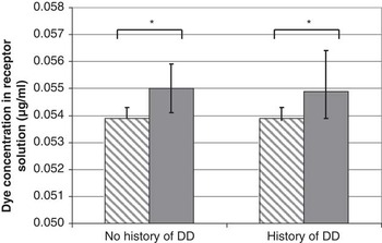
Keeping the skin samples for 24 h (without slurry treatment) increased the permeability of the samples by a mean (±s.d.) of 0.007 (±0.0066) absorbance units, and results from slurry-treated samples were adjusted accordingly. This value was subtracted from the absorbance values of all slurry-treated samples before the concentration of dye was calculated. There was no difference in the concentration of methylene blue dye in the receptor solution between DD+ and DD− animals in either the samples not treated with slurry (P = 0.560) or those treated with slurry (P = 0.999; Figure 1). Skin samples treated with slurry had significantly higher concentrations of dye in the receptor solution than samples from the same animal that were not treated with slurry (P = 0.010; Figure 1). The difference in dye concentration in the receptor solution between slurry-treated and untreated samples was the same for DD+ and DD− animals (P = 0.991). There was no difference between animals of different breeds in the dye concentration in the receptor solution from untreated skin (median (Q1, Q3); Holstein–Friesian = 0.054 (0.054, 0.054); Jersey × Holstein–Friesian =0.054 (0.054, 0.054); Norwegian and Norwegian × Holstein–Friesian = 0.054 (0.054, 0.054); P = 0.509). There was also no difference between animals of different breeds in the dye concentration in the receptor solution from slurry-treated skin (median (Q1, Q3); Holstein–Friesian = 0.055 (0.054, 0.056); Jersey × Holstein–Friesian = 0.056 (0.055, 0.056); Norwegian and Norwegian × Holstein–Friesian =0.055 (0.054, 0.057); P = 0.266).
The median concentration of methylene blue dye in the receptor solution for samples without slurry treatment  $$$$
and with slurry treatment
 $$$$
from cows with (n = 17) and without (n = 12) a history of DD. The error bars represent the interquartile range. *P < 0.05. DD = digital dermatitis.

Morphology
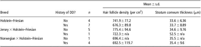
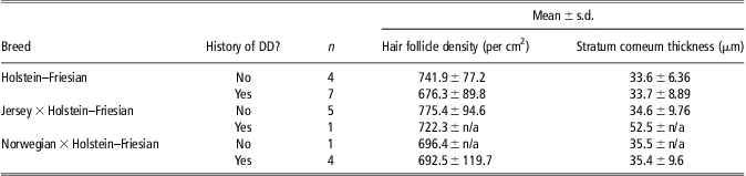
The number of DD+ and DD− animals of each breed, mean number of hair follicles per cm2 and stratum corneum thickness is shown in Table 1. No relationship was found between hair follicle density and DD susceptibility (P = 0.288) or breed (P = 0.722), and no interaction was found between the effects of breed and susceptibility (P = 0.881). There was also no relationship found between the thickness of the stratum corneum and DD susceptibility (P = 0.418) or breed (P = 0.538), and no interaction was found between breed and susceptibility (P = 0.285). No linear association was found between the thickness of the stratum corneum and the dye concentration in the receptor solution (r s = 0.115, P = 0.612).
The mean and standard deviation (s.d.) of hair follicle density and stratum corneum thickness for skin samples from animals of three breed groups with and without a history of digital dermatitis
Discussion
The small sample size used in the study caused a number of problems, not least the fact that there were small numbers of animals in some categories, meaning that results were more likely to be influenced by chance. The sample size was limited by a number of factors. First, only animals from the single herd that had been carefully monitored for DD could be used. Second, samples had to be collected and tested within a restricted period of time, in order to minimise the time when the samples were frozen. Finally, samples were taken from animals culled for herd management reasons so the number, breed and susceptibility characteristics of these animals could not be controlled.
The method used here of examining the feet for DD in the milking parlour is similar to that used by Thomsen et al. (Reference Thomsen, Klaas and Bach2008) and Stokes et al. (Reference Stokes, Leach, Main and Whay2012). When compared with the gold standard of lifting and examining feet in a crush, Stokes et al. (Reference Stokes, Leach, Main and Whay2012) found a sensitivity of 1.0 and a specificity of 0.99 for determination of the presence or absence of DD lesions, however, they had up to 7 min to examine each animal as the whole herd was not scored at once. Thomsen et al. (Reference Thomsen, Klaas and Bach2008) scored animals more quickly (15 s/cow) and found a more moderate and more variable sensitivity (0.65) and specificity (0.84) at the three farms included in their study. In addition to the imperfect sensitivity and specificity of the in-parlour detection method used, it is possible that the regular foot bathing regimen that the cows experienced could have led to some early lesions healing before detection. Although frequent observations were used to minimise incorrect classifications due to methodological factors and foot bathing frequency, it was not possible to completely eliminate this possibility.
Thickness of the stratum corneum and susceptibility to DD
The skin samples used to create the sections for morphological examination came from a position 3.5 to 4.5 cm above the top of the interdigital cleft. This is above the level where DD lesions were normally seen in this herd of dairy cows, so the skin sampled was unlikely to have been previously affected by DD.
No previous measurements of the thickness of the stratum corneum in bovine heel skin could be found, but values in this study broadly agree with previous findings for other sampling sites. Stahl et al. (Reference Stahl, Niedorf and Kietzmann2009) found that the mean thickness of bovine udder stratum corneum was 10.1 μm, smaller than the values found in the present study, but areas subject to more physical challenges generally do have a thicker stratum corneum (Egawa et al., Reference Egawa, Hirao and Takahashi2007). The lack of relationship in our data between stratum corneum thickness and skin permeability does not agree with the proposal of Holbrook and Odland (Reference Holbrook and Odland1974), that there is an inverse relationship between stratum corneum thickness and skin permeability in humans. However, that proposal was based on comparing stratum corneum thickness and permeability data from various sites in the body rather than from comparing the same site in different individuals, as was done here. Our findings do agree with Elias et al. (Reference Elias, Cooper, Korc and Brown1981), who found no relationship between permeability and stratum corneum thickness in humans and suggested that the skin permeability may be more closely linked to its lipid content than to the thickness of the stratum corneum.
Hair follicle density and susceptibility to DD
The stratum corneum layer is not completely continuous around the base of hair follicles, so there are areas of the hair follicle where it is easier for molecules (and presumably pathogens) to pass through (Wosicka and Cal, Reference Wosicka and Cal2010). Indeed, electron micrographs of tissues infected with DD have shown treponemes in the hair follicles and radiating out towards deeper parts of the tissues (Evans et al., Reference Evans, Brown, Demirkan, Singh, Getty, Timofte, Vink, Murray, Blowey, Birtles, Hart and Carter2009). Despite this, no difference was found between the number of hair follicles in animals with and without a history of DD in this study.
Skin permeability of animals with and without a history of DD
Franz diffusion chambers are widely used in dermatology and pharmaceutical research for the measurement of permeability in samples of human and animal skin (e.g. Bronaugh et al., Reference Bronaugh, Stewart and Simon1986; Dragicevic-Curic et al., Reference Dragicevic-Curic, Scheglmann, Albrecht and Fahr2009). The choice of methylene blue dye as a substance to use in the permeability testing was based on its successful in vitro use with pig skin (Johnson et al., Reference Johnson, Gallo, Hui and Oseroff1998) and rat skin (Genina et al., Reference Genina, Bashkatov and Tuchin2008) in Franz diffusion chambers and the simple detection method in the acceptor fluid using a spectrophotometer. Although the passive movement of the dye through the skin may be different to the movement of a larger, motile bacterium, the basic permeability of the epidermis does appear to be a valid representation of the skin barrier function, no matter what particle is used to test it. For example, the skin permeability of humans with atopic dermatitis has been measured using a number of different methods and all of them have found similar results (Yoshiike et al., Reference Yoshiike, Aikawa, Sindhvananda, Suto, Nishimura, Kawamoto and Ogawa1993; Sakurai et al., Reference Sakurai, Sugiura, Matsumoto and Uehara2002; Jakasa et al., Reference Jakasa, de Jongh, Verberk, Bos and Kezic2006).
The skin used for the permeability tests was not living, and is therefore not an exact representation of the skin of a living dairy cow. However, the most important layer of the skin for maintaining the skin's barrier function is the stratum corneum (Madison, Reference Madison2003), which is composed of ‘dead’ cells called corneocytes filled with keratin (Ersser et al., Reference Ersser, Getliffe, Voegeli and Regan2005). As the stratum corneum cells are ‘dead’ even in vivo, postmortem samples of human, pig and cattle tissues are frequently used as a model for live human tissue in pharmaceutical studies examining skin permeability (Pitman and Rostas, Reference Pitman and Rostas1982; Netzlaff et al., Reference Netzlaff, Schaefer, Lehr, Meiers, Stahl, Kietzmann and Niedorf2006). The skin samples used in this study were frozen for a maximum of 4.5 months before being defrosted and tested for permeability. Freezing the skin for this relatively short period of time should not have affected its permeability (Bronaugh et al., Reference Bronaugh, Stewart and Simon1986; Dragicevic-Curic et al., Reference Dragicevic-Curic, Scheglmann, Albrecht and Fahr2009).
No differences were found between the concentration of dye in the receptor solutions of skin from animals with and without a history of DD in this study, implying that differences in susceptibility to DD are unlikely to be caused by differences in the basic skin barrier function. There are, however, other aspects of skin biology which could affect susceptibility to infection, for example the strength of the innate immune response (Arikawa et al., Reference Arikawa, Ishibashi, Kawashima, Takagi, Ichikawa and Imokawa2002; McGirt and Beck, Reference McGirt and Beck2006), which have not been investigated with respect to DD.
The effect of slurry on skin permeability
Treatment of the skin with slurry for 24 h substantially increased the concentration of methylene blue dye in the receptor solution, indicating an increase in the permeability of the skin to methylene blue dye. This increase in permeability is likely to be caused in part by the high water content of slurry. Contact with water disrupts the stratum corneum structure of human skin in vivo after only 4 h (Warner et al., Reference Warner, Stone and Boissy2003) and contact with water for 48 h increases human skin permeability and susceptibility to bacterial colonisation (Farage et al., Reference Farage, Miller, Berardesca and Maibach2007). Other components of slurry may also have a damaging effect on the skin. Gregory (Reference Gregory2004) found that urea and sulphide anions could cause the heel horn to swell when used at concentrations found in cattle urine. This is because urea disrupts hydrogen bonds and sulphide ions uncouple disulphide bridges in keratin, causing the keratin to unfold and swell as it takes up water (Gregory, Reference Gregory2004). As keratin is also a major component of the stratum corneum layer of the skin (Nemes and Steinert, Reference Nemes and Steinert1999), these mechanisms of softening and swelling of keratin may also occur in heel skin when in contact with slurry, which could result in an increase in skin permeability. In addition to the damaging affect that it has on keratin, urea in urine is converted to ammonia, which with prolonged contact raises the skin pH and destroys the acid mantle (Farage et al., Reference Farage, Miller, Berardesca and Maibach2007). The acid mantle contributes to the skin's resistance to microbial invasion (Ersser et al., Reference Ersser, Getliffe, Voegeli and Regan2005) and its loss allows irritants and pathogens to penetrate the skin more easily (Farage et al., Reference Farage, Miller, Berardesca and Maibach2007).
Conclusions
Individual differences in the natural permeability or morphological characteristics of the skin of dairy cows did not appear to be related to susceptibility to DD; however, other characteristics of the skin including the innate immune response have not yet been investigated. Contact with slurry increased skin permeability, which could facilitate the entry of pathogens. Further work is needed to clarify the role played by slurry in the pathogenesis of DD.
Acknowledgements
Assistance from staff at AFBI Hillsborough (particularly Mr B. Dawson and Mr S. Martin), staff and students working in the laboratory of Dr R. Donnelly and the Tissue Core Technology Unit at Queen's University Belfast is gratefully acknowledged. M. Palmer acknowledges DARDNI for receipt of a Postgraduate Studentship.