In dairy farms high humidity, elevated temperature, poor airflow in parlours, and other animal-housing related problems have been associated with decreased milk production and increased mastitis prevalence. Regarding the effect of HS on immune system, the concensus conclusion is that cows exposed to HS have depressed immunity thus reducing resistance to intramammary infections (IMI) (Nardone et al. Reference Nardone, Ronchi, Lacetera, Ranieri and Bernabucci2010; Smith et al. Reference Smith, Smith, Rude and Ward2013). Work conducted in our and other labs showed that, at cellular level, HS has an important effect on intra- and extracellular signals (Kaufman et al. Reference Kaufman, Kassube, Baravalle and Ríus2015; Li et al. Reference Li, Sun, Wu, Li, Luo and Wang2015). This alters the cell cycle progression, regulation of the cytoskeleton and ultimately the cellular adaptation in response to stress settings. Interaction of host cells with microbial pathogens imposes a stress condition in which the host cell response is crucial for the success or failure of the infection. It is possible that host cells under HS react differently from cells in normal physiological temperature, thus leading to the increased susceptibility to infection observed in HS-animals. To gain insight on the effect of HS on the pathogenesis of S. uberis IMI, HS bovine mammary epithelial cells (BMEC) were co-incubated with Streptococcus uberis and the viability of the epithelial cells, development of membrane damage, as well as number of bacteria attached to and/or internalised into mammary epithelial cells were evaluated.
Previous studies showed that under normal temperature S. uberis adhered to and internalised into bovine mammary epithelial cells (Almeida & Oliver, Reference Almeida and Oliver2006; Batcha et al. Reference Batcha, Almeida, Dunlap and Oliver2006), without significantly affecting BMEC viability. Further studies showed that S. uberis exploit preferential intracellular trafficking pathways to achieve persistence into the host (Almeida & Oliver, Reference Almeida and Oliver2006; Batcha et al. Reference Batcha, Almeida, Dunlap and Oliver2006). However, there is no data on the outcome of S. uberis cocultured with HS BMEC, and such information could provide additional understanding on the effect of HS on the pathogenesis of S. uberis IMI. Thus, the objective of this investigation was to assess initial events on the interaction of S. uberis with HS BMEC.
Material and methods
Streptococcus uberis UT888, originally isolated from a cow with mastitis, was plated onto blood agar plates (BAP) and incubated overnight at 37 °C. For adherence and internalisation, 3 isolated colonies were transferred into a Todd Hewitt broth (THB), incubated for 16 h at 37 °C in a 5% CO2: 95% air incubator. After incubation, the bacterial suspension was washed 3 times diluted 1: 100 in Dulbecco's Modified Eagle's medium (DMEM) to ~106 CFU/ml, and cocultured with a bovine mammary epithelial cell line (MAC-T cells; Huynh et al. Reference Huynh, Robitaille and Turner1991). The adherence and internalisation assays were performed as described (Almeida & Oliver, Reference Almeida and Oliver2006), with the exceptions that for HS, BMEC were pre-cultured without foetal bovine serum for 10 h at 43 °C in a 5% CO2: 95% air incubator. As control, BMEC were cultured at 37 °C in a similar culture medium and conditions as described for HS BMEC cells. After pre-incubation at 43 °C or 37 °C, HS and non-HS BMEC were cocultured for 60 min at 37 °C with S. uberis at multiplicity of infection of 10 : 1 (bacteria : BMEC).
Cell viability was determined by Trypan blue exclusion. HS and non-HS BMEC cocultured with or without S. uberis, were incubated with Trypan-blue (0·2% final cc) for 4 min. After incubation, dye exclusion was determined by inspecting 20 fields at 200× using inverted light microscope.
Cell damage was determined by measuring lactate dehydrogenase activity in the supernatant of co-cultures at 1 and 2 h post-infection and comparing them with LDH activity of untreated BMEC as described (Almeida & Oliver, Reference Almeida and Oliver1995). Total activity was measured from BMEC lysate of HS and non-HS monolayers. The LDH activity was expressed as Berger–Broida units per millilitre and determined by a colorimetric assay for pyruvate, with the amount of residual pyruvate inversely proportional to the amount of LDH activity (Sigma-Aldrich; https://www.sigmaaldrich.com/content/dam/sigmaaldrich/docs/Sigma/Bulletin/1/mak066bul.pdf).
Unless otherwise noted, assays were performed three times with each condition tested in triplicate. Means from each experiment were analysed by two-way analysis of variance and those showing statistically significant differences (P ≤ 0·05) were further analysed by Student's t test using ProStat (Poly Software International, Salt Lake City, UT) statistical software.
Results and discussion
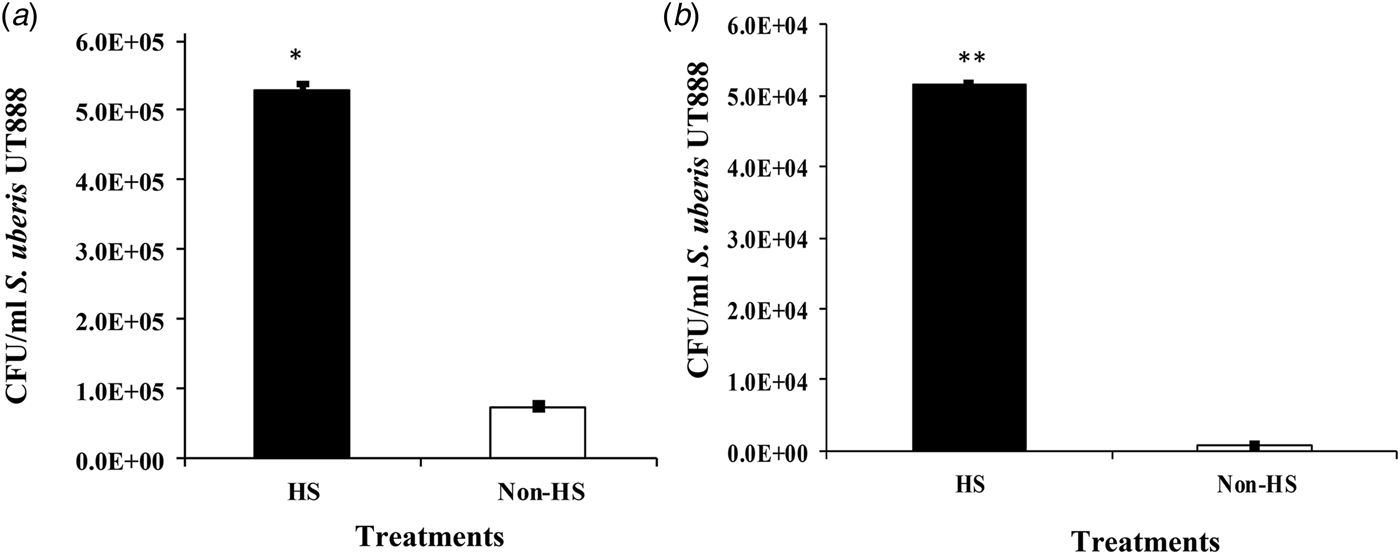
Average adherence of S. uberis to BMEC cultured at 37 °C was 7·4 × 104, which was consistent with previous observations (Almeida & Oliver, Reference Almeida and Oliver2006). However, when S. uberis was cocultured with HS BMEC, the average adherence was 5·3 × 105 CFU/ml, which was 0·85 log10 higher than the non-HS controls (Fig. 1a). These differences were statistically significant (P < 0·05). Averaged values for intracellular S. uberis for non- or HS BMEC were 7·4 × 102 and 5·2 × 104 CFU/ml, respectively, representing 1·85 log10 difference (Fig. 1b). The number of intracellular S. uberis was higher when cocultures were conducted with HS BMEC as compared with non-HS controls. However, different from adherence, differences between treatments more noticeable (differences of 0·85 vs. 1·85 log10 for adherence and internalisation, respectively).
Adherence and internalisation of Streptococcus uberis into HS and non-HS BMEC. Streptococcus uberis UT888 was cocultured with HS and non-HS BMEC for 60 min and adherence (A) and internalisation (B) calculated. Data are presented as colony forming units per ml (CFU/ml) and error bars are the standard error of the mean (sem) of three independent observations run in triplicate. *P ≥ 0·05.

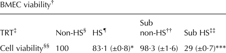
Results presented in Table 1, showed a moderate reduction (P ≥ 0·05) of viability in HS-treated as compared to non-HS BMEC (83·1 vs. 100%, respectively). Still, no significant reduction in viability was observed between non-HS BMEC and non-HS BMEC cocultured with S. uberis. This data indicated that adherence and internalisation of S. uberis to BMEC increased significantly when the assay was conducted with HS-BMEC. However, when the rate of adherence and internalisation were compared to the corresponding to non-HS BMEC controls, it was observed that differences were more marked with internalisation than with adherence. In other words, it seems that the ratio internalised: adhered S. uberis was higher in HS than in non-HS BMEC. A possible explanation for this observation may be that S. uberis exploits the host cell signal transduction and cytoskeleton to induce its own uptake and trafficking in the host cell cytoplasm (Almeida et al. Reference Almeida, Calvinho and Oliver2000). The uptake of S. uberis into BMEC occurred through the classical endocytosis and the caveolae/lipid raft pathways (Almeida & Oliver, Reference Almeida and Oliver2006). Streptococcus uberis internalised into BMEC through the classical endocytosis pathways face intracellular bactericidal mechanisms, which cause reduction in the number of internalised S. uberis. Conversely, S. uberis internalised through the caveolae/lipid raft pathway survives, escapes from the caveloae vesicle becoming free in the BMEC cytoplasm, and therefore transcytosing and internalising into adjacent BMEC (Almeida & Oliver, Reference Almeida and Oliver2006). Since it has been reported that the activity of the cytoskeleton is affected by HS, it is possible that the translocation of endocytic vesicles, as well as their progressive pH reduction and fusion with lysosome is negatively affected, thus leading to increased number of S. uberis in the BMEC cytoplasm. In this project, we did not generate data supporting this conjecture, therefore, this theory requires additional research in order to be validated.
Cell viability and cell damage of bovine mammary epithelial cells under heat stress and when cocultured with S. uberis UT888

† BMEC viability under HS and non-HS conditions was evaluated by trypan-blue exclusion.
‡ BMEC were cultured with CGM with insulin, but without hydrocortisone or foetal bovine serum at 37 °C in 5% CO2: 95% air (vol/vol) at 37 °C (non-HS) or at 43 °C and cocultured with S. uberis UT888.
§ BMEC incubated at 37 °C.
¶ BMEC pre-incubated at 43 °C for 10 h.
†† BMEC incubated at 37 °C and cocultured with S. uberis UT888.
‡‡ BMEC pre-incubated at 43 °C for 10 h previous to coculture with S. uberis UT888.
§§ Numbers are the percentage of cell viability taking as 100% the corresponding non-heat stress (non-HS) treatments. Numbers in parenthesis are the standard deviation (sd) of 20 independent observations.
*P ≥ 0·05.
***P ≥ 0·001.
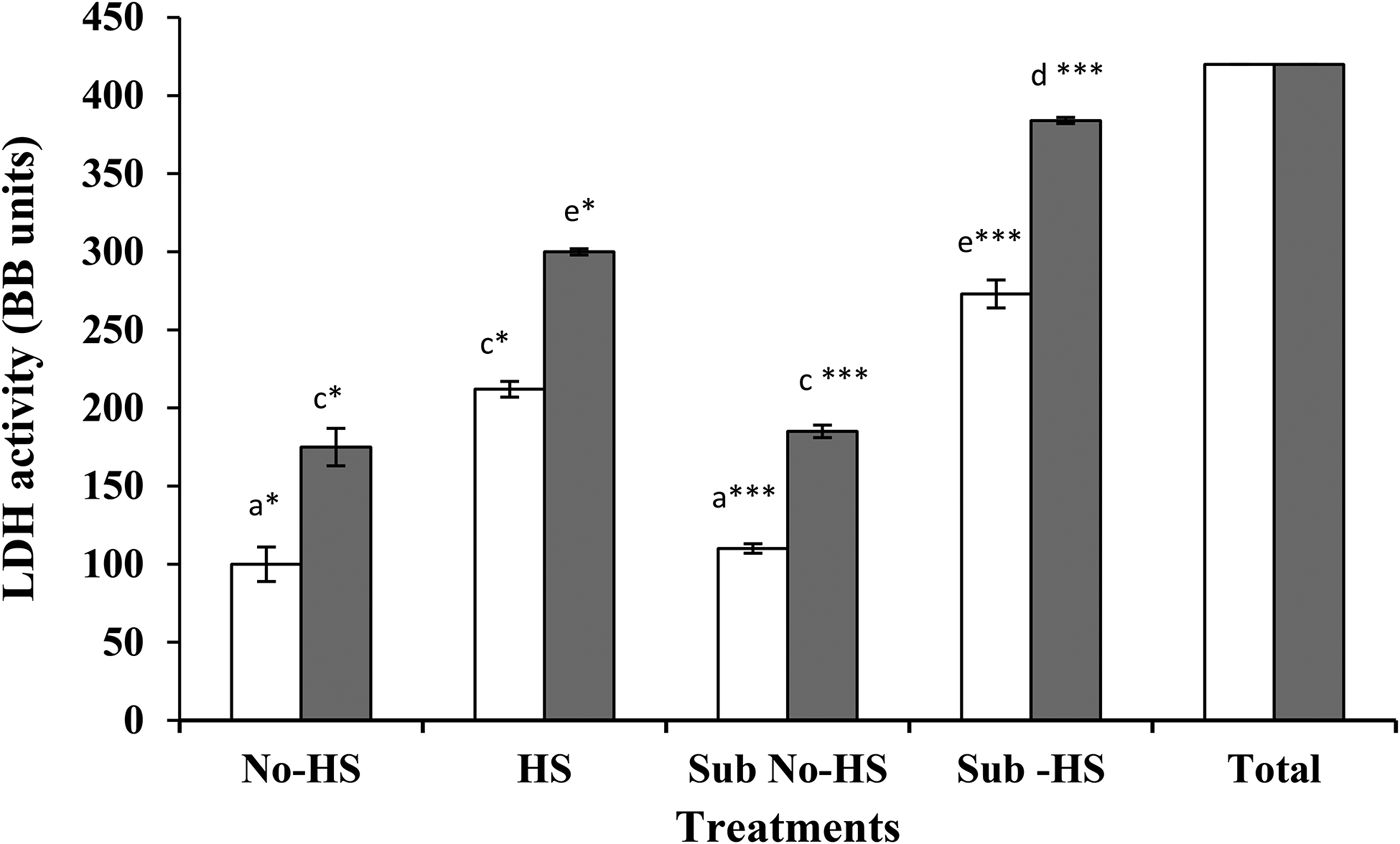
LDH activity in the BMEC supernatant was taken as an indication of membrane damage. LDH values, which followed the pattern described for trypan blue exclusion, were higher in HS-treated as compared with non-treated BMEC. For all the treatments, LDH activity increased with time. Significant differences were found in LDH activity in non-HS BMEC with or without coculture with S. uberis. However, high significant values were detected in the supernatant of HS BMEC cocultured with S. uberis (Fig. 2).
Lactate dehydrogenase (LDH) released by BMEC heat stressed and cocultured with Streptococcus uberis UT888. LDH was measured after 60 (white bars) and 120 min (grey bars) of incubation in the supernatant of heat stressed (HS) BMEC cocultured with S. uberis (Sub-HS). Controls were not heat stressed BMEC (No-HS), w/o coculturing with S. uberis (Sub No-HS). Total LDH was measured from untreated lysed BMEC and expressed in Berger-Broida Units (BB). Error bars are the sem of 3 independent experiments run in triplicate. Bars with different caption letters were statistically different. *P ≥ 0·05, ***P ≥ 0·001.

Data from this research showed a slight reduction (although significant) in cell viability and BMEC membrane damage when cultured under HS conditions. Conversely, interaction between S. uberis and BMEC did not induce apparent pathological changes on BMEC. However, when HS BMEC were cocultured with S. uberis, adherence and internalisation of this pathogen into BMEC increased dramatically. Parallel with the increased number of BMEC-associated S. uberis the percentage of non-viable BMEC as well as membrane damage, increased significantly. At this point, we cannot pinpoint the mechanisms involved in the damage of BMEC. Previous results showed that HS impaired the translation of protein by altering the phosphorylation activity of mTOR signalling factors in MAC-T cells (Kaufman et al. Reference Kaufman, Kassube, Baravalle and Ríus2015) as well as affecting gene regulation and expression in HS-BMEC. It could be that the effect of HS on cell regulation and metabolism are enhanced by the additional stress imposed by the internalisation of S. uberis, thus leading to further detriment on BMEC and diminution of cell viability and membrane damage. However, more research is needed to define specific altered functions and linked metabolic activities affected by the internalisation of S. uberis on HS-BMEC.
In conclusion, HS conditions induced increased adherence to and internalisation into BMEC as well as reduction on BMEC viability. The negative effect of HS and S. uberis combined was higher than that induced by each condition alone, suggesting synergistic interaction between these conditions. This interaction together with the negative effect of HS on the immune system may explain the increased susceptibility to IMI, observed in HS cows.
This work was funded by a grant from the Innovation Grant programme of The University of Tennessee Agricultural Experiment Station.
Disclosure
The authors declare no conflict of interest.