Introduction
Microsporidial Gill Disease of Salmon (MGDS), caused by the microsporidian Loma salmonae, once considered an emerging disease in Canadian aquaculture, is now best considered as an endemic disease with a strong seasonal trend favoring late summer and early fall of each year. The first reported case of MGDS in British Columbia, Canada was in coho salmon (Oncorhynchus kisutch) smolts from a hatchery on Vancouver Island in 1987 (Magor, Reference Magor1987). Globally, MGDS is reported among rainbow trout (Oncorhynchus mykiss) on farms in Scotland and England and in hatcheries in Georgia, USA (Poynton, Reference Poynton1986; Markey et al., Reference Markey, Blazer, Ewing and Kocan1994; Bruno et al., Reference Bruno, Collins and Morrison1995; Gandhi et al., Reference Gandhi, Locatelli and Feist1995; Bader et al., Reference Bader, Shotts, Steffens and Lom1998). Since its initial discovery in Canada, L. salmonae has been identified as an important salmonid pathogen for the British Columbia Pacific salmon industry; MGDS is characterized as a severe inflammatory gill disease, with variable (generally lesser) degrees of systemic organ involvement, high mortality rates, and prolonged recovery periods during which production efficiency is severely affected (Kent et al., Reference Kent, Elliot, Groff and Hedrick1989, Reference Kent, Dawe and Speare1995; Speare et al., Reference Speare, Arsenault and Buote1998a; Constantine, Reference Constantine1999). Until recently, little pertinent information on the transmission of this pathogen was available. Accordingly, management techniques to minimize MGDS on fish farms were limited and were not linked to specific attributes of the causative agent. The earlier reports of L. salmonae from freshwater hatcheries initially led to the concept that the pathogen infected young salmon while they were in their juvenile freshwater production phase and that clinical expression of the disease (MGDS) arising later in the saltwater production phase of salmon stemmed from recrudescence following prolonged latency of the pathogen within an infected host. Strategies to screen juvenile salmon for L. salmonae, prior to the transfer of these salmon to marine net-pen sites, were therefore considered a key management tool. Current findings reverse this early assumption and demonstrate the ease with which this parasite transmits horizontally within environments of widely different salinities. Understanding the extracorporeal and corporeal persistence of the spore stages of this pathogen, and developing models to better evaluate the efficiency of horizontal transmission provide a strategic basis to limit the effect of MGDS on high host density salmon farms.
Impact of L. salmonae on the Canadian Pacific salmon industry
Salmon farming in British Columbia began in the early 1970s and the industry has grown to become the fourth largest producer of farmed salmon in the world after Norway, Chile and the United Kingdom (Ministry of Agriculture and Lands (MAL), 2005). In 2003, the industry produced 72,700 tonnes of salmon, resulting in over (CDN) $255 million for the provincial economy (MAL, 2005). The main fish species cultured in British Coulmbia are Atlantic salmon (Salmo salar), chinook salmon and coho salmon, representing 76, 22 and 2% of the production, respectively (MAL, 2005). Although it appears that the chinook salmon production is minor compared to Atlantic salmon, chinook salmon are the third most cultured aquaculture species in Canada (based on tonnage) and the second most valuable cultured species (after Atlantic salmon). Following a brief hatchery phase, Chinook salmon postsmolts are reared in marine-based net-pens and this approach is economical because of the relatively low construction cost involved and the absence of a requirement to pump water (Kent, Reference Kent, Kent and Poppe1998). However, this form of aquaculture allows for the exacerbation of infections with certain disease agents, such as L. salmonae (Georgiadis et al., Reference Georgiadis, Gardner and Hedrick2001) and presents some unique fish health problems, particularly interactions with wild and feral fish, because of the shared ocean environment. Since 1998, the production of farmed chinook salmon has risen from 6,600 tonnes to over 15,500 tonnes in 2003 (MAL, 2005), coinciding with increased losses attributed to L. salmonae, with cumulative mortality reported to be as low as 3% and as high as 13% (Hauck, Reference Hauck1984; Constantine, Reference Constantine1999; J. Lovy, personal communication). Presumably, the observed increase in MGDS outbreaks is indirectly related to the more than doubling in the production of chinook salmon and this upward production trend will maintain L. salmonae as an important pathogen for the future salmon industry in Canada. Chinook salmon production, although not interchangeable with Atlantic salmon production on any particular farm site, is poised for further growth due to diminishing returns from Atlantic salmon farming, and the emergence of several diseases for which Atlantic salmon are highly susceptible in their non-native environment of coastal British Columbia.
Biology of L. salmonae
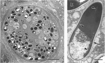
Until recently, microsporidians have been viewed somewhat as a biological oddity; as their host range is most typically within lower vertebrates and invertebrates, their study has not benefited from the larger interests of medical sciences. The recent emergence of microsporidia as agents of disease of humans, especially immunosuppressed AIDS patients has spurred a huge increase in research into the basic and applied biology of these organisms. Recently reclassified from protozoa to fungi (although in this review they will be referred to as protozoans), many aspects of their life cycle, restricted host range, intracellular life style, transmission and environmental persistence remain an enigma. As a group they are known to be obligate intracellular protozoan parasites of eukaryotes with the transmissible stage being a resistant spore, which is small, possesses a thick wall and contains a characteristic polar tube apparatus (Fig. 1) (Canning and Lom, Reference Canning and Lom1986; Wittner, Reference Wittner, Wittner and Weiss1999; Didier et al., Reference Didier, Didier, Snowden and Shadduck2000; Lom and Nilsen, Reference Lom and Nilsen2003). Microsporidians are unusual in that they lack mitochondria and presumably rely on their host cell for obtaining cellular energy (Canning and Lom, Reference Canning and Lom1986). Additionally, members of this phylum have a Golgi apparatus possessing an accumulation of small, opaque vesicles enclosed by a single membrane forming a meshwork, which eventually forms the individual parts of the spore extrusion apparatus, notably consisting of the polar tube which is responsible for transferring the sporoplasm (the parasite genetic material) to the host cell (Fig. 1B) (Vávra and Larsson, Reference Vávra, Larsson, Wittner and Weiss1999; Keeling and Fast, Reference Keeling and Fast2002). Microsporidians, especially those infecting fish hosts, are embedded directly in the cytoplasm of the host cell causing enormous hypertrophy of the cell to transform it into a special structure called the xenoma, in which the developing parasite and host cell represent a physiologically integrated unit (Fig. 1A) (Lom and Nilsen, Reference Lom and Nilsen2003).
(A) Xenoma within the primary gill filament of a chinook salmon containing a hypertrophied nucleus (N), meronts (arrowhead) and spores. Bar=8 μm. (B) L. salmonae spore with the electron-dense exospore (arrow) adjacent to the translucent endospore and characteristic coiled polar tube (arrowheads). Bar=700 nm. (Photos courtesy of J. Lovy.)

Recent morphological evidence has suggested that the host endoplasmic reticulum is the source of membranes forming the parasitophorous vacuole during xenoma formation (Lovy et al., Reference Lovy, Wright, Wadowska and Speare2006). Xenoma size is variable amongst the genera, for example xenomas during a L. salmonae infection are approximately 0.4 mm, whereas the xenomas produced during a Glugea infection can be up to 13 mm (Canning and Lom, Reference Canning and Lom1986). Eventually the xenoma becomes too large and ruptures, releasing spores into the environment to infect new hosts. L. salmonae infects endothelial cells, causing the formation of xenomas throughout vascularized organs, including kidney, heart, spleen and liver but the majority of the infection occurs in the gills (Fig. 1A) (Shaw and Kent, Reference Shaw, Kent, Wittner and Weiss1999; Rodriguez-Tovar et al., Reference Rodriguez-Tovar, Wright, Wadowska, Speare and Markham2002; Lom and Nilsen, Reference Lom and Nilsen2003).
The life cycle for microsporidians is marked by three phases: the infective phase, the proliferative phase and the sporogonic phase. There are several reviews (Canning and Lom, Reference Canning and Lom1986; Didier, Reference Didier1998; Bigliardi and Sacchi, Reference Bigliardi and Sacchi2001; Dunn and Smith, Reference Dunn and Smith2001) on the life cycle of microsporidians and only specific information pertaining to L. salmonae is noted. The infective phase involves mature spores in the environment (e.g. ocean net-pen) which are ingested by the host (chinook salmon), next the spore germinates in the gut with the sporoplasm being injected into a host cell and finally the infected host cells are transported to other parts of the body to complete the life cycle (Cali and Takvorian, Reference Cali, Takvorian, Wittner and Weiss1999; Sánchez et al., Reference Sánchez, Speare, Markham, Wright and Kibenge2001d). There has been considerable study into the events leading to polar tube eversion, and in summary these would suggest that the low pH environment of the gut is a necessary step for extrusion of the polar filament. However, recent work has also shown the ability of at least some microsporidian pathogens to transmit vertically (transovarial), through the skin, or following experimental intraperitoneal injection. It is possible that subsequent to intracellular uptake of an intact spore, pH declines within a phagosome may create a condition that allows for polar tube extrusion, thus affording a non-conventional route for transmission.
Based on experimental per os infection studies, and using PCR and in situ hybridization (ISH), L. salmonae can be detected in the gut mucosal epithelium of rainbow trout as early as 24 h post exposure (PE) and becomes localized in the lamina propria of the intestine within this time period (Sánchez et al., Reference Sánchez, Speare, Markham, Wright and Kibenge2001d). The proliferative phase (also known as merogony) includes all cell growth and division from the sporoplasm through the parasite's commitment to spore formation (Cali and Takvorian, Reference Cali, Takvorian, Wittner and Weiss1999). During a L. salmonae infection, ISH shows that dividing merogonic stages in infected cells are detectable in the heart as early as 2 days PE (Sánchez et al., Reference Sánchez, Speare, Markham, Wright and Kibenge2001d). The infective sporoplasm of L. salmonae seems to initiate early merogonic development within 5 days following infection (Sánchez et al., Reference Sánchez, Speare, Markham, Wright and Kibenge2001d) and by the third week of the infection, meronts are the first recognizable parasite stage (Rodriguez-Tovar et al., Reference Rodriguez-Tovar, Wright, Wadowska, Speare and Markham2002). L. salmonae meronts are uninucleate or binucleate structures up to 3 μm in diameter, which develop into elongate plasmodia with at least five nuclei (Canning and Lom, Reference Canning and Lom1986).
The proliferative phase during a L. salmonae infection is most frequently completed in the gills, where the final phase begins. The sporogonic phase or sporogony culminates in the production of sporoblasts, which eventually undergo morphogenesis to develop into mature spores (Canning and Lom, Reference Canning and Lom1986). Sporoblasts mature into a spore with the complete formation of all the internal structures, notably including the extrusion apparatus, which consists of the anchoring disc, polar tube, polaroplast membranes and the posterior vacuole (Cali and Takvorian, Reference Cali, Takvorian, Wittner and Weiss1999). L. salmonae DNA can be found in the gills beginning at 2 weeks PE (Sánchez et al., Reference Sánchez, Speare and Markham2001a).
An unexplored feature of the life cycle of microsporidians is the process by which an obligate intracellular pathogen is able to migrate from initial sites of infection (usually the gut) to what are in some cases highly specific organ and cell locations. In the case of L. salmonae, it appears that translocation to the gill takes place within a host inflammatory cell which is taken up by the gill pillar cells; development to the xenoma stage occurs subsequently (Rodriguez-Tovar et al., Reference Rodriguez-Tovar, Wright, Wadowska, Speare and Markham2002). This has potential significance for treatment, in that targeting of pre-xenoma stages of the parasite has been shown to be the only effective approach when various compounds have been evaluated.
Treatment
Historically, it has proven difficult to develop effective control strategies and therapies against disease-causing microsporidia. This has mainly been attributed to their intracellular localization in host cells and to the resistant infectious spore (Canning and Lom, Reference Canning and Lom1986); another roadblock has been the failure to develop models of infection in which levels of infection can be quantified. Several drugs have been used to treat microsporidial infections in fish and humans, but mainly on an experimental basis. The major therapies described center on the use of the antibiotic, fumagillin, and the antihelminthic, albendazole (Chinabut et al., Reference Chinabut, Tonguthai, Kamlerd, Shariff, Subasinghe and Arthur1992; Didier, Reference Didier1998; Higgins et al., Reference Higgins, Kent, Moran, Weiss and Dawe1998; Speare et al., Reference Speare, Athanassopoulou, Daley and Sánchez1999a; Conteas et al., Reference Conteas, Berlin, Ash and Pruthi2000; Costa and Weiss, Reference Costa and Weiss2000; Didier et al., Reference Didier, Didier, Snowden and Shadduck2000).
In contrast to the problems facing therapeutic models for microsporidial diseases, studies on MGDS have several advantages: the disease is easily reproduced using standardized challenge techniques, and xenomas on the gill can be quantified, thus making MGDS an excellent general model for evaluating therapeutic strategies. For example, the sodium ionophore monensin was examined (Speare et al., Reference Speare, Daley, Dick, Novilla and Poe2000); this agent modifies intracellular ion channels and selectively acts on post-Golgi endosomes which are key for polar tube formation (Dinter and Berger, Reference Dinter and Berger1998). Blocking this development may halt spore formation, and this approach represents a unique strategy. A pilot study revealed that monensin-treated rainbow trout exposed to an oral dose of L. salmonae spores showed a 93% reduction in xenoma production (Speare et al., Reference Speare, Daley, Dick, Novilla and Poe2000). Subsequently, a study investigated the minimum monensin dose and treatment time required for therapeutic success. Rainbow trout offered monensin-treated feed at a concentration of 1000 ppm (fed daily at a rate of 2% of their body weight) showed the greatest reduction in xenoma formation of 69% at week 7 PE compared to the similarly exposed non-treated fish, while maintaining similar growth rates as non-exposed fish (Becker et al., Reference Becker, Speare, Daley and Dick2002). Additionally, at a dose of 1000 ppm of monensin, treatment must be started at the time of exposure or one week before to be effective at reducing the numbers of branchial xenomas. As described above, L. salmonae is localized in the gut and heart during the first week of the parasite life cycle, which was in concordance with most beneficial therapy period, indicating that once the parasite has localized in the gills, the therapy was no longer effective. By reducing xenoma formation, and thus reducing the production of infectious spores, the effectiveness of treatment is not only aimed at the exposed fish, but also the fish which would otherwise become exposed when spores are liberated from an infected fish. This appears to be the central theme for treatment. Whereas it is unlikely that the disease will be completely blocked by treatment, reducing the xenoma burden and consequently the numbers of spores being produced and liberated, the infectious dose in the surrounding water should be directly diminished.
Host factors
Generally, transmission factors that fall under the umbrella of host factors are considered constantly present, such as host species, fish size, population size or nutritional status (Hedrick, Reference Hedrick1998). Experimental infection models have been developed using rainbow trout (Speare et al., Reference Speare, Arsenault and Buote1998a), chinook (Kent et al., Reference Kent, Dawe and Speare1995) and coho salmon (Shaw et al., Reference Shaw, Kent and Adamson1998; Ramsay et al., Reference Ramsay, Speare, Dawe and Kent2002) as hosts; however experimental transmission has not been demonstrated in Atlantic salmon or Arctic charr (Salvelinus alpinus) (Speare et al., Reference Speare, Arsenault and Buote1998a; Shaw et al., Reference Shaw, Kent and Adamson2000) despite repeated attempts. Furthermore, non-salmonids are not susceptible to L. salmonae (Shaw et al., Reference Shaw, Kent and Adamson2000). Brook trout (Salvelinus fontinalis) as a host, are susceptible to MGDS caused by a Salvelinus-infecting variant of L. salmonae (discussed further under the Pathogen factors section) (Sánchez et al., Reference Sánchez, Speare, Markham and Jones2001c) which is apparently carried by Chinook salmon.
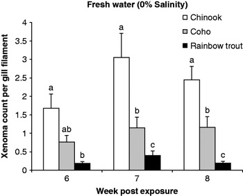
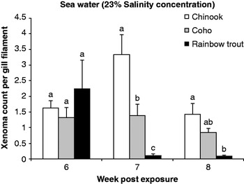
Potential exists for MGDS to serve as a useful model to further examine the role of innate immunity in the differential susceptibility of various salmonid species to this microsporidian. Early reports of MGDS outbreaks reported that rainbow trout generally experienced lower rates of mortality and developed fewer branchial xenomas compared to chinook and coho salmon (Poynton, Reference Poynton1986; Bruno et al., Reference Bruno, Collins and Morrison1995). Since this time, a laboratory study confirmed this and revealed that chinook salmon showed significantly higher numbers of xenomas, which persisted for longer time periods in both fresh and sea water compared to rainbow trout while coho salmon demonstrated an intermediate level of disease between these two fish hosts (Figs. 2 and 3) (Ramsay et al., Reference Ramsay, Speare, Dawe and Kent2002). Interestingly, although onset of xenomas occurred at weeks 5 and 6 PE for all three species, rainbow trout exhibited rapid xenoma clearance with complete recovery by week 9 PE compared to both chinook and coho, which both still showed at least on average one xenoma per gill filament at week 9 (Figs. 2 and 3) (Ramsay et al., Reference Ramsay, Speare, Dawe and Kent2002). Kent et al. (Reference Kent, Dawe and Speare1999) reported that chinook salmon exposed to an oral dose of L. salmonae spores developed high numbers of branchial xenomas by week 7 PE and the infection was cleared by week 20 PE, with no xenomas visible at this time. Additionally, differences in disease susceptibility to L. salmonae were demonstrated amongst the three strains of chinook salmon used by the aquaculture industry in British Columbia, Canada. Chinook salmon endemic to the Yukon river in the Yukon Territory, Canada were observed to have higher numbers of xenomas following a laboratory oral challenge to L. salmonae spore compared to the strain of salmon from Big Qualicum River, Vancouver Island, Canada, and a third strain that was a hybrid of the two strains (Shaw et al., Reference Shaw, Kent and Adamson2000). It was suggested that the increased mortality and xenoma levels observed in the northern Yukon strain of chinook indicated a naïve strain of salmon that may be unable to mount an effective immune response to the parasite compared to strains of chinook from areas where L. salmonae is found in the wild populations (Shaw et al., Reference Shaw, Kent and Adamson2000). Whether the phenomenon relates to adaptive immunity or innate immunity has yet to be determined, but remains a crucial question.
Mean number of branchial xenomas observed in chinook and coho salmon and rainbow trout held in fresh water during an experimental L. salmonae infection. Within each week post exposure, columns with different letters represent statistically significant differences (P<0.05).

Mean number of branchial xenomas observed in chinook and coho salmon and rainbow trout held in sea water during an experimental L. salmonae infection. Within each week post exposure, columns with different letters represent statistical significance (P<0.05).

An exciting area of study relates to the high degree of adaptive immunity that develops following recovery from infection, and following the use of experimental vaccines; differences between Loma-susceptible salmonids deserve further investigation and this could be a useful window through which to compare the dynamics of cellular immune mechanisms between salmonid species. Both chinook salmon (Kent et al., Reference Kent, Dawe and Speare1999) and rainbow trout (Speare et al., Reference Speare, Beaman, Jones, Markham and Arsenault1998b) are resistant to re-infection following recovery from a L. salmonae infection; however brook trout do not develop this resistance (Speare and Daley, Reference Speare and Daley2003). Rainbow trout recovered from a L. salmonae infection were protected from MGDS in that they did not develop xenomas when challenged 36 weeks after an initial exposure to either live or dead spores, suggesting a strong potential for the development of a vaccine to prevent MGDS (Speare et al., Reference Speare, Beaman, Jones, Markham and Arsenault1998b; Kent et al., Reference Kent, Dawe and Speare1999). A prototype vaccine, based on frozen (inactivated) spores injected in the intraperitoneal (IP) cavity of naïve rainbow trout induced protection against MGDS (Speare et al., Reference Speare, Beaman, Jones, Markham and Arsenault1998b). Further research revealed that rainbow trout injected with a spore-based vaccine against L. salmonae developed strong protection by week 4, with complete protection observed 6 weeks after vaccination (Rodriguez-Tovar et al., Reference Rodriguez-Tovar, Becker, Markham and Speare2006).
More recently, it was also reported that fish size was found to be a significant host-related factor for MGDS caused by L. salmonae (Becker et al., Reference Becker, Speare and Dohoo2005b). Using a cohabitation experimental challenge model, rainbow trout ranging from 17 to 23 g had a significantly faster rate of development of xenomas with the median onset time ranging from 7 to 11 days sooner compared to the two larger size groupings of 32–38 and 57–63 g. Moreover, generally the smallest group had significantly higher numbers of xenomas observed on the gill filaments with a maximum average of one xenoma per filament compared to an average of less than one xenoma per filament for the other two size groups. Interestingly, MGDS was first reported in smolts and this research would indicate that smaller fish are more susceptible to L. salmonae; however at the aquaculture sites, typically larger (almost market size) salmon succumb to disease. The conflicting observation indicates that fish size as a host factor is most likely interacting with other transmission factor(s) that are present at the sea cage but absent from the laboratory. Size-related susceptibility has been reported in other significant salmonid pathogens. Notably, it has been demonstrated that small (1.7 and 0.2 g, respectively) rainbow trout and kokanee salmon (O. nerka) are more susceptible to infectious hematopoietic necrosis virus (IHNV) compared with larger sizes (7.4 and 7.2 g, respectively) (Lapatra et al., Reference Lapatra, Groberg, Rohovec and Fryer1990; Lapatra, Reference Lapatra1998).
Becker et al. (Reference Becker, Speare and Dohoo2005b) also reported that feeding ratio (of 1, 2 and 4% of fish biomass daily) did not alter the onset or resulting intensities of branchial xenomas in rainbow trout exposed to L. salmonae via a cohabitation challenge model. Additionally, growth suppression was not observed in L. salmonae-infected trout (exposed using a low dose cohabitation challenge) compared to naïve control fish (Becker et al., Reference Becker, Speare and Dohoo2005b). However, a reduction in specific growth rates has been reported in rainbow trout challenged with a high dose oral exposure of spores (Speare et al., Reference Speare, Daley, Markham, Beaman and Sánchez1998c). Becker et al. (Reference Becker, Speare and Dohoo2005b) suggested that the lower parasite burdens observed in trout exposed to L. salmonae via a cohabitation challenge may not influence growth rate; however additional studies are required to decipher the mechanism at work.
Pathogen factors
Transmission factors associated with the pathogen generally include the infective dose or the number of pathogens available, how they are delivered to the host, strain variability and duration of exposure, which directly influences the severity of the resulting infection (Hedrick, Reference Hedrick1998; Lapatra, Reference Lapatra1998). Several experimental infection models have been developed for L. salmonae with the most popular being per os and cohabitation (Kent et al., Reference Kent, Dawe and Speare1995; Shaw et al., Reference Shaw, Kent and Adamson1998; Speare et al., Reference Speare, Arsenault and Buote1998a; Ramsay et al., Reference Ramsay, Speare, Sánchez and Daley2001). Compared with the per os model, the cohabitation model exposes naïve fish to a chronic low dose of spores over a longer time period and is considered to be more representative of the actual challenges occurring in the sea cage (Becker et al., Reference Becker, Speare and Dohoo2003). Ramsay et al. (Reference Ramsay, Speare, Sánchez and Daley2001) reported a difference in the transmission potential when using either high dose per os or presumed low dose cohabitation challenge models. Rainbow trout that were exposed using the per os model developed xenomas faster and with greater intensity than those exposed using the cohabitation model (Ramsay et al., Reference Ramsay, Speare, Sánchez and Daley2001).
A difference has been observed in the amount of time required for xenoma clearance between these two infection models, with rainbow trout exposed via cohabitation taking approximately two weeks longer to clear the infection compared to per os exposed fish (Ramsay et al., Reference Ramsay, Speare, Becker and Daley2003). However, xenoma clearance time was reported to be a more intermittent event compared to onset time, and high variability has been recorded with regard to xenoma clearance (Beaman et al., Reference Beaman, Speare and Brimacombe1999a; Ramsay et al., Reference Ramsay, Speare, Becker and Daley2003).
More recently, a non-contact horizontal transmission model using only L. salmonae-infected-effluent water transmitted the pathogen to naïve rainbow trout without the need for physical contact with the infectious fish (Becker and Speare, Reference Becker and Speare2004b). Interestingly, the median xenoma onset time was reportedly delayed by approximately one week compared to the contact cohabitation model (Becker and Speare, Reference Becker and Speare2004b), which also shows a delay of one to three weeks for xenoma onset compared to the per os model (Ramsay et al., Reference Ramsay, Speare, Sánchez and Daley2001). These results indicated the potential for L. salmonae transmission between fish that share the same ocean environment, although they are not in direct contact (e.g. migrating wild salmon and reared salmon, within a salmon farm with several net-pens). In a subsequent study, naïve rainbow trout did not develop xenomas following 80 days of exposure to ultraviolet (UV) light-sterilized L. salmonae-infected effluent water (Becker and Speare, Reference Becker and Speare2004b). This emphasizes the importance of biocontrol of incoming water to a salmon or rainbow trout hatchery and that UV sterilization can increase this control.
Naturally-infected chinook salmon from British Columbia, Canada, carry two variants of L. salmonae, with the majority of spores belonging to the typical Oncorhynchus-infecting L. salmonae (known as OA – Oncorhynchus associated – strain) and a small percentage belonging to the Salvelinus-infecting variant (known as SV strain) (Sánchez et al., Reference Sánchez, Speare, Markham and Jones2001c). Due to the marked host preference, it is possible that the SV strain is Loma fontinalis, as described by Morrison and Sprague (Reference Morrison and Sprague1983) ; however a reliable source of L. fontinalis is not available for direct comparison (Lovy et al., Reference Lovy, Wadowska, Wright and Speare2004). A detailed characterization for both the OA and SV strains of L. salmonae revealed morphological distinctions between the two variants, suggesting that the SV strain may belong to a separate species; however molecular studies and further characterization are needed to verify the taxonomy (Lovy et al., Reference Lovy, Wadowska, Wright and Speare2004). Using chinook salmon gill material as the initial inoculum, followed by serial passages in either rainbow trout or brook trout only will select for the species-specific strain of L. salmonae (Sánchez et al., Reference Sánchez, Speare, Markham and Jones2001c; Speare and Daley, Reference Speare and Daley2003).
The SV strain has low virulence for Oncorhynchus species and has been used as an experimental vaccine with preliminary success (Sánchez et al., Reference Sánchez, Speare, Markham and Jones2001b). Rainbow trout exposed to the SV strain as a vaccine and subsequently exposed to the virulent OA strain, developed on average six xenomas per gill arch compared to trout exposed only to the OA strain, which developed on average 22 xenomas (Speare and Daley, Reference Speare and Daley2003). Interestingly, brook trout initially exposed to either the OA or SV strain and subsequently challenged with the SV strain failed to develop a protective immune response (Speare and Daley, Reference Speare and Daley2003).
The minimum infective dose required for transmission between naïve rainbow trout and L. salmonae-infected cohabitants was investigated by varying exposure time. This was initially evaluated based on adding one, five or ten infectious rainbow trout into a tank of naïve fish for a period of 21 days and, secondly, it was studied by limiting the cohabitation period amongst five infectious trout and naïve fish to 1, 12, 24 and 96 h, with subsequent evaluations on xenoma development (Becker et al., Reference Becker, Speare and Dohoo2005a). This study demonstrated the remarkable ability for this pathogen to initiate a full disease cycle with the minimum infective dose for only one hour exposure time between cohabiting infectious and naïve rainbow trout (RBT). Adding five L. salmonae-infected fish (at week 7 PE) to a group of naïve RBT for a period of one hour resulted in 95% disease prevalence (Becker et al., Reference Becker, Speare and Dohoo2005a). This was the shortest exposure duration investigated in this experiment and that has been reported for MGDS. Additionally, the 21 day cohabitation period with one L. salmonae-infected fish was sufficient to cause over 95% disease prevalence in a group of 45 naïve fish (Becker et al., Reference Becker, Speare and Dohoo2005a). Similarly, Jones and Groman (Reference Jones and Groman2001) reported increasing mortality rates during experimental infectious salmon anemia (ISA) cohabitation challenge with increasing numbers of infectious cohorts. Presumably, there was an increase in the infective dose with an increase in the number of infectious cohorts added to a naïve population (Jones and Groman, Reference Jones and Groman2001). Applying these results, there are indications that in a net-pen scenario, a few L. salmonae-infected pen-mates will probably transmit MGDS to the entire pen. Moreover, these few infected fish have the potential to transmit L. salmonae to neighboring pens or farms because fish-to-fish contact is not required for horizontal transmission (Becker and Speare, Reference Becker and Speare2004b). The ease of transmission, with relatively few donor fish showing the capacity to efficiently infect naïve fish, poses a daunting challenge to the fish farming community; even brief exposure to infected fish, as could arise when a wild or feral infected fish approaches a cage stocked with naïve farmed fish, appears capable of generating a significant infection in the latter.
Environmental factors
Water temperature is considered to be an important environmental variable in the transmission of many fish diseases because it can act directly on the development of the pathogen, on the immune system of the fish or both (Antonia and Hedrick, Reference Antonia and Hedrick1995). Typically, an increase in water temperature leads to a reduction in the number of days until disease onset, increased disease prevalence and intensity of disease for many key pathogens to the salmonid industry, including Aeromonas salmonicida, the causative agent for furunculosis (Nordmo and Ramstad, Reference Nordmo and Ramstad1999) and L. salmonae, the causative agent for MGDS (Beaman et al., Reference Beaman, Speare and Brimacombe1999a). As a result of the overall importance of water temperature to fish disease modeling, this factor has been the subject of intense investigation during L. salmonae infections; whereas temperature manipulation is possible for hatchery stocks of fish, it is rarely possibly within the marine net-pen environment. Nevertheless using water temperature history as a means of predicting the timing of outbreaks could be put to use with respect to use of therapeutic agents in prophylactic or metaphylactic regimes.
Temperature has been identified as having a defining role in the life cycle of L. salmonae (Beaman et al., Reference Beaman, Speare and Brimacombe1999a). The permissible temperature range for this parasite to proceed to sporogony and xenoma formation is between 9 and 20°C (Beaman et al., Reference Beaman, Speare and Brimacombe1999a). Rainbow trout that were held at 11, 15 and 19°C and exposed to an oral high dose of spores showed mean xenoma onset times of 70, 36.5 and 30.4 days, respectively (Beaman et al., Reference Beaman, Speare and Brimacombe1999a). Rainbow trout exposed to L. salmonae spores via the cohabitation model and held at 11, 15 and 19°C were examined for xenoma onset and intensity. Similar to the results reported using a per os exposure (Beaman et al., Reference Beaman, Speare and Brimacombe1999a), fish held at 19°C had the least number of days to the development of branchial xenomas (Becker et al., Reference Becker, Speare and Dohoo2003). However, unlike the per os exposure results, there was no difference in the time to onset for the fish held at 15 and 11°C using the cohabitation challenge model. The fish held at 11°C were expected to have a lagged onset time because of the delayed parasite development reported by Beaman et al. (Reference Beaman, Speare and Brimacombe1999a). Further research suggested that the regulatory effects of water temperature on xenoma onset during a L. salmonae infection were dependent on the experimental challenge model (Becker et al., Reference Becker, Speare and Dohoo2006). The effect of temperature appeared to be dampened when using the cohabitation exposure model, which is considered to give a low dose of spores compared to the per os challenge (Becker et al., Reference Becker, Speare and Dohoo2006). The dependency of the effect of water temperature on the challenge model used was presumed to be related to the fact that per os exposures led to significantly higher numbers of xenomas compared to a cohabitation exposure (Becker et al., Reference Becker, Speare and Dohoo2006). The cohabitation model results are likely to be a better reflection of the dynamics occurring at farm sites where cohabitation transmission is more likely to be the dominant method of infection.
Water temperature manipulation was key during the initial studies investigating the resistance to re-infection that develops following a L. salmonae infection. Rainbow trout initially exposed to L. salmonae spores outside of the permissible temperature range (e.g. below 7°C or above 21°C) are subsequently resistant to a secondary challenge at a permissible temperature (e.g. 15°C) (Beaman et al., Reference Beaman, Speare, Brimacombe and Daley1999b). Using PCR, parasite DNA can be detected in the heart, gills and intestine of rainbow trout held at 5°C three weeks after exposure to L. salmonae spores (Sánchez et al., Reference Sánchez, Speare and Markham2000). It was hypothesized that the parasite was unable to complete sporogony but may remain dormant within the host for some period of time. To investigate this, rainbow trout were exposed to spores at 5°C and within four weeks after exposure were transferred to 15°C. These fish develop xenomas along a predictable time course and had as many xenomas as fish constantly held at 15°C (Speare et al., Reference Speare, Beaman and Daley1999b).
Using data collected from various temperature experiments, a polynomial model based on the number of degree-days above 7°C was developed, leading to accurate predictions of xenoma onset with water temperatures between 11 and 17°C (Beaman et al., Reference Beaman, Speare and Brimacombe1999a; Speare et al., Reference Speare, Beaman and Daley1999b). Additionally, the thermal unit model was able to predict the onset of xenomas if infected rainbow trout were transferred from cooler temperatures (e.g. 5 or 11°C) to warmer water at 15°C; however the model was unable to accurately predict onset if fish were moved from warmer (e.g. 15°C) to cooler water (e.g. 5°C) (Speare et al., Reference Speare, Beaman and Daley1999b). The ability to predict xenoma onset under fluctuating water temperatures is very important at the farm level because the presumed exposure of chinook salmon to L. salmonae is in April through June, when water temperatures are cooler and the resulting xenomas are typically observed in late August, when water temperatures are the highest. In addition to xenoma onset, the clearance of xenomas from the gills is moderated by water temperature. Rainbow trout per os exposed to L. salmonae were held at 15°C until peak xenoma formation (about week 4 PE) and then were transferred to tanks held at 11, 15 or 19°C (Becker and Speare, Reference Becker and Speare2004a). As the water temperature of the transfer tank increased, the amount of time required for the dissolution of all branchial xenomas decreased. In other words, fish shifted up from 15 to 19°C, cleared their xenomas faster than fish shifted down to 11°C or maintained at 15°C (Becker and Speare, Reference Becker and Speare2004a). This indicated that the temperature during the exposure period or during the early developmental stages of merogony did not fix the xenoma dissolution rate of this parasite (Becker and Speare, Reference Becker and Speare2004a).
In addition to water temperature, other major environmental transmission factors studied were flow rate and salinity. Flow rate manipulation is a routine husbandry suggestion to increase overall fish health and presumably is a key factor when dealing with pathogens that are horizontally transmitted. Flow rate is easily manipulated in a laboratory setting (or possibly in a hatchery) by either increasing water flow or by reducing the habitable volume of the tanks to achieve an increase in water turnover rates. Rainbow trout held in tanks with either low, medium or high flow rates, resulting in 1, 2 or 3 water exchanges per hour, respectively were cohabitated with L. salmonae-infected trout for three weeks and evaluated for the onset time and numbers of branchial xenomas (Becker et al., Reference Becker, Speare and Dohoo2003). Fish held in a low flow tank developed xenomas the fastest with consistently higher intensity levels compared to the rainbow trout held in medium and high flow tanks (Becker et al., Reference Becker, Speare and Dohoo2003). Although flow rate manipulation is not feasible in an ocean net-pen aquaculture situation, it could be relatively practical if the industry moves towards land-based rearing facilities.
Additionally, L. salmonae was initially identified as a hatchery-related disease (Magor, Reference Magor1987) and subsequently has been reported to cause severe disease with high mortalities at rainbow trout hatcheries (Markey et al., Reference Markey, Blazer, Ewing and Kocan1994; Bader et al., Reference Bader, Shotts, Steffens and Lom1998), where increasing flow rates can be a practical approach. As described under the Host factors section, L. salmonae is readily transmitted in both fresh and sea water in all of the Oncorhynchus species. However, there was no difference in the onset time and intensity of branchial xenomas when rainbow trout, chinook and coho salmon were exposed to L. salmonae spores and held in either fresh water or partial sea water (23%) (Ramsay et al., Reference Ramsay, Speare, Dawe and Kent2002).
Conclusions
A single L. salmonae-infected chinook salmon has the potential to release tens of thousands of spores in the local environment. Factors mitigating the transmission of L. salmonae include host species, strain and size, the duration and amount of physical contact with exposed fish, water temperature and flow rates. Although L. salmonae was first reported at a hatchery on Vancouver Island in 1987, it was not until the mid 1990s that it was declared a major pathogen to the chinook and coho salmon aquaculture industry. Additionally, as there are no efficacious drug therapies available to treat MGDS, the control measures for this disease have focused on pathogen avoidance through changes in fish husbandry practices. The combination of the more than doubling of the British Columbia chinook salmon industry since 1998 and the high cumulative percent mortality associated with L. salmonae infections, will place MGDS in the forefront of emerging diseases in Canadian aquaculture.