Introduction
The Nlrp gene family contains 20 members and plays a key role in the innate immune and reproductive systems in the mouse (Tong et al., Reference Tong, Gold, Pfeifer, Dorward, Lee, Bondy, Dean and Nelson2000; Hamatani et al., Reference Hamatani, Falco, Carter, Akutsu, Stagg, Sharov, Dudekula, Buren and Ko2004; Boyden & Dietrich, Reference Boyden and Dietrich2006; Guarda et al., Reference Guarda, Dostert, Staehli, Cabalzar, Castillo, Tardivel, Schneider and Tschopp2009; Nakahira et al., Reference Nakahira, Haspel, Rathinam, Lee, Dolinay, Lam, Englert, Rabinovitch, Cernadas, Kim, Fitzgerald, Ryter and Choi2011; Peng et al., Reference Peng, Chang, Lu, Su, Wu, Lv, Wang, Liu, Zhang, Quan, Guo and Zhang2012). In silico methods have revealed that NLRP2, NLRP4, NLRP5, NLRP7, NLRP8, NLRP9, NLRP11, NLRP13 and NLRP14 are mammalian reproduction-related genes (Tian et al., Reference Tian, Pascal and Monget2009). In the mouse, Nlrp7, Nlrp8, Nlrp11 and Nlrp13 have been lost; however, Nlrp2, Nlrp5 and Nlrp14 accumulate during oogenesis and are essential for the normal development of preimplantation embryos (Tong et al., Reference Tong, Gold, Pfeifer, Dorward, Lee, Bondy, Dean and Nelson2000; Hamatani et al., Reference Hamatani, Falco, Carter, Akutsu, Stagg, Sharov, Dudekula, Buren and Ko2004; Peng et al., Reference Peng, Chang, Lu, Su, Wu, Lv, Wang, Liu, Zhang, Quan, Guo and Zhang2012). Meanwhile, Nlrp4a–Nlrp4g and Nlrp9a–Nlrp9c display specific or preferential ovary expression patterns (Dade et al., Reference Dade, Callebaut, Paillisson, Bontoux, Dalbies-Tran and Monget2004; Hamatani et al., Reference Hamatani, Falco, Carter, Akutsu, Stagg, Sharov, Dudekula, Buren and Ko2004; Peng et al., Reference Peng, Zhuang, Wu, Li, Hong, Zhang, Lin and Zhang2013, Reference Peng, Zhang, Xiao and Zhang2014a). In addition, the latest studies have indicated that Nlrp4g is an oocyte-specific gene in the mouse (Peng et al., Reference Peng, Zhang, Xiao and Zhang2014b). However, the expression pattern of Nlrp4g (also known as Nalp4g) in preimplantation embryo development has not been reported.
We report here that Nlrp4g was highly expressed in mature oocytes and zygotes, then downregulated and not detected after the 2-cell embryo stage. NLRP4G protein remained present through the blastocyst stage and localized in the cytoplasm. Furthermore, overexpression of Nlrp4g in zygotes did not affect normal development in terms of the rate of blastocyst formation.
Experimental procedures
Animals
The experimental procedure was approved by the Animal Care Commission of the College of Animal Science, Fujian Agriculture and Forestry University. Adult male and female ICR strain mice were purchased from the Experimental Animal Center of Fujian Medical University (Fuzhou, China). They were kept in a 14–10 h light–dark cycle in the Laboratory Animal Facility of the College of Animal Science with free access to food and water according to the institutional guidelines on the care and use of laboratory animals.
Collection and culture of oocytes and embryos
Metaphase II oocytes were recovered from the oviducts of female mice superovulated by intraperitoneal injections of 10 IU of pregnant mare serum gonadotropin and human chorionic gonadotropin (hCG) 48 h apart. Cumulus masses were treated with hyaluronidase (1 mg/ml) to release the oocytes. Zygotes (1-cell embryos) were obtained from females who had been induced to superovulate as described above and then mated with males immediately after the hCG injection. Zygotes were collected from the oviducts 16 h later, after which they were microinjected with expression plasmids and cultured in groups of 30–50 in microdrops (100 μl) of KSOMaa medium (Lawitts & Biggers, Reference Lawitts and Biggers1991) supplemented with 4 mg/ml bovine serum albumin (KSOMaa-BSA) under sterile liquid paraffin oil in a humidified atmosphere of 5% CO2/95% air at 37°C. Embryos (2-cell, 4-cell, 8-cell, morulae and blastocysts) were collected by flushing oviducts with H-KSOM (Lawitts & Biggers, Reference Lawitts and Biggers1993).
In situ hybridization
Oocytes and preimplantation embryos at various stages were fixed in 4% paraformaldehyde and used to perform whole-mount in situ hybridization (ISH) as described (Yoshikawa et al., Reference Yoshikawa, Piao, Zhong, Matoba, Carter, Wang, Goldberg and Ko2006). ISH Detection Kits (Boster-Bio, MK-3530-m, Wuhan, China) were used to detect Nlrp4g mRNA according to the manufacturer's instructions. The 46-mer digoxigenin (DIG)-labelled oligonucleotide probe (antisense) used for detecting Nlrp4g was 5′-GGGTTTCCTGAGTCAGTGAGTCCAAGAGGGGAAGAATCTACAACAT-3′. Negative controls consisted of identical reactions with Nlrp4g sense probes. Imaging was done using a Nikon Eclipse Ti-S microscope equipped with a 198 Nikon DS-Ri1 digital camera (Nikon, Tokyo, Japan).
Reverse transcription polymerase chain reaction (RT-PCR)
Pooled oocytes or embryos (10 per group) were lysed and first-strand cDNA was synthesized directly using SuperScript® III CellsDirect cDNA Synthesis kits (Invitrogen/Life Technologies, Carlsbad, CA, USA) according to the manufacturer's protocol. Sequences of the primer pairs for RT-PCR are listed below. Nlrp4g, 5′-GCCTGGGATATGACCTTCAACA-3′ and 5′-TGGAGCAATCATGGGTCAG-3′; Hprt1, 5′-GCTTGCTGGTGAAAAGGACCTCTCGAAG-3′ and 5′-CCCTGAAGTACTCATTATAGTCAAGGGCAT-3′. Each primer pair's specificity was confirmed by performing electrophoresis and melting temperature analysis of the PCR DNA product to ensure its correct size and the absence of primer dimers. RT-PCR was performed using 30 cycles of 94°C for 30 s, annealing temperature for 30 s and elongation at 72°C for 1 kb/min. Reactions were performed using 1.25 units ExTaq DNA polymerase (TaKaRa, Otsu, Japan), 1× ExTaq buffer, 2.5 mM dNTP, and 40 pmol of primers in a final reaction volume of 50 μl.
Western blotting
A KLH-coupled NLRP4G peptide (C terminus, SLQYLDLGCSFLTHSEVK) was used to immunize rabbits to produce the primary antibody (Boster-Bio, PA1687). The NLRP4G-specific antibody was purified by peptide affinity chromatography. To determine protein levels, 50 embryos at the appropriate stage of development were transferred into a 0.5-ml microtube to perform western blotting as described (Peng et al., Reference Peng, Chang, Lu, Su, Wu, Lv, Wang, Liu, Zhang, Quan, Guo and Zhang2012).
Immunofluorescence
Preimplantation embryos at various stages were fixed in Immunol Staining Fix Solution (Beyotime, P0098, Jiangsu, China) for 45 min and permeabilized in phosphate-buffered saline (PBS)/0.2% Triton X-100 for 20 min at room temperature. Following three 5 min washes in PBS/0.3% polyvinylpyrrolidone (PVP), they were blocked in the Immunol Staining Blocking Solution (Beyotime, P0102) for 4 h at room temperature and then incubated with a rabbit anti-NLRP4G antibody (1:50, Boster-Bio) overnight at 4°C. After washing in PBS containing 0.3% polyvinylpyrrolidone (PBS/0.3% PVP), samples were incubated for 1 h at room temperature with Cy3-labelled goat anti-rabbit-IgG (Beyotime, A0516). The secondary antibodies were diluted 1:500 with PBS containing 1% BSA. After washing in PBS/0.3% PVP, nuclei were stained with DAPI (Beyotime, C1005) for 5 min. Negative controls consisted of samples not incubated with the primary antibody. Samples were observed using a Zeiss LSM510 confocal microscope equipped with differential interference contrast optics (Carl Zeiss, Inc., Thornwood, NY, USA).
Construction of NLRP4G expression vector
Nlrp4g full-length cDNAs with EcoRI and SmaI sites at the 5′ and 3′ ends (underlined in the sequences below) were obtained by PCR amplification (sense primer 5′-CCGGAATTCGCCACCATGGCATCTTTCATTTCAG-3′, antisense primer 5′-TCCCCCGGGTTACACAAGTTTTTCTATGTTGC-3′). The PCR product was digested with EcoRI and SmaI and inserted to the EcoRI/SmaI-digested pIRES2–AcGFP1–Nuc (Clontech, Otsu, Japan) eukaryotic expression vector. The recombinant expression plasmid was then sequenced.
Zygote microinjection with NLRP4G expression vectors
Zygotes were collected and washed in H-KSOM, then placed into a microculture dish at room temperature and microinjected with NLRP4G expression vectors. In overexpression experiments, 5 pl of 0.8 μg/μl solution of pIRES2–AcGFP1–Nuc–Nlrp4g (pIRES2–Nlrp4g) was injected into the cytoplasm of each zygote. This is a bicistronic vector independently expressing NLRP4G and green fluorescent protein (GFP). Zygotes injected with sterile deionized water or pIRES2–AcGFP1–Nuc (pIRES2) were used as microinjection and vector controls, respectively.
Microinjection was performed using a Narishige microinjector connected to a micromanipulation system (Nikon, Tokyo, Japan). Zygotes were kept in H-KSOM during injection and then cultured in KSOMaa-BSA. Embryos were collected for western blotting assays at different stages.
Statistical analysis
Each experiment was performed at least three times. Outcomes were tested by one-way analysis of variance (ANOVA) and least significant difference (LSD) tests using the SPSS 13.0 software. The rates of blastocyst formation are presented as the mean ± standard error of the mean (SEM). Statistical significance was assumed at P-value < 0.05.
Results
Nlrp4g expression during preimplantation embryo development
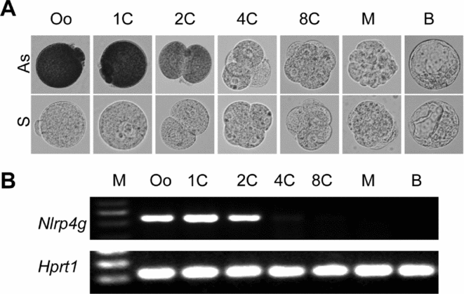
To evaluate the expression pattern of Nlrp4g transcripts, mature oocytes and preimplantation embryos were subjected to ISH and RT-PCR analyses. The transcripts were detected in oocytes, zygotes and 2-cell embryos (Fig. 1 A). The signals of the transcripts in 2-cell embryos were weaker than those in oocytes and zygotes and no signals were detected from the 4-cell embryo through to the blastocyst stages (Fig. 1 A), confirmed by RT-PCR analysis (Fig. 1 B).
Nlrp4g expression in preimplantation embryos. (A) Expression of Nlrp4g in mature oocytes and preimplantation embryos by in situ hybridization (ISH). Mature oocytes (Oo) and preimplantation embryos were hybridized in the same reaction and stained for the same time with DIG-labelled Nlrp4g oligonucleotide antisense and sense probes. The embryonic stages are: 1-cell (1C), 2-cell (2C), 4-cell (4C), 8-cell (8C), morula (M) and blastocyst (B) (all ×200 magnification). (B) Expression analysis of Nlrp4g in oocytes and preimplantation embryos by RT-PCR using cDNA synthesized from Oo, 1C, 2C, 4C, 8C, M and B (lanes 1–7, respectively). Hprt1 was used as a control.

Cellular localization of NLRP4G
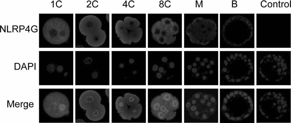
To determine the localization of NLRP4G in preimplantation embryos, we performed confocal microscopy analysis. This revealed that the NLRP4G proteins were mainly localized in the cytoplasm and persisted in cleavage-stage embryos, while a dramatic decrease was observed in morulae and blastocysts (Fig. 2). In addition, the area of nucleus without the fluorescence is smaller in the merged photographs than DAPI-stained ones, suggesting that NLRP4G proteins may also expressed on the outer and/or inner nuclear membranes (Fig. 2).
Cellular localization of the NLRP4G protein showing confocal microscopy images of preimplantation embryos. Each sample was counterstained with DAPI to visualize DNA (blue). The embryonic stages are: 1-cell (1C), 2-cell (2C), 4-cell (4C), 8-cell (8C), morula (M) and blastocyst (B). The original magnification was ×200.

Overexpression of Nlrp4g in zygotes did not affect normal development
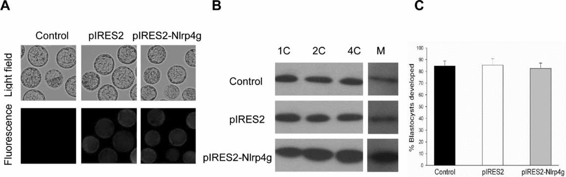
As described above, Nlrp4g transcripts were not detected after zygotic genome activation. To determine the effect of Nlrp4g overexpression on the preimplantation development, zygotes were microinjected with pIRES2–Nlrp4g. Zygotes microinjected with sterile deionized water (Control), pIRES2 or pIRES2–Nlrp4g reached the blastocyst stage and expressed GFP (Fig. 3 A). The level of NLRP4G protein in 2-cell and 4-cell embryos derived from the pIRES2–Nlrp4g-microinjected group was increased obviously compared with the control group (Fig. 3 B). However, the rates of blastocyst formation were similar between the three groups (Fig. 3 C).
Overexpression of Nlrp4g in preimplantation embryos. (A) Representative bright field micrographs of blastocysts developing from zygotes that had been microinjected with sterile deionized water (Control), pIRES2 or pIRES2-Nlrp4g. Fluorescence images show the expression of GFP. The original magnification was ×100. (B) Western blot of extracts of mouse 1-cell (1C), 2-cell (2C), 4-cell (4C) embryos and morula (M) after microinjection. (C) The blastocyst formation rate of zygotes microinjected with sterile deionized water, pIRES2 or pIRES2-Nlrp4g after being cultured for 3.5 days (P > 0.05). Data are presented as the mean ± standard error of the mean (SEM).

Discussion
Preimplantation embryo development is largely reliant on maternal RNA transcripts and proteins that are synthesized and stored during oogenesis (Latham, Reference Latham1999). Here we demonstrated that Nlrp4g was highly expressed in mature oocytes and zygotes, then downregulated and not detected after the 2-cell stage, suggesting that the source of embryonic Nlrp4g transcripts is maternal and not from expression of the zygotic genome during preimplantation development. NLRP4G proteins remained present through the blastocyst stage, although Nlrp4g transcripts were undetectable after the 2-cell stage, implying that this protein might play a role during zygotic genome activation. The expression pattern of NLRP4G is similar to NLRP2 and NLRP5 in the mouse (Tong et al., Reference Tong, Gold, De Pol, Vanevski, Dorward, Sena, Palumbo, Bondy and Nelson2004; Peng et al., Reference Peng, Chang, Lu, Su, Wu, Lv, Wang, Liu, Zhang, Quan, Guo and Zhang2012). The only different point between them is that NLRP4G was degraded sharply at the morula stage, whereas NLRP2 and NLRP5 were degraded at the blastocyst stage. The cytoplasmic localization of NLRP4G protein suggested that the protein might participate in intracellular functions.
To determine the effect of replenishing Nlrp4g transcripts supplies after zygotic genome activation on preimplantation embryo development, we employed an overexpression approach using pIRES2–Nlrp4g vector. Microinjection with the pIRES2–Nlrp4g vector into zygotes did not affect preimplantation development in terms of the rate of blastocyst formation, suggesting that an excessive accumulation of NLRP4G protein has no appreciable detrimental effect on the viability of blastomeres.
The knock-out of maternal-effect genes blocks early embryogenesis (Burns et al., Reference Burns, Viveiros, Ren, Wang, DeMayo, Frail, Eppig and Matzuk2003; Payer et al., Reference Payer, Saitou, Barton, Thresher, Dixon, Zahn, Colledge, Carlton, Nakano and Surani2003; Wu et al., Reference Wu, Viveiros, Eppig, Bai, Fitzpatrick and Matzuk2003; Bultman et al., Reference Bultman, Gebuhr, Pan, Svoboda, Schultz and Magnuson2006; Ma et al., Reference Ma, Zeng, Schultz and Tseng2006; Zheng & Dean, Reference Zheng and Dean2009). In the mouse, Nlrp2, Nlrp5 and Nlrp14 are the maternal-effect genes required for the normal development of preimplantation embryos (Tong et al., Reference Tong, Gold, Pfeifer, Dorward, Lee, Bondy, Dean and Nelson2000; Hamatani et al., Reference Hamatani, Falco, Carter, Akutsu, Stagg, Sharov, Dudekula, Buren and Ko2004; Peng et al., Reference Peng, Chang, Lu, Su, Wu, Lv, Wang, Liu, Zhang, Quan, Guo and Zhang2012). To further study the functions of Nlrp4g in preimplantation embryos, knock-out models or other targeted inhibition experiments on this gene should be carried out. In addition, NLRP proteins are comprised of an amino-terminal pyrin domain (PYD), a central NACHT domain and carboxy-terminal leucine-rich repeats (LRRs) (Ting et al., Reference Ting, Lovering, Alnemri, Bertin, Boss, Davis, Flavell, Girardin, Godzik, Harton, Hoffman, Hugot, Inohara, Mackenzie, Maltais, Nunez, Ogura, Otten, Philpott, Reed, Reith, Schreiber, Steimle and Ward2008). The PYD and LRRs domain present in many proteins that mainly participate in protein–protein interactions (Bertin & DiStefano, Reference Bertin and DiStefano2000; Kobe & Kajava, Reference Kobe and Kajava2001). Identification of the proteins interacting with NLRP4G, therefore, will facilitate an analysis of Nlrp4g function during zygotic genome activation.
In summary, our data indicate that Nlrp4g mRNA appeared to degrade sharply after zygotic genome activation, while the cytoplasmic protein persisted until the blastocyst stage. Furthermore, overexpression of Nlrp4g in zygotes did not obviously affect early embryo development in terms of the rate of blastocyst formation. Knock-out models or identifying the proteins interacting with NLRP4G will help in analysing the function of Nlrp4g in preimplantation embryo development.
Acknowledgements
The authors thank Prof. Xu Wu for helpful discussions and editorial assistance. This work was supported by Foundation of Fujian Educational Committee (grant number JA13110), Fujian Provincial Natural Science Foundation (grant number 2014J01071) and the National Key Technology R&D program, People's Republic of China (grant number 2011BAD19B04-8).
Competing interests
The authors have declared that no competing interests exist.