




No CrossRef data available.
Article contents
Right ventricular function and ventricular perfusion defects in adults with congenitally corrected transposition: correlation of echocardiography and nuclear medicine
Published online by Cambridge University Press: 20 January 2005
Abstract
We undertook our study in order to evaluate right ventricular function and perfusion by conventional and contrast echocardiography in adults with congenitally corrected transposition who had not undergone cardiac surgery, comparing the echocardiographic findings with those obtained using equilibrium radionuclide ventriculography and gated single-photon emission computed tomography with Technetiumc-99 m sestamibi.
We discovered severe tricuspid regurgitation in 8 patients (61%). Right ventricular ejection fraction, as calculated by nuclear medicine, had a correlation of 0.67 (p = 0.059) with area fractional shortening and 0.84 (p = 0.01) with ejection fraction calculated by the method depending on descent of the tricuspid ring. All patients with severe tricuspid regurgitation also had right ventricular dysfunction. Of these 8 patients, 7 had persistent perfusion defects, while 6 also had ischemic perfusion defects.
Echo contrast had a high sensitivity, at 91%, and also specificity and positive predictive value, both at 100%, for persistent defects, and a negative predictive value of 66% compared to methods depending on nuclear medicine. The sensitivity of contrast echocardiography for detection of ischemic defects was 66%, the specificity 100%, the positive predictive value 100%, and the negative predictive value 77% compared to the methods involving nuclear medicine.
The method depending on descent of the tricuspid ring had the highest correlation with equilibrium radionuclide ventriculography in evaluation of right ventricular function in patients with congenitally corrected transposition.
We conclude that contrast echocardiography is extremely valuable when assessing right ventricular myocardial perfusion, having high sensitivity and specificity for detecting persistent defects, although sensitivity was less for detection of ischemic defects than that of gated single-photon emission computed tomography with Technetium-99 m Sestamibi.
Persistent and ischemic perfusion defects, together with chronic volume overload from tricuspid regurgitation, are the determining factors of right ventricular dysfunction.
Information
- Type
- Original Article
- Information
- Copyright
- © 2004 Cambridge University Press
Congenitally corrected transposition, comprising the combinations of discordant connections at both atrioventricular and ventriculo-arterial junctions, is an uncommon congenital anomaly that leads to right ventricular dysfunction. The mechanism of such dysfunction is still unknown, but it is believed to be the result of structural alterations of the myocardium, or myocardial ischemia, since the supply of oxygen provided by the right coronary artery is inadequate when the right ventricle is hypertrophic or hyperplastic.1 Other authors have suggested, however, that tricuspid regurgitation is the most important risk factor.2
There are studies in the literature that have evaluated right ventricular function using gated equilibrium radionuclide ventriculography,3, 4 along with isolated case reports of unoperated patients with congenitally corrected transposition studied with resting/stress single-photon emission computed tomography with Technetium-99 m Sestamibi5 that suggest myocardial perfusion defects as a possible mechanism for the dysfunction. Advances in techniques using contrast echocardiography have now made it possible to examine left ventricular perfusion.6 When looking for perfusion defects in patients with concordant atrioventricular and ventriculoarterial connections, the latter technique has high sensitivity, high negative predictive value, and a 92% agreement with single-photon emission computed tomography.7
The aim of our study was to evaluate right ventricular function and perfusion in unoperated patients with congenitally corrected transposition using conventional and contrast echocardiography, and to correlate the findings with those obtained using equilibrium ventriculography and gated resting/stress single-photon emission computed tomography using Technetium-99 m Sestamibi.
Materials and methods
We studied 13 adults with congenitally corrected transposition, 7 being men and 6 women. The mean age was 31.5 ± 14.88 years. All patients were seen in the Adult Congenital Heart Disease out-patient clinic of the Instituto Nacional de Cardiología “Ignacio Chávez” in the period between March, 2000 and December 2001. Clinical histories were taken from all patients, and all underwent conventional transthoracic and contrast echocardiography, gated equilibrium radionuclide ventriculography, myocardial perfusion studies using gated resting/stress single-photon emission computed tomography using Tecnetium-99 m Sestamibi, and coronary angiography. Coronary arterial obstruction was considered significant when it was more than 50%.
Echocardiographic study
Standard transthoracic cross-sectional and colour coded Doppler echocardiography was performed in all patients utilizing the Agilent Technologies Sonos 5500 ultrasound system in order to assess tricuspid valvar morphology and function, and right ventricular function. Echocardiograms were recorded on videotape for subsequent off-line analysis.
Tricuspid valvar and morphologically right ventricular function. The state of the tricuspid valve was determined by transthoracic echocardiography. The degree of tricuspid insufficiency was determined qualitatively in a four-chamber view by taking into account the ratio of area of the jet of tricuspid insufficiency to the area of the left atrium, and the distance the jet extended into the left atrium. If there was evidence of reversal of systolic flow in the pulmonary veins, the ratio of the area of the jet was more than 40%, and the jet extended 4.5 cm into the left atrium, tricuspid insufficiency was considered to be severe.8, 9
The tricuspid valve itself was classified as being morphologically abnormal if there was echocardiographic evidence of a structural abnormality, such as thickened or redundant leaflets, or an Ebstein-like malformation.10 Right ventricular function was evaluated by echocardiography and radionuclide angiography and classified as either normal or abnormal. The right ventricular area was calculated from apical four chamber images in telediastole and telesystole and area fractional shortening by the formula:

The right ventricular area fractional shortening was considered normal when it was equal to or greater than 45%. The right ventricular ejection fraction was calculated by measuring the descent of the tricuspid ring in diastole and systole from the same apical four-chamber projection. The systolic value was subtracted from the diastolic value, and the difference was multiplied by 3.2. A normal ejection fraction was considered to be equal to or greater than 44%.11
Radionuclide technique
Ventricular function was calculated using equilibrium radionuclide ventriculography with ultra-tag in vitro red blood cell technique with 20 mCi of Technetium-99 m. Imaging was performed with a one single head orbiter Siemens gammacamera equipped with a high-resolution, low-energy, parallel-hole collimator and recorded on a dedicated nuclear machine computer system. The study was acquired in left anterior oblique position to obtain maximal separation of ventricular chambers. The R wave of the electrocardiogram served as the synchronizing impulse. The cardiac cycle was divided into 8 frames per cycle, with an acquisition time of 10 min. Statistically reliable information was obtained by summing the radioactivity in the ventricle during many beats. Ejection fraction was averaged from all beats recorded.
Processing. Each gated study was displayed as an endless-loop movie on a video display to visualize the ventricles. A region of interest was outlined over the right and left ventricles, and a time/activity curve was obtained. End-diastolic and end-systolic frames were identified, and the activity over the aorta and pulmonary trunk was excluded. Ventricular ejection fractions were calculated as 100 × (end-diastolic counts – end-systolic counts)/end diastolic counts. A study was defined as abnormal if the ejection fraction was less than 45% for the morphologically right ventricle, and 50% for the morphologically left ventricle.
Right ventricular myocardial perfusion
Right ventricular myocardial perfusion was evaluated by two methods: contrast echocardiography at rest and at peak pharmacological stress with dobutamine, and nuclear scanning using a one day gated single photon emission computed tomography sestamibi rest and stress protocol, gating only the stress images.
Myocardial contrast echocardiography
Myocardial perfusion was assessed using equipment with perfusion harmonics software and power Doppler. Apical four and two chamber views were used, and each dose of the contrast agent (Optison) was intravenously administered as a bolus of 0.4 ml into a cubital vein, followed by a rapid flush of 5 ml of saline. Optison (Mallinckrodt Medical Inc, St. Louis, MO) is a perfluoropropane filled albumin shell with a diameter of 3.7 μm. The injection rate did not exceed 1 ml/s. Optison was injected at baseline and at peak stress with dobutamine. The protocol of dobutamine stress echocardiography was performed as previously described.12 Triggered imaging at end-systole at intervals of 1 to 3, and 1 to 5, was performed. Visual judgement mainly relies on the signal intensities of myocardial contrast. When no contrast enhancement was observed, it was graded as 0, 0.5 when contrast enhancement was patchy, and 1 when strong contrast enhancement was observed. A perfusion defect is a relative decrease in contrast enhancement in one region compared with other regions with the same or worse imaging conditions. Fixed perfusion defects appear at rest, with no or patchy contrast enhancement. Reversible perfusion defects were defined as those defects that appeared during stress after images that were normal at rest. They showed a relative decrease in signal intensity compared to adjacent normal myocardium.
Single photon emission computed tomography Sestamibi study
Myocardial perfusion was assessed with the Technetium-99 m Sestamibi (Technetium-99 m methoxy isobutyl isonitrile) using a one day rest-stress, either physical or pharmacological. Patients exercised on a treadmill to maximum exercise capacity according to a standard Bruce protocol. At peak exercise, 20 mCi of Sestamibi was injected intravenously. Single-photon emission computed tomographic images were acquired one hour after rest and stress injection using an orbiter Siemens single head camera, undergoing rotation to 180°, for 32 frames with a 64 × 64 matrix. Stress studies were obtained with gated technique; images were gated dividing the cardiac cycle into 8 frames using an R wave trigger. Quantitative gated single photon emission computed tomography software was employed automatically to evaluate the ejection fraction of the morphologically right ventricle. A high resolution, low energy, parallel-hole collimator was used.
Perfusion scans were read by two expert observers blinded to the clinical state and echocardiographic data of each patient. We divided the heart into 20 segments to evaluate perfusion: anterior, anteroseptal, inferoseptal, inferior, inferolateral, and anterolateral at apical, mid-ventricular and basal levels, and anteroapical and inferoapical segements in the vertical long axis view. Tracer uptake in each segment was scored on a 5-point scale, with 0 being normal and 4 showing absence of uptake. Gated single-photon emission computed tomographic images were read in one mode to assess mural motion and thickening, and in a 3-dimensional view.
Statistical analysis
Quantitative data were expressed as means plus or minus the standard deviation. A t-test or U Mann-Whitney test was used as indicated to compare two independent mean values. When factors associated with ventricular function were sought, the t-test, or Kruskal-Wallis analysis of variance, were used. The correlation of anatomical and functional characteristics with age were each tested by Cox's univariate proportional hazard regression modeling. A p value of less than .05 was set as the level of statistical significance. Spearman's correlation coefficient was used to determine the degree of agreement between the values for the ejection fraction obtained by echocardiography and radionuclide ventriculography. Kappa's index was used to evaluate the degree of concordance between contrast echocardiographic and single-photon emission computed tomographic Sestamibi studies.
Results
Clinical characteristics
We evaluated 13 adults, including 7 (53.8%) men and 6 (46.2%) women. The mean age of the patients was 31.5 ± 14.88 years, with a range from 17 to 72 years. The heart was right-sided in 5 of the patients, with four of these having usual atrial arrangement and the other a mirror-imaged arrangement. The heart was left-sided in the 8 remaining patients, 6 of whom had usual arrangement and 2 mirror-imagery. Of the 13 patients, 7 (53.8%) had small ventricular septal defects.
The morphologically tricuspid valve, guarding the junction between the left atrium and the morphologically right ventricle, was normal in only one case (7.7%), while 9 demonstrated some degree of dysplasia, and 3 showed overt evidence of Ebstein's malformation. Mild tricuspid regurgitation was found in 4, and severe tricuspid regurgitation in 8 (Fig. 1). Only one patient had no tricuspid regurgitation.

Figure 1. Apical echocardiographic view of a patient with atrioventricular discordant connections. Colour flow mapping detects a severe tricuspid regurgitation (arrows) and mild mitral regurgitation (arrow head). LV: left ventricle; RA: right atrium; RV: right ventricle.
In 7 patients (53.8%), we also found abnormalities of the aortic valve and the outlet from the morphologically right ventricle, with 4 of the patients having aortic valvar stenosis, 3 with subvalvar stenosis, and one with stenosis of mixed origin.
Functional class. All patients were in functional classes II (77%) and III (23%) of the grading system of the New York Heart Association.
Ventricular function. The mean of ejection fractions calculated by the regression equation method was 45% ± 11%, with the comparable value calculated by area fractional shortening method being 43% ± 11%. When we used equilibrium ventriculography, the mean was 45% ± 11%. Right ventricular systolic function was depressed in 7 patients (53.8%) using area fractional shortening, in 8 (61%) by ejection fraction as calculated using the descent of the tricuspid ring, and in 9 (69%) with equilibrium ventriculography (Table 1).
Table 1. Echocardiographic and nuclear medicine results.

Right ventricular ejection fraction calculated by nuclear medicine had a Pearson correlation of 0.67 (p = 0.05) with area fractional shortening, and of 0.84 (p = 0.01) with the ejection fraction calculated by the regression equation. The correlation coefficient of the two echocardiographic methods was 0.87.
Nuclear medicine versus contrast echocardiography. A total of 11 (84%) nuclear medicine studies were positive for persistent perfusion defects (Table 2). Of these, 10 (76%) were also positive in resting contrast echocardiographic studies, giving a kappa index of diagnostic agreement of 0.76 (p = 0.005) (Figs 2a–c).
Table 2.


Figure 2. Contrast echocardiographic study using harmonic power Doppler at rest. A, B. In the four-chamber and two apical images a patched perfusion defect in the basal and medial portion of the ventricular septum (arrow heads) and a perfusion defect in the infero-lateral wall (black arrows) of the right ventricle in its basal and medial portion are visualized. C. Short axis view of right ventricular perfusion at rest and stress with sestamibi single-photon emission computed tomography in the medial and basal portion. Fixed non-transmural inferolateral perfusion defect and mild septal perfusion abnormality with mild reversibility are observed.
The sensitivity of echo perfusion for detection of persistent defects was 91%, the specificity 100%, the positive predictive value 100%, and the negative predictive value 66% in comparison to nuclear medicine studies.
In 9 (61%) of the nuclear medicine studies, we found evidence of ischemic defects, of which 7 (53.8%) were also positive by dobutamine contrast echocardiography (Table 3). This gave a kappa index of diagnostic agreement of 0.68 (p = 0.009).
Table 3.

In comparison with nuclear medicine, the sensitivity of the perfusion echo for detection of ischemic defects was 66%, the specificity 100%, the positive predictive value 100%, and the negative predictive value 77%.
All of the 8 patients with severe tricuspid regurgitation also had right ventricular dysfunction, with 7 of these patients having persistent perfusion defects, and 6 also having ischemic perfusion defects (Figs 3a–c).

Figure 3. Contrast echocardiography images with Doppler angio at rest. A. In the four-chamber and two apical views a patched perfusion defect in the basal and medial portion of the interventricular septum (arrows) and a perfusion defect in the apical portion of antero-lateral wall are observed. B. With dobutamine also a perfusion defect in the basal portion of the anterior wall was found (arrow). C. Short axis view of right ventricular perfusion at rest and stress with sestamibi single photon emission computed tomography. Fixed non-transmural perfusion defect in the basal and medial portion of the ventricular septum and in the apical portion of the anterolateral wall is visualized. Also a mild ischemic perfusion defect in the basal portion of the anterior wall is observed.
Coronary angiography. No significant atherosclerotic lesions were found in any patients.
Discussion
Dysfunction of the systemic morphologically right ventricle is a part of the natural history of congenitally corrected transposition. It is associated with advanced stages of clinical deterioration, and carries an ominous prognosis.2, 13, 14 For this reason, adequate evaluation of right ventricular function is particularly important. The gold standard for evaluation in the setting of the normal heart is equilibrium ventriculography. Studies of right ventricular function using this technique in patients with congenitally corrected transposition subsequent to surgical correction have demonstrated its value in the diagnosis of right ventricular systolic dysfunction.15, 16 Cineangiography has also been used. Thus, Graham and colleagues17 studied 19 patients using cineangiography, and found that the right ventricular ejection fraction was abnormal in only 15% of patients less than 10 years of age. In patients older than 17 years, in contrast, the incidence of right ventricular dysfunction was as high as 33%. These data suggest that right ventricular function assessed by ejection fraction deteriorates with age because of the limited capacity of the morphologically right ventricle to support the systemic circulation. Right ventricular ejection fraction obtained by cineangiography in their patients showed excellent correlation with equilibrium ventriculography, with an r value of 0.98.17 Cineangiography, nonetheless, is an invasive procedure. Another consideration is that a substantial percentage of these patients have dysplasia of the leaflets of the morphologically tricuspid valve that leads to regurgitation and right ventricular volume overload, with dilation and right ventricular dysfunction over time. Nowadays, magnetic resonance imaging dobutamine stress testing can be used to assess systemic right ventricular function in patients with congenitally corrected transposition either prior to surgery, or after physiological correction. Larger systemic right ventricular volumes, diminished ejection fraction, and an appropriate response to dobutamine were demonstrated in relation to the left ventricles of healthy controls.18 In our study, we evaluated right ventricular function in 13 adults with congenitally corrected transposition by transthoracic echocardiography using two methods: areas fractional shortening and ejection fraction with descent of the tricuspid ring. Right ventricular ejection fraction calculated by radionuclide ventriculography showed good correlation with ejection fraction (r = 0.76) and area fractional shortening (r = 0.67). These results indicate that transthoracic echocardiography is a useful technique to evaluate the function of the morphologically right ventricle in patients with congenitally corrected transposition. Its great advantages include low cost, and the fact that it can be used at the bedside. The calculation of ejection fraction using descent of the tricuspid ring has a better correlation with equilibrium ventriculography.
The literature includes nuclear medicine studies that assessed defects in right ventricular myocardial perfusion in patients with congenitally corrected transposition who have not undergone cardiac surgery. They suggest that these defects may play an important role in the development of progressive right ventricular dysfunction.19–21
We examined right ventricular microvascular myocardial perfusion with contrast echocardiography (Optison), and compared our findings with those obtained with single-photon emission computed tomography. High levels of sensitivity, specificity and positive predictive value were achieved for detection of persistent defects, although the negative predictive value was less. Sensitivity for ischemic defects was lower than that for persistent defects, but specificity and positive predictive value were unchanged.
We found that the three patients with Ebstein's malformation developed right ventricular dysfunction. Eidem and colleagues19 showed that such patients had a tendency to develop severe dilation and right ventricular dysfunction. The Tei Doppler index is significantly higher compared to the control group and to patients with atrial septal defects and pulmonary stenosis.19, 20
In summary, over nine-tenths of our patients with congenitally corrected transposition had dysplasia of the tricuspid valve that caused tricuspid regurgitation, with significant hemodynamic repercussion in almost two-thirds. Our findings suggest that persistent and ischemic perfusion defects, together with chronic volume overload from tricuspid regurgitation, are the determining factors of right ventricular dysfunction.
References
Losekoot TG, Becker AE. Discordant atrioventricular connection and congenitally corrected transposition. In: Anderson RH, Macartney FJ, Shinebourne EA, Tynan M (eds). Paediatric Cardiology. Churchill Livingstone, Edinburgh, 1987, pp 867–887.
Prieto LR, Hordof AJ, Secic M, Rosenbaum MS, Gersony WN. Progressive tricuspid valve disease in patients with congenitally corrected transposition of the great arteries. Circulation 1998; 98: 997–1005.Google Scholar
Berger HJ, Matthay RA, Loke J, Marshall RC, Gottschalk A, Zaret BL. Assessment of cardiac performance with quantitative radionuclide angiocardiography: right ventricular ejection fraction with special reference to findings in chronic obstructive pulmonary disease. Am J Cardiol 1978; 41: 897–905.Google Scholar
Benson LN, Burns R, Schwaiger M, Schelbert HR, Lewis AB, Freedom RM, Olley PM, McLaughlin, Rowe RD. Radionuclide angiographic evaluation of ventricular function in isolated congenitally corrected transposition of the great arteries. Am J Cardiol 1986; 58: 319–324.Google Scholar
Hornung TS, Bernard EJ, Jaeggi ET, Howman-Giles RB, Celermajer DS, Hawker RE. Myocardial perfusion defects and associated systemic ventricular dysfunction in congenitally corrected transposition of the great arteries. Heart 1998; 80: 322–326.Google Scholar
Kaul S. Myocardial contrast echocardiography. 15 years of research and development. Circulation 1997; 96: 3745–3760.Google Scholar
Kaul S, Senior R, Ditrich H, Raval U, Khattar R, Lahiri A. Detection of coronary disease with myocardial contrast echocardiography: Comparison with 99 m Tc-sestamibi single-photon emission computed tomography. Circulation 1997; 96: 785–792.Google Scholar
Miyatake K, Okamoto M, Kinoshita N, Mitsushige O, Takahiro K, Hiroshi S, Yasuharu N. Evaluation of tricuspid regurgitation by pulsed Doppler and two-dimensional echocardiography. Circulation 1982; 66: 777–789.Google Scholar
Sakai K, Nakamura K, Satomi G, Kondo M, Kirosawa K. Hepatic vein blood flow pattern measured by Doppler echocardiography as an evaluation of tricuspid valve insufficiency. J Cardiogr 1983; 13: 33.Google Scholar
Becker AE, Becker MJ, Edwards JE. Pathologic spectrum of dysplasia of the tricuspid valve: features in common with Ebstein's malformation. Arch Pathol 1971; 91: 167–178.Google Scholar
Kaul S, Tei C, Hopkins, Shah PM. Assessment of right ventricular function using two-dimensional echocardiography. Am Heart J 1984; 107: 526–531.Google Scholar
Secknus MA, Marwick TH. Evolution of dobutamine echocardiography protocols and indications: Safety and side effects in 3011 studies over 5 years. J Am Coll Cardiol 1997; 29: 1234–1240.Google Scholar
Graham TP, Bernard YD, Mellen BG, Celermajer D, Baumgartner H, Cetta F, Connolly HM, Davidson WR, Dellborg M, Foster E, Gersony WN, Gessner IH, Hurwitz RA, Kaemmerer H, Kugler JD, Murphy DJ, Noonan JA, Morris C, Perloff JK, Sanders SP, Sutherland JL. Long-term outcome in congenitally corrected transposition of the great arteries. A Multi-Institutional Study. J Am Coll Cardiol 2000; 36: 255–261.Google Scholar
Connelly MS, Liu PP, Williams WG, Webb GD, Robertson P, McLaughlin PR. Congenitally corrected transposition of the great arteries in the adult: Functional status and complications. J Am Coll Cardiol 1996; 27: 1238–1243.Google Scholar
Peterson RJ, Franch RH, Fajman WA, Jones RH. Comparison of cardiac function in surgically corrected and congenitally corrected transposition of the great arteries. J Thorac Cardiovasc Surg 1988; 96: 227–236.Google Scholar
Lubiszewska B, Gosiewska E, Hoffman P, Teresinska A, Rozanski J, Piotrowski W, Rydlewska-Sadowska W, Kubicka K, Ruzylo W. Myocardial perfusion and function of the systemic right ventricle in patients after atrial switch procedure for complete transposition: long-term follow-up. J Am Coll Cardiol 2000; 36: 1365–1370.Google Scholar
Graham TP, Parrish MD, Boucek RJ, Boerth RC, Breitweser JA, Thompson S, Robertson RM, Morgan JR, Friesinger GC. Assessment of ventricular size and function in congenitally corrected transposition of the great arteries. Am J Cardiol 1983; 51: 244–251.Google Scholar
Dodge-Khatami A, Tulevski II, Bennink GBWE, Hitchcock JF, De Mol BAJM, Van der Wall EE, Mulder BJM. Comparartive systemic function in healthy adults with unoperated congenitally corrected transposition using MRI dobutamine stress testing. Ann Thorac Surg 2002; 73: 1759–1764.Google Scholar
Eidem BW, O'Leary PW, Tei C, Seward JB. Usefulness of the myocardial performance index for assessing right ventricular function in congenital heart disease. J Am Coll Cardiol 2000; 86: 654–658.Google Scholar
Tei C, Nishimura RA, Seward JB, Tajik J. Noninvasive Doppler-derived myocardial performance index: Correlation with simultaneous measurements of cardiac catheterization measurements. J Am Soc Echocardiogr 1997; 10: 169–178.Google Scholar
Hornung TS, Bernard EJ, Celermajer DS, Jaeggi E, Howman-Giles RB, Chard RB, Hawker RE. Right ventricular dysfunction in congenitally corrected transposition of the great arteries. J Am Coll Cardiol 1999; 84: 1116–1119.Google Scholar

Apical echocardiographic view of a patient with atrioventricular discordant connections. Colour flow mapping detects a severe tricuspid regurgitation (arrows) and mild mitral regurgitation (arrow head). LV: left ventricle; RA: right atrium; RV: right ventricle.

Table 1.
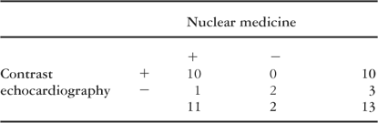
Table 2.

Contrast echocardiographic study using harmonic power Doppler at rest. A, B. In the four-chamber and two apical images a patched perfusion defect in the basal and medial portion of the ventricular septum (arrow heads) and a perfusion defect in the infero-lateral wall (black arrows) of the right ventricle in its basal and medial portion are visualized. C. Short axis view of right ventricular perfusion at rest and stress with sestamibi single-photon emission computed tomography in the medial and basal portion. Fixed non-transmural inferolateral perfusion defect and mild septal perfusion abnormality with mild reversibility are observed.

Table 3.

Contrast echocardiography images with Doppler angio at rest. A. In the four-chamber and two apical views a patched perfusion defect in the basal and medial portion of the interventricular septum (arrows) and a perfusion defect in the apical portion of antero-lateral wall are observed. B. With dobutamine also a perfusion defect in the basal portion of the anterior wall was found (arrow). C. Short axis view of right ventricular perfusion at rest and stress with sestamibi single photon emission computed tomography. Fixed non-transmural perfusion defect in the basal and medial portion of the ventricular septum and in the apical portion of the anterolateral wall is visualized. Also a mild ischemic perfusion defect in the basal portion of the anterior wall is observed.
 You have
Access
- 11
- Cited by