Introduction
Healthy tissues are continually producing and destroying cells, resulting in the presence of cell-free DNA (cfDNA) fragments in the blood derived from these dead cells. However, the plasma levels of free circulating cfDNA increase in some pathologies such as cancer, cardiovascular and autoimmune disorders, neurodegenerative syndromes such as spinocerebellar ataxias and infectious diseases (Swarup et al., Reference Swarup, Srivastava, Padma and Rajeswari2011; Yi et al., Reference Yi, Zhang, Zhang, Ma, Zhang, Li, Liu, Liu, Liu, Zhang, Zhuang and Jin2014). A study performed by Czamanski-Cohen et al. (Reference Czamanski-Cohen, Sarid, Cwikel, Lunenfeld, Douvdevani, Levitas and Har-Vardi2013) also described an association between higher plasma cfDNA levels and low pregnancy rates among women who were undergoing in vitro fertilization embryo transfer.
Some previous investigations have described correlations among parameters linked to normal sperm function and cfDNA levels present in semen. Li et al. (Reference Li, Huang, Zhou, Liao and Xiong2009) observed higher cfDNA levels in azoospermia than normospermia, whereas Chou et al. (Reference Chou, Jacobson, Patton, King and Chan2004) observed an association between low cfDNA quantity with rapid progression, curvilinear velocity, normal strict morphology and capacitation index. These authors also described that the sperm freezing process did not influence the cfDNA sperm concentration. Recent investigation also detected testis and epididymis-specific methylate promoters in human cfDNA (Xie et al., Reference Xie, Hou, Li, Gao and Cheng2004). Despite the great relevance of these studies, most techniques used to quantify seminal cfDNA are expensive, need a short time to be performed and present high sensitivity to detect dsDNA. This outcome is the case with the polymerase chain reaction assay to detect cfDNA fragments. Moreover, for many clinical laboratories the use of these techniques is not realistic. Therefore, the present investigation evaluated the use of DNA PicoGreen assay to determine seminal cfDNA because this dye is an ultrasensitive fluorescent nucleic acid stain for quantitating double-stranded DNA (dsDNA), with which it forms very stable complexes under alkaline conditions instead of single-stranded DNA (ssDNA), proteins, sodium dodecyl sulphate (SDS) and urea (Levy et al., Reference Levy, Loptian, Kennedy, Lo-Yim and Shamlou2000). We also evaluated in a large semen sample the potential association between cfDNA seminal levels with sperm parameters used to estimate the fertilization capacity announced by the World Health Organization (WHO) (2010).
Materials and methods
Ethics
The study was approved by the Ethics Committee on Human and Animal Research at the Federal University of Santa Maria, Brazil, number 0332.0.243.000-11. All procedures occurred in accordance with the principles set out in the Declaration of Helsinki. Participants signed informed consent terms.
Study design and sample collection
To determine the use of seminal cfDNA quantification using PicoGreen dye, two complementary protocols were performed in vivo and in vivo. The in vivo protocol determined cfDNA levels using PicoGreen staining of semen samples from male patients that had undergone a spermiogram in a commercial clinical analysis laboratory (Pasteur, Santa Maria-RS, Brazil) to evaluate their fertility condition. The samples were obtained via masturbation after 2–5 days of ejaculatory abstinence and were analyzed within 1 h following collection. After seminal liquefaction, a routine semen analysis was performed using computer-aided equipment, and sperm samples were classified according to the WHO (2010) specifications: concentration ≥ 15×106 sperm/ml; motility ≥ 40%; viability ≥ 58%; normomorphology ≥ 4% and leukocytes in semen (LeS) < 1 × 10⁶ cells/ml. Two groups were found in the sperm evaluation: 27 (16%) considered fertile to all WHO criteria and 142 (84%) with at least one infertile WHO criteria. After collection and determination of the WHO parameters, the sperm samples that were discarded were cryopreserved (~1 ml) to subsequently perform genetic and biochemical analyses related to oxidative stress parameters. The semen samples were frozen at −80°C in the absence of a cryopreservative reagent to avoid interference in the evaluated biochemical variables. A brief protocol using sperm sample frozen with and without cryopreservative reagent did not show any significant differences between cfDNA levels. Prior to analysis, the frozen samples were left at room temperature for 1 min before further processing.
In vivo cfDNA determination by fluorimetric PicoGreen assays
The seminal cfDNA was determined in a black, 96-well plate using a Quant-iT™ PicoGreen® kit purchased from Invitrogen (Eugene, OR, USA) and diluted in Tris–EDTA (TE) buffer (10 mM Tris–HCl, 1 mM EDTA, pH 7.5) with reagents of the highest purity/grade purchased from Sigma-Aldrich (St. Louis, MO, USA). The fluorimetric analyses were performed using a SpectraMax M2/M2e Multi-mode Plate Reader (Molecular Devices Corporation, Sunnyvale, CA, USA) at an excitation of 480 nm and an emission of 520 nm recorded at room temperature. To improve the experiment by avoiding oxidative light effects on the reaction, the incubation periods of H2O2 and PicoGreen® were conducted in darkness. As the fluorimeter equipment is highly sensitive, three repeated measurements were carried out on each sample. A DNA quantification curve was performed using calf DNA purchased from Sigma-Aldrich.
The semen samples were centrifuged at 300 g for 10 min. Ninety microlitres of TE buffer were added to each well of a 96-well black microplate, into which 10 µl of sperm plasma sample was diluted. Then, 100 µl of the PicoGreen dye diluted 1:200 in TE buffer were added to the microplate in order to make a final volume of 200 µl per well. Following incubation in the dark for 10 min at room temperature, the fluorescent signal of the sample was measured at 480 nm excitation and 520 nm emission using Gemini XPS (Molecular Devices, Sunnyvale, USA). Each sample was assessed in triplicate. The SoftMax Pro 5.4.1 program was used to establish the standard curve and calculate the concentrations of the semen samples. Double-stranded DNA quantified by PicoGreen is highly stable in the first 120 min reaction (Cadona et al., Reference Cadona, Manica-Cattani, Machado, Oliveira, Flores, Assmann, Algarve and Cruz2014). The unit of measure was fluorescence (cfDNA).
In vitro cfDNA determination by fluorimetric PicoGreen assays
To confirm the cfDNA quantification by the PicoGreen assays, an in vitro test was also performed using three or four semen samples obtained in conditions similar to those described in the in vivo approach. The fresh sperm sample (seminal plasma plus sperm cells) was exposed with different paraquat concentrations (0.1, 1, 3 and 10 µM). Paraquat is an organic compound that interferes with electron transfer by reduction of an electron donor such as NADPH in the mitochondria and increasing the superoxide anion levels (Bus & Gibson, Reference Bus and Gibson1984). After a 60-min exposure, the samples were centrifuged at 300 g for 10 min, and seminal cfDNA levels were determined as previously described, as well as sperm viability. To confirm that paraquat treatment increased the oxidative stress in the sperm samples, ROS, lipoperoxidation and protein carbonylation levels were evaluated in seminal plasma.
Sperm viability was determined by flow cytometry using annexin-V−FITC and a propidium iodide (PI) staining kit (BD Pharmingen™). Annexin-V−FITC can detect early apoptotic processes, whereas PI staining allows identification of late-stage or necrotic apoptotic cells. The detection is realized through bonding between the cell receptors with these fluorescent compounds, annexin-V and PI. The analysis was performed according to the manufacturer's instructions. Briefly, cells were seeded in 6-well plates at 1 × 10⁶ cells per well in 2 ml of the different treatments. Following incubation, the sperm cells were washed twice with cold PBS and then resuspended in 1× Binding Buffer at a concentration of 1 × 10⁶ cells/ml. The sperm cells were then transferred in 100 µl of solution (1 × 10⁵ cells) to a 5-ml culture tube. Resuspended sperm cells were gently vortexed and stained with 5 µl of Annexin-V–FITC and 5 µl of PI. Following a short incubation for 15 min in the dark at room temperature, 400 µL of 1× Binding Buffer was added to each tube, and the cell fluorescence was analyzed by flow cytometry according to the manufacturer's specifications (Zhao et al., Reference Zhao, Huang, Luo, Wan, Gui, Li and Wu2014)
The ROS, lipoperoxidation and protein carbonylation levels of the sperm cells were determined. The ROS levels were determined by dichlorofluorescein diacetate fluorometric assays (DCF-DA) (Halliwell & Whiteman, Reference Halliwell and Whiteman2004) with fluorescence measured at an excitation of 485 nm and an emission of 520 nm. Lipoperoxidation was spectrophotometrically estimated by the formation of thiobarbituric acid reactive substances (TBARS) as described by Jentzsch et al. (Reference Jentzsch, Bachmann, Fürst and Biesalski1996) and protein carbonylation by the method of Levine (Levine, Reference Levine1983). All measurements were performed in triplicate.
Statistical analysis
All analyses were carried out using the SPSS software program (Version 21.0), and the results were expressed as the mean ± standard deviation (SD) or percentage (%). In the in vivo analysis, association between seminal cfDNA levels and WHO sperm fertility criteria was determined by Student's t-tests. The percentile analysis of cfDNA levels was determined and the sample cut-off was the 75th percentile. The frequency distribution of higher cfDNA levels, sperm fertility criteria and other oxidative stress variables were analyzed by chi-squared tests. A multivariate logistic regression (backward Wald) was also performed to evaluate the influence of age and other oxidative stress variables on the association between cfDNA levels and sperm fertility parameters. Data from in vitro analysis were compared by one-way analysis of variance followed by Tukey's post hoc tests. Pearson correlation and kappa concordance tests were performed to determine if seminal cfDNA levels were representative of sperm viability evaluated by flow cytometry. All statistical comparisons with P < 0.05 were considered significant.
Results
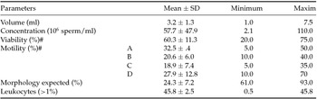
In total, 163 semen samples from men of an average age of 35.89 ± 7.33 (minimum = 19; maximum = 63) were initially collected. The general sperm fertility parameters of the samples are presented in Table 1. The prevalence of low viability was 27.6% (n = 45); low concentration was 16% (n = 26); altered morphology was 9.8% (n = 16) and high leukocyte frequency was 36.2% (n = 59) (Table 1).
General parameters evaluated in the semen samples

SD, standard derivation; #, determined by hyposmotic test.
To evaluate the potential association between cfDNA levels determined by PicoGreen fluorescence and sperm fertility parameters, we initially compared this parameter between fertile group with the group that presented at least one infertile parameter. The mean ± SD cfDNA seminal plasm in fertile group was significantly lower (0.869 ± 0.79 nm) than in the group with infertile parameters (3.657 nm) (P = 0.006).
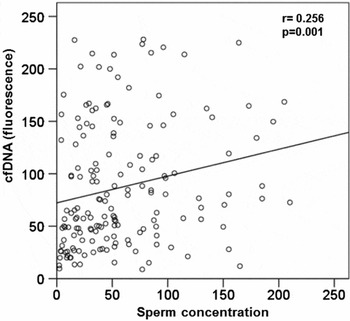
As the volume or sperm concentration are two fertile parameters we evaluated their influence on cfDNA levels. The results showed that semen volume was not associated with cfDNA levels, whereas sperm concentration interfered with fluorescence values (Fig. 1). For this reason, seminal cfDNA values of the samples were corrected to sperm concentrations and estimated to be 111.5 ± 95.7 nm (minimum = 9.6; maximum = 820.6 nm) (Fig. 1).
Association between seminal cfDNA fluorescence and sperm concentration determined by linear regression.

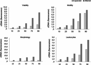
All four sperm fertility criteria were significantly associated with higher seminal cfDNA levels: Vexp = 1.2 ± 1.2; Valt = 4.0 ± 5.4; Mexp = 2.8 ± 4.3; Malt= 5.2 ± 6.6; MOexp = 2.5 ± 3.1; MOalt = 9.9 ± 10.0; Lexp = 1.9 ± 2.5; Lalt= 5.4 ± 6.8 nm. However, due to the high variance observed and non-parametric distribution of cfDNA values a second analysis was performed that initially evaluated the cfDNA percentile distribution in each sperm fertility criteria (Fig. 2).
Percentile distribution of seminal cfDNA fluorescence values corrected by sperm concentrations considering four expected and altered sperm fertility parameters: viability; motility; morphology and presence of leukocytes.

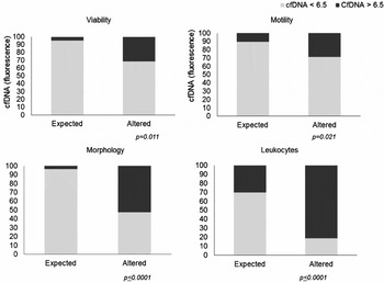
The 75th percentile cfDNA values of altered variables were: viability = 5.0; motility = 6.6; morphology = 7.7 and leukocytes = 6.6. From these results a mean cfDNA value was calculated and used as a cut-point to classify the sample into the following two groups: higher cfDNA values of > 6.5 and moderate or low values of < 6.5 fluorescence. The frequency of higher cfDNA values was significantly related to all criteria investigated here (Fig. 3).
Frequency distribution of seminal cfDNA levels in sperm fertility altered and expected parameters. P-values derive from chi-squared statistical tests or Fisher Exact tests that compared each sperm fertility parameter with low and moderate cfDNA levels (<6.5 fluorescence) and high cfDNA levels (≥6.5 fluorescence).

The cfDNA categorization allowed for the calculating odds ratios between altered cfDNA levels and sperm fertility parameters. Sperm samples with cfDNA > 6.5 fluorescence presented a 1.39 (95% CI = 1.20–1.6) higher chance of altered viability; 2.72 (95% CI = 1.19–6.24) more chance to have altered mobility; 14.98 (95% CI = 5.78–38.4) more chance to have altered morphology and 2.69 (95%CI = 1.94–3.73) when compared with samples that presented expected parameters announced by the WHO.
From these results, we analyzed if seminal cfDNA levels could be associated with other seminal plasma oxidative stress variables such as ROS, lipoperoxidation, and protein carbonylation. To avoid misinterpretation, we also corrected these variables to the sperm concentration of each sample. All oxidative variables evaluated here were significantly higher in samples that presented high seminal cfDNAs (> 6.5 fluorescence) (Table 2).
Comparison of oxidative stress variables in semen samples categorized with low/moderate seminal cfDNA fluorescence levels (<6.5) and high cfDNA fluorescence levels (>6.5)

ROS, reactive oxygen species; SD, standard deviation. Groups were statistically compared by non-parametric Mann−Whitney tests as the values did not present a normal distribution.
Considering the potential use of seminal cfDNA quantification by PicoGreen dsDNA assays to detect sperm samples with altered fertility parameters, a complementary in vitro analysis was performed to determine levels in controlled conditions (Table 3).
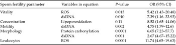
Multivariate analysis to test the influence of Val16Al-SOD2 SNP and age on odds ratios related to association between WHO sperm fertility criteria and oxidative stress variables

Multivariate analysis was performed for each sperm fertility parameter by logistic regression (backward Wald method). Only variables that maintained significance to each sperm fertility parameter are presented.
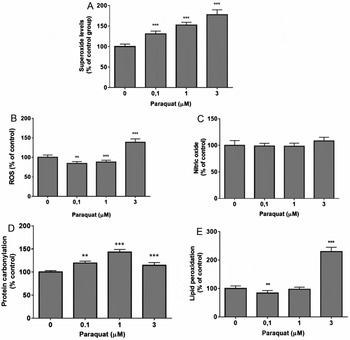
Initially, analysis of oxidative stress indicators was performed to confirm the toxicological effects of different paraquat concentrations on sperm samples (Fig. 4). The superoxide levels presented a dose-dependent increase in the sperm samples confirming the generation of ROS by paraquat exposure. Nitric oxide levels did not change in the samples exposed to different paraquat concentrations. ROS and lipo-peroxidation presented higher levels only in sperm samples exposed to 3 µM paraquat. However, all paraquat concentrations increased the levels of protein carbonylation when compared to the control group (Fig. 4).
Oxidative indicator levels of sperm samples after a 1-h exposure to various paraquat concentrations. The treatments were compared with untreated groups by analysis of variance (ANOVA) followed by Dunnet post hoc tests. **P = 0.01; ***P = 0.0001. (A) Superoxide. (B) ROS. (C) Nitric oxide. (D) Protein carboxylation. (E) Lipid peroxidation.

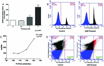
The in vitro analysis confirmed the association between dsDNA levels and sperm viability. Sperm obtained from samples and mixed with buffer media with and without varying paraquat concentrations presented different dsDNA concentrations after one-hour exposures (Fig. 5 A). Maximal dsDNA concentrations were observed after exposure to 3 µM paraquat when compared to the control group and other low doses. Flow cytometry analysis confirmed higher sperm mortality after exposure to 3 µM paraquat (Fig. 5 B). A significant correlation between high dsDNA levels and dead cells levels by flow cytometry indicated that dsDNA concentrations reflect sperm viability (Fig. 5 C). An additional analysis showed higher numbers of apoptotic cells in the presence of paraquat (Fig. 5 D).
Comparison of sperm samples after 1-h exposures to varying paraquat concentrations. (A) Supernatant dsDNA levels were measured by PicoGreen fluorescence in sperm. The treatments were compared with untreated groups by ANOVA followed by Dunnet post hoc tests. ***P = 0.0001. (B) Representative flow cytometry of living and dead samples determined by propidium iodide staining of control sperm and those exposed to 3 µM paraquat. (C) Correlation between dsDNA levels and dead cells determined by flow cytometry and statistically analyzed by Pearson's correlation tests. (D) Representative flow cytometry of living and dead samples determined by annexin V immunohistochemistry to determine the frequency of apoptosis in control sperm and those exposed to 3 µM paraquat.

Discussion
Previous evidence has highlighted the central role of oxidative stress on the etiology of sperm DNA damage (Bus & Gibson, Reference Bus and Gibson1984; Swarup & Rajeswari, Reference Swarup and Rajeswari2007; Zhu et al., Reference Zhu, Ye, Dong, Lu, Sun, Liu, McCormack, Gu and Liu2015) and the potential role in the etiology of male fertility (Bus & Gibson, Reference Bus and Gibson1984; Wu et al., Reference Wu, Zhang, Chia, Tsao, Sun and Wu2002). For this reason, some authors consider that microscopic assessment including the concentration, motility and morphology of sperm could be insufficient to diagnose male infertility. However, it has not yet been clarified how the parameters used to determine the potential fertility of sperm account for altered states of oxidative stress. The whole results showed that the DNA PicoGreen assay can be used to detect seminal cfDNA quantification and these values presented association with WHO sperm fertility parameters and higher levels of at least one oxidative stress variable analyzed here. The results confirmed the Li et al. (Reference Li, Huang, Zhou, Liao and Xiong2009) study that described association with cfDNA seminal plasma and normozoospermia and azoospermia using PCR amplification analysis. Despite the high specificity of PCR technique used by these authors, and the use of this method by a large number of clinical laboratories, an assay using fluorescent DNA quantification, such as DNA PicoGreen dye could be more feasible.
Therefore, the present study investigated semen samples obtained from men who had undergone clinical laboratory sperm analyses (Fig. 5).
To test the potential association between WHO sperm fertility parameters and oxidative stress variables, we chose to analyze only seminal plasma, because this reflects the natural oxidative microenvironment conditions in which sperm are immersed. Seminal plasma is easy to collect and analyze without requiring additional procedures, such as sperm membrane rupture to access the internal cell contents, which increases the complexity and cost of evaluation, whether we consider the possible incorporation of these biochemical tests on traditional spermiogram analysis. A complementary in vitro protocol was performed to confirm the association between dsDNA levels, sperm oxidative stress and viability. The results showed that dsDNA evaluation reflects sperm viability as well as oxidative stress status.
Considering previous studies that suggested a potential influence of Val16Al-SOD2 SNP on male fertility, we also determined the genotypes of this polymorphism using DNA extracted from sperm samples (Cadona et al., Reference Cadona, Manica-Cattani, Machado, Oliveira, Flores, Assmann, Algarve and Cruz2014; Zhang et al., Reference Zhang, Zhang, Jia, Zhang, Fu, Li, Wang, Sun, Shi and Zhang2014). Our results showed an association between the VV genotype and at least one altered sperm fertility criteria, but not with one specific criteria. This result was not consistent with those of Yan et al. (Reference Yan, Liu, Wu, Ji and Gu2014) who reported an association between the AA genotype and Chinese male infertility. However, both homozygous genotypes cause oxidative imbalance and are influenced by other lifestyle variables such as diet (Cadona et al., Reference Cadona, Manica-Cattani, Machado, Oliveira, Flores, Assmann, Algarve and Cruz2014), and therefore this apparent controversy is not so crucial. Even though this polymorphism can contribute to some modulation of biochemical oxidative stress variables, the results together suggest that this genetic variable is not a good marker associated with sperm fertility parameters.
Conversely, our results are consistent with those reported by Iommiello et al. (Reference Iommiello, Albani, Di Rosa, Marras, Menduni, Morreale, Levi, Pisano and Levi-Setti2015) of an association between oxidative stress caused by superoxide anions, sperm DNA fragmentation and high concentrations of round cells. Even though we did not determine the superoxide anion levels (due to limitations of seminal plasma volume to perform this and complementary analyses), our results showed an association between higher lipoperoxidation levels and all sperm fertility parameters evaluated. The negative effects of high superoxide anion levels were confirmed in the in vitro protocol performed in the present investigation. This association can reflect a superoxide anion imbalance that in high levels can react with nitric oxide producing peroxynitrite, an important reactive nitrogen species (RNS). Nitrosative stress causes nitration and oxidation of lipids, protein and DNA. A recent in vitro investigation performed by Uribe et al. (Reference Uribe, Boguen, Treulen, Sánchez and Villegas2015) showed that sperm, when exposed to a molecule that generates peroxynitrite, exhibited decreased progressive and total motility and mitochondrial membrane potential without decreasing sperm viability. The authors suggested that nitrosative stress could cause male infertility.
An independent association between higher levels of protein carbonylation and altered sperm morphology was found in our study. These results corroborate previous results describing a significant association between higher protein carbonylation with altered sperm morphology (Shiva et al., Reference Shiva, Gautam, Verma, Shivgotra, Doshi and Kumar2011). Higher ROS levels were independently associated with a higher frequency of leukocytes.
One new marker used in the present study was the quantification of free dsDNA in seminal plasma. In fact, dsDNA is present only in viable cells, and therefore the detection of dsDNA fragments in blood or seminal plasma may reflect the occurrence of cell death. Surprisingly, dsDNA levels were the marker that presented an independent association with multiple sperm fertility variables (low viability and alterations of motility and morphology). To our knowledge, this work is the first to report an association between free dsDNA levels and sperm alterations. Therefore, dsDNA levels correlated with alterations in all three sperm fertility patterns, and the analysis of this marker is relatively easy.
Despite the methodological limitations of our study design, including the lack of some lifestyle and health information of males when semen samples were obtained, the results suggest that the traditional WHO sperm fertility parameters reflect oxidative stress imbalance. Another important consideration from our results is that different oxidative variables are independently associated with different sperm fertility parameters.
Endogenous and exogenous factors that contribute to oxidative stress are involved and in many cases are easily modifiable. Antioxidants play a protective role, although a delicate balance of reduction and oxidation is required for essential sperm function, including fertilization. Reducing oxidative stress may improve a couple's chances of conception either naturally or via assisted reproduction treatment. Sources of oxidative stress, therefore, should be thoroughly examined in men with high levels of DNA fragmentation and modified where possible.