INTRODUCTION
The classification of the zoonotic cestode genus Echinococcus is presently undergoing a major revision. The recognition of E. granulosus, E. multilocularis, E. vogeli and E. oligarthra Footnote † as species was, among other criteria, inspired by the growth form of the metacestode and the family of definitive hosts. Within E. granulosus, the morphological diversity of the adults and distinct biological features of various ‘strains’ have long been recognized (Thompson and McManus, Reference Thompson and McManus2002), but this name was still retained for all taxa with cystic metacestodes due to uncertainty about the variability of separating characters, specifically host-induced variation (Rausch, Reference Rausch1967). By now, a wealth of genetic data has confirmed the distinctness of some of these strains, and phylogenetic approaches based on extensive sequence data of mitochondrial and nuclear genes in combination with morphological and biological characters lead to the separation of E. equinus, E. ortleppi, E. canadensis and E. felidis from E. granulosus sensu stricto (Thompson and McManus, Reference Thompson and McManus2002; Nakao et al. Reference Nakao, McManus, Schantz, Craig and Ito2007; Hüttner et al. Reference Hüttner, Nakao, Wassermann, Siefert, Boomker, Dinkel, Sako, Mackenstedt, Romig and Ito2008; Saarma et al. Reference Saarma, Jogisalu, Moks, Varcasia, Lavikainen, Oksanen, Simsek, Andresiuk, Denegri, Gonzalez, Ferrer, Garate, Rinaldi and Maravilla2009).
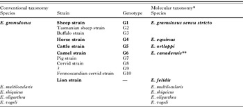
The species of this ‘Echinococcus granulosus-complex’ (Table 1) are maintained in a variety of life cycles. Most of these are based on domestic animals, with the domestic dog as definitive host and various species of livestock (sheep, goats, cattle, yak, buffaloes, camels and pigs) as intermediate hosts. In many instances, wild mammals are marginally involved in transmission. Wild cycles independent from transmission in domestic animals are rare. In Australia E. granulosus sensu stricto has shifted from the dog-sheep transmission to a cycle involving wild dogs (dingoes) and wallabies, kangaroos and feral pigs (Jenkins and Macpherson, Reference Jenkins and Macpherson2003; Jenkins and Morris, Reference Jenkins and Morris2003). In southern Argentina a cycle between an indigenous fox species (Pseudalopex culpaeus) and introduced European hares (Lepus europaeus) has been described, which, however, might not be independent from the sheep-dog cycle in the same area (Schantz et al. Reference Schantz, Lord and de Zavaleta1972). In the northern hemisphere, a taxon of Echinococcus is transmitted between wolf and cervids which was originally known as the ‘northern form’ of E. granulosus. For a long time, this wildlife form was considered ancestral to the various domestic forms, which were thought to have derived from it after the domestication of livestock (Rausch, Reference Rausch1967). Molecular data tell a different story (Table 1): the ‘northern form’ is part of a species, or species complex, – E. canadensis – which also includes domestic forms (the pig and camel strains)Footnote ‡. Based on sequence divergence of mtDNA, the separation of the E. canadensis lineage from other Echinococcus taxa was estimated to have occurred more than two and a half million years ago (McManus, Reference McManus2006). In the absence of a molecular clock calibrated for cestodes this figure might not be precise, but it is clear that the separation had occurred long before the domestication of livestock, which only began in the Neolithic period, approximately 9000 years ago. This strongly suggests that also the various cyst-forming Echinococcus species, or their ancestors, had existed long before the domestication of livestock. Except for E. canadensis, their ancestral wildlife cycles are apparently extinct, as there are few places in the world where stable ecosystems with large wild ungulates and their predators still exist. The one major exception is Africa.
Summary of the taxonomic status of the ‘Echinococcus granulosus complex’ and other Echinococcus species
(Taxa recorded from sub-Saharan Africa are highlighted in bold)

* Combined from Nakao et al. (Reference Nakao, McManus, Schantz, Craig and Ito2007) and Hüttner et al. (Reference Hüttner, Nakao, Wassermann, Siefert, Boomker, Dinkel, Sako, Mackenstedt, Romig and Ito2008).
** The camel and pig strain might be split from E. canadensis as a separate species (Thompson, Reference Thompson2008; Saarma et al. Reference Saarma, Jogisalu, Moks, Varcasia, Lavikainen, Oksanen, Simsek, Andresiuk, Denegri, Gonzalez, Ferrer, Garate, Rinaldi and Maravilla2009).
ECHINOCOCCUS IN SUB-SAHARAN AFRICA
Most data on echinococcosis in sub-Saharan Africa have been gathered in the second half of the 20th century. There is no doubt, however, that these parasites were present much earlier. As an example, Gough (Reference Gough and Schönland1908) put the following remark in his ‘Notes on South African parasites’: ‘Taenia echinococcus, v. Siebold. – Cysts of this cestode from a horse, from sheep and from man are in my possession. Echinococcus almost appears to me to be common in South Africa. I have been able to breed specimens from sheep in the saddle-backed jackal’. This indicates the existence of vigorous domestic life cycles, at least in South Africa, in the 19th century already. Which strains and species were involved at that time, whether they were indigenous, or from where and when they had perhaps been introduced, is not known. We can only assume that ‘domestic’ forms were introduced together with their hosts at various times in history, perhaps starting with the introduction of the first domesticated ruminants in ancient times, and continuing up to the present via the modern livestock trade. From what we know today, those invaders had met indigenous African forms, which were parasites of various species of wild animals.
The first historic record of an infected wild African animal seems to be the above cited report from a silver-backed jackal (Canis mesomelas), although as the result of an experimental infection with unspecified material. Interestingly, the first Echinococcus from Africa to be honoured with a detailed scientific description was of wildlife origin, recovered from an African wild dog (Lycaon pictus) in South Africa. On account of the long handle (or manubrium) of the small hooks, it was described as E. longimanubrius (Cameron, Reference Cameron1926). The African wild dog, almost extinct today, must have been widely available for parasitological research at that time, as some years later a different species, E. lycaontis, was described (Ortlepp, Reference Ortlepp1934) owing to the differences in the lengths of the large rostellar hooks. Another discovery by the same author was E. felidis from South African lions (Panthera leo) (Ortlepp, Reference Ortlepp1937). He collected the parasites from a lion's intestine which was ‘literally felted over’ by these worms. He described that species on the basis of the marked rugosity of the rostellar hooks and its occurrence in a felid as definitive host. Lastly, experimental infections (Verster, Reference Verster1965) showed that the ‘subspecies’ E. g. granulosus, E. g. africanus and E. g. ortleppi (see below) were infectious to silver-backed jackals, and E. g. africanus to Cape foxes (Vulpes chama). Therefore, of the 25 species and subspecies of Echinococcus that had ever been described, not less than 6 were reported to occur in African wild carnivores (Verster, Reference Verster1965; Rausch and Bernstein, Reference Rausch and Bernstein1972; Kumaratilake and Thompson, Reference Kumaratilake and Thompson1982; Xiao et al. Reference Xiao, Qiu, Nakao, Li, Yang, Chen, Schantz, Craig and Ito2005).
As most descriptions were based on structural characters of adult worms whose variation within a parasite population, or between worms in different host species, was unknown, a period of reviews and synonymizations set in. The first was done in 1963, where the differences between most of these species were considered to be within the limits of normal variation (Rausch and Nelson, Reference Rausch and Nelson1963). The authors only retained E. granulosus, E. multilocularis, E. oligarthra, E. patagonicus and E. felidis. In the latter case, it was the unique preference of felids as final hosts rather than convincing morphological differences that led to the retention of that species (Rausch and Nelson, Reference Rausch and Nelson1963). Shortly after, a major revision of Echinococcus was published (Verster, Reference Verster1965). Criteria for classification were the morphological characters of the adult worms like number of segments, arrangement of the sexually mature segment, number and distribution of the testes and, in addition, host specificity. Size and shape of the rostellar hooks, the foundation of some early descriptions, were not considered sufficiently stable to warrant taxonomic significance. By this approach, only E. granulosus and E. multilocularis retained their species status, and E. oligarthra from South America was considered a species inquirendae. All species which had been described by then from the African continent either were reduced to the rank of subspecies of E. granulosus (E. felidis and E. lycaontis, together with E. ortleppi, described for the first time 1943 from a domestic dog in the South of Spain (Lopez-Neyra and Soler Planas, Reference Lopez-Neyra and Soler Planas1943)), or were included in E. g. granulosus due to the lack of separating characters. An additional subspecies E. g. africanus was created for worms recovered from a dog from unknown locality and from 3 additional experimental infections of dogs and jackals, which had been fed with hydatid cysts of bovine and ovine origin. This subspecies was characterized by the difference in the number of testes and the shape of the seminal receptacle (Verster, Reference Verster1965). Just 2 years later, the concept of Echinococcus subspecies was refuted, because at least some of these occurred sympatrically in the same definitive host species without any obvious reproductive isolation (Rausch, Reference Rausch1967). It was proposed to provisionally retain all these taxa in E. granulosus and suggested to designate the different forms of E. granulosus informally as strains until new information would become available to decide otherwise. Up to the present, this system eventually led to the distinction of 11 strains of E. granulosus (G1 to G10, and the ‘lion strain’). Five of them, namely the sheep, horse, cattle, camel and the lion strains, were reported from sub-Saharan Africa. The recent availability of reliable genetic data, showing clustering of some strains and rather distant relationship of others, has brought about the next major reshuffle of the genus (Table 1) (Nakao et al. Reference Nakao, McManus, Schantz, Craig and Ito2007). The horse and cattle strains were the first to be split from E. granulosus as E. equinus and E. ortleppi, respectively (Thompson and McManus, Reference Thompson and McManus2002), followed by the camel, pig and cervid strains (as E. canadensis) (Nakao et al. Reference Nakao, McManus, Schantz, Craig and Ito2007) and finally the lion strain as E. felidis (Hüttner et al. Reference Hüttner, Nakao, Wassermann, Siefert, Boomker, Dinkel, Sako, Mackenstedt, Romig and Ito2008). Awaiting further biological and epidemiological data, the pig and camel strains might have to be split from E. canadensis, but there is still uncertainty on this (Thompson, Reference Thompson2008; Saarma et al. Reference Saarma, Jogisalu, Moks, Varcasia, Lavikainen, Oksanen, Simsek, Andresiuk, Denegri, Gonzalez, Ferrer, Garate, Rinaldi and Maravilla2009).
Of all the species originally described from African wild carnivores, only E. felidis has survived the storms of time. It was therefore suggestive to attribute all previous reports of Echinococcus cysts from wild herbivores in natural African ecosystems to that species. Is this justified?
Looking at Echinococcus records from African wildlife, it is intriguing to note the long list of species which are known to act as hosts for these worms. Definitive hosts are quite few: silver-backed jackal (Canis mesomelas), golden jackal (Canis aureus), Cape fox (Vulpes chama), African wild dog (Lycaon pictus), spotted hyena (Crocuta crocuta), African wild cat (Felis silvestris lybica) and lion (Panthera leo) (Ortlepp, Reference Ortlepp1937; Nelson and Rausch, Reference Nelson and Rausch1963; Verster and Collins, Reference Verster and Collins1966; Troncy and Graber, Reference Troncy and Graber1969; Dinnik and Sachs, Reference Dinnik and Sachs1972; Young, Reference Young1975a; Graber and Thal, Reference Graber and Thal1980; Macpherson et al. Reference Macpherson, Karstad, Stevenson and Arundel1983). In contrast, hydatid cysts could be isolated from a large number of different ungulates: 11 species of bovids, warthogs (Phacochoerus sp.), bushpig (Potamochoerus larvatus, P. porcus), hippopotamus (Hippopotamus amphibius), giraffes (Giraffa camelopardalis), zebras (Equus quagga, E. zebra), cape mole rats (Georychus capensis), baboons (Papio sp.) and colobus monkeys (Colobus sp.) (Dinnik and Sachs, Reference Dinnik and Sachs1969; Graber et al. Reference Graber, Troncy, Tabo, Service and Oumatie1969; Woodford and Sachs Reference Woodford and Sachs1973; Young, Reference Young1975a, Reference Youngb; Graber and Thal, Reference Graber and Thal1980; Macpherson et al. Reference Macpherson, Karstad, Stevenson and Arundel1983, Reference Macpherson, Else and Suleman1986; Krecek et al. Reference Krecek, Boomker, Penzhorn and Scheepers1990; Macpherson and Wachira, Reference Macpherson, Wachira, Andersen, Ouhelli and Kachani1997).
The numerous species of antelopes (wild bovids other than buffalo) appear to play a minor role as intermediate hosts. The prevalence of hydatid cysts in these animals is low compared to other ungulates in the same surveys, whether due to low susceptibility or behavioural factors. In the Serengeti of Tanzania, 4 ‘antelopes’ (sic) out of 350 were found infected (Dinnik and Sachs, Reference Dinnik and Sachs1969), in the Maasai Mara of Kenya 1 out of 17 wildebeest (Connochaetes taurinus) and none out of 47 other antelopes of 10 species had hydatid cysts (Nelson and Rausch, Reference Nelson and Rausch1963), a survey in Uganda found no hydatids in 122 examined antelopes of 5 species (Woodford and Sachs, Reference Woodford and Sachs1973), and in South Africa only 3 of 600 impalas (Aepyceros melampus) were infected (Young, Reference Young1975a). This is different with buffalos and warthogs, for which prevalences of 17% and 10–40%, respectively, were reported from Tanzania and Uganda. Cysts from warthogs contained protoscolices, but those from buffaloes were mostly non-fertile (Dinnik and Sachs, Reference Dinnik and Sachs1969; Woodford and Sachs, Reference Woodford and Sachs1973). At least in South Africa, zebras seem to be an important host species: 60% of an unspecified number of Burchell's zebras (E. quagga) were found to be infected there (Young, Reference Young1975a).
In the absence of any recognized diagnostic feature of the cysts, it is unknown for any of these records if they reflect infections by E. felidis, by another as yet uncharacterized wildlife species (like those uncertain species described early on from Lycaon pictus), or constitute spill-overs from domestic cycles. In many parts of eastern and southern Africa, livestock and wildlife coexist in the same area or in close neighbourhood. Interaction between wildlife and domestic lifecycles could occur under several circumstances. Livestock grazed in or near conservation areas might acquire a form present in wildlife, and wild carnivores preying on livestock or scavenging carcasses might acquire ‘domestic’ forms. Even where large wild herbivores are extinct, jackals or hyenas can still partake in domestic cycles. In parts of Kenya it could even be shown that wild carnivores scavenge on human corpses. In the Turkana district of Kenya, where human infection is extremely frequent, the consumption of undercooked jackal intestines (which are eaten) was proposed as one of several important infection sources (Nelson and Rausch, Reference Nelson and Rausch1963; Macpherson, Reference Macpherson1983; Macpherson et al. Reference Macpherson, Karstad, Stevenson and Arundel1983, Reference Macpherson, French, Stevenson, Karstad and Arundel1985, Reference Macpherson, Craig, Romig, Zeyhle and Watschinger1989a).
In the past, some infection experiments were done to shed light on the epidemiology in wildlife. As mentioned above, silver-backed jackals were successfully infected in South Africa with hydatid material from human, cattle, pig and sheep; the worms obtained could be characterized as E. g. granulosus, E. g. africanus and E. g. ortleppi (Verster, Reference Verster1965). N. F. Viljoen, in 1937 infected the same species successfully with protocolices from cattle (cited by Macpherson and Wachira, Reference Macpherson, Wachira, Andersen, Ouhelli and Kachani1997). African wild dogs were also confirmed as definitive hosts of E. g. lycaontis (larvae derived from sheep) and E. g. africanus (larvae derived from cattle). An experimental infection with the latter larval material resulted also in the development of adult worms in Cape foxes (Verster, Reference Verster1965). In the 1980s, an attempt to infect spotted hyenas with unspecified larval material of human origin in Kenya was unsuccessful (Macpherson et al. Reference Macpherson, Karstad, Stevenson and Arundel1983). Another experiment was carried out by feeding beagles with cysts from warthog (Phacochoerus africanus) and red river hog (Potamochoerus porcus) from the Central African Republic. Two months later no adult Echinococcus could be found in the dogs (Graber and Thal, Reference Graber and Thal1980). Negative results of infection experiments, however, should be taken with caution, as numerous factors could adversely influence the trial. The only experimental infection between two wildlife species was done in South Africa (Young, Reference Young1975a), where hydatid cysts of zebra origin were fed to a lion which was found to be highly susceptible. An additional infection experiment with cysts from zebra yielded E. equinus in dogs (see below) (Kumaratilake et al. Reference Kumaratilake, Thompson and Eckert1986).
In summary, the results of these experiments, some done in a rather unsystematic way, or using parasite material which was neither morphologically nor genetically characterized, gave us a few pieces of the puzzle, but are not sufficient to resolve the epidemiological situation. This experimental approach is unlikely to be a practical method in future either, due to the effort, cost, biohazard and ethical considerations involved (most wildlife species are endangered and legally protected). In the absence of distinctive morphological features of the cysts, and of uncertainty about the taxonomic value of characters of the adults, only genetic characterization of isolates can solve this riddle. To provide some firm ground from where to begin this exploration, we summarize those records from sub-Saharan Africa which are unequivocally characterized either morphologically or genetically.
Echinococcus granulosus s. s.
The frequent occurrence of the common sheep strain (G1) of this species was confirmed by 2 molecular studies. In Kenya, a total of 248 samples from goat, cattle, camel, sheep, dogs and humans were examined by RFLP-PCR of the its1 gene. Echinococcus granulosus s. s. could be diagnosed in all host species (Wachira et al. Reference Wachira, Bowles, Zeyhle and McManus1993). The second investigation was done with samples from Kenya and Sudan. A species- and strain-specific PCR-system based on the mitochondrial 12SrRNA gene was used, and the examination of 520 cyst samples from cattle, camels, sheep, goats, pigs and humans revealed the existence of E. granulosus s. s. in all these species (Dinkel et al. Reference Dinkel, Njoroge, Zimmermann, Wälz, Zeyhle, Elmahdi, Mackenstedt and Romig2004). In addition to this, we have the morphologically based records of ‘E. g. granulosus’ from various domestic animals in South Africa, and from successful infection experiments with jackals, wild dogs and foxes (Verster, Reference Verster1965).
This is, worldwide, the most widespread form of Echinococcus in domestic animals and it is well known that various wild animals can be involved in the transmission. If a true wildlife cycle of E. granulosus s.s. exists in Africa, is not known. The potential for this is shown by the above-mentioned experimental infections and by a recently confirmed natural E. granulosus s. s. infection of a wild warthog from the Queen Elizabeth National Park in Uganda (Hüttner et al. Reference Hüttner, Siefert, Mackenstedt and Romig2009).
Echinococcus equinus
E. equinus represents the ‘horse strain’ of E. granulosus, or G4, which seems to use exclusively equids as intermediate hosts (Thompson et al. Reference Thompson, Lymbery and Constantine1995). There are no molecular data confirming its presence in Africa. However, in south-western Africa adult worms were recovered from the intestine of a dog which had been fed with a hydatid cyst from a mountain zebra (Equus zebra). The worms were identified morphologically as E. equinus (Kumaratilake et al. Reference Kumaratilake, Thompson and Eckert1986). Disregarding some unspecified records of cysts from horses and donkeys, this is the only firm evidence that E. equinus occurs in Africa in wild or domestic animals.
Echinococcus ortleppi
The ‘cattle strain’, or G5, was first recorded in South Africa (Verster, Reference Verster1965) by re-examining material from dogs, which was previously identified as E. granulosus (Ortlepp, Reference Ortlepp1934). Apart from dogs, silver-backed jackals were found susceptible for this species (Verster, Reference Verster1965). The infection of cattle with hydatid cysts is no indication for the presence of this species, as cattle can also harbour cysts of E. granulosus s.s., although those are usually infertile (Thompson et al. Reference Thompson, Kumaratilake and Eckert1984; Dinkel et al. Reference Dinkel, Njoroge, Zimmermann, Wälz, Zeyhle, Elmahdi, Mackenstedt and Romig2004). Its existence was proven with molecular techniques in an unspecified species of zebra from Namibia, which constitutes the only proven record in a naturally infected wild animal (Obwaller et al. Reference Obwaller, Schneider, Walochnik, Gollackner, Deutz, Janitschke, Aspöck and Auer2004), and in cattle from Sudan and a domestic pig from Kenya (Dinkel et al. Reference Dinkel, Njoroge, Zimmermann, Wälz, Zeyhle, Elmahdi, Mackenstedt and Romig2004).
Echinococcus canadensis
There are important camel-raising regions in the north of sub-Saharan Africa, and the presence of the ‘camel strain’ (G6) is therefore not surprising. However, camels are also infected with other forms of Echinococcus, and molecular characterization is needed to confirm the nature of the parasite. The species was found to be frequent in Kenya and in Sudan, where goat, cattle, camel, sheep and pig, as well as dogs, were found infected (Wachira et al. Reference Wachira, Bowles, Zeyhle and McManus1993; Dinkel et al. Reference Dinkel, Njoroge, Zimmermann, Wälz, Zeyhle, Elmahdi, Mackenstedt and Romig2004). Human patients are also known from these regions, but cases appear to be much rarer than with E. granulosus s.s., despite its frequency in livestock (Dinkel et al. Reference Dinkel, Njoroge, Zimmermann, Wälz, Zeyhle, Elmahdi, Mackenstedt and Romig2004).
Echinococcus felidis
The first Echinococcus specimens found in lions were described as a new species (Ortlepp, Reference Ortlepp1937). Later, 5 of 7 South African lions were found harbouring these tapeworms (Verster and Collins, Reference Verster and Collins1966), and a similar situation was reported from Uganda where lions were found to be commonly infected (Dinnik and Sachs, Reference Dinnik and Sachs1972; Woodford and Sachs, Reference Woodford and Sachs1973). A later report of 4% of 75 faecal samples from lions found to be positive for Echinococcus spp. in the Serengeti and Ngorongoro Crater, Tanzania, needs confirmation, as the animals were examined with an immunodiagnostic test of unclear specificity (Müller-Graf, Reference Müller-Graf1995). Recent studies on the genome of this parasite, based on Echinococcus eggs from Ugandan lion faeces, and preserved South African worms that had been determined by A. J. Verster in the 1960s, confirmed the distinctiveness of E. felidis, and established its status as a sister taxon of E. granulosus s. s. (Hüttner et al. Reference Hüttner, Nakao, Wassermann, Siefert, Boomker, Dinkel, Sako, Mackenstedt, Romig and Ito2008). The faecal prevalence of E. felidis in the small lion population from this park was 72% and confirmed the persistence of this parasite at least in this refuge. In addition to lions, E. felidis eggs could be identified in 1 of 5 faecal samples from spotted hyenas (Hüttner et al. Reference Hüttner, Siefert, Mackenstedt and Romig2009). Concerning intermediate hosts, there is only 1 molecularly characterized infection in a warthog from the Queen Elizabeth National Park in Uganda (Hüttner et al. Reference Hüttner, Siefert, Mackenstedt and Romig2009).
From the data summarized above it is evident that the epidemiology of Echinococcus spp. in African wildlife is only known in sketches. The value of morphological descriptions of the past is acknowledged, but future research priorities have to include the molecular and biological characterization of isolates from wild host species, and the comparison with isolates from domestic animals and human patients living in close neighbourhood. This will serve to identify the host range and geography of the various species, the possible identification of ‘cryptic’ forms which had been described from wild carnivores (e.g. E. lycaontis), and the putative linkage between domestic and sylvatic life cycles. An important aspect will be the appraisal of the potential of wild carnivores as transmitters of zoonotic agents. E. felidis is most closely related to E. granulosus s. s., the major agent for human echinococcosis worldwide. The pastoralist peoples living in the vicinity of East Africa's conservation areas (e.g. the Maasai) show a mean prevalence of cystic echinococcosis higher than 1% (Macpherson et al. Reference Macpherson, Spoerry, Zeyhle, Romig and Gorfe1989b), and not all these cases might be caused by the domestic forms of Echinococcus. A recent re-examination of 412 cyst samples from humans, cattle, camels, sheep and goats from eastern Kenya only found E. granulosus s. s. and E. canadensis (G6/7), but isolates from the vicinity of conservation areas were few in that sample (Hüttner et al. Reference Hüttner, Siefert, Mackenstedt and Romig2009). Vice versa, there might be a possible detrimental impact of domestic Echinococcus forms to endangered wild mammals, as has been reported from Australia (Barnes et al. Reference Barnes, Goldizen, Morton and Coleman2008). Large African wildlife species, even in conservation areas, vanish rapidly due to population pressure, bad governance and civil strife. This is especially true for the large predators, some of which are even on the verge of extinction in the wild (cheetah and wild dog). Parasites are not only agents of disease, but an integral part of the ecology of these areas, and those with complex life cycles, involving various host species, are even more vulnerable than their hosts. If we don't look now, we might not have another chance.
FINANCIAL SUPPORT
This work was partially supported by the Eiselen Foundation Ulm; by the graduated grant (LGFG) of Baden-Württemberg (M. H.) and by the DFG project R03753 1-1 (M. H., T. R.).