Syncope is defined as a transient loss of consciousness, which happens suddenly and recovers quickly. 1 The pathophysiological change of syncope is transient global cerebral hypoperfusion. Syncope is a common emergent problem, experienced by up to 15% of children before the end of adolescence,Reference Friedman and Alexander 2 and the chief complaint of syncope accounts for 1% of all paediatric emergency department visits.Reference Bo, Carano and Agnetti 3 – Reference McLeod 5
Neurally mediated syncope is the most common type of syncope in children and adolescents. Neurally mediated syncope accounts for 75–80% of syncopal patients and is rarely life threatening.Reference Bo, Carano and Agnetti 3 – Reference McLeod 5 It is composed of postural orthostatic tachycardia syndrome, vasovagal syncope, and orthostatic hypertension. Neurally mediated syncope must be differentiated from cardiac syncope, which is more dangerous and associated with an increased risk for sudden cardiac death. Cardiac syncope can be grouped according to arrhythmic and structural causes. Common causes of arrhythmic syncope include catecholaminergic polymorphic ventricular tachycardia, long QT syndrome, and sick sinus syndrome, 1 , Reference Zhang, Zhu and Wang 6 whereas structural causes of syncope include acute myocardial infarction, hypertrophic cardiomyopathy, pulmonary hypertension, and pericardial diseases. A vital task of the primary-care physicians is to differentiate neurally mediated syncope from cardiac syncope by history, physical signs, and accessory tests.Reference Friedman and Alexander 2 , Reference Tretter and Kavey 7 Looking for useful markers to differentiate neurally mediated syncope from arrhythmic syncope is particularly important, especially in the paediatric emergency department.
Recently, reports have indicated that low iron storage and vitamin B12 deficiency both are involved in the pathophysiology of neurally mediated syncope.Reference Jarjour and Jarjour 8 – Reference Stewart 11 Compared with controls, patients with neurally mediated syncope have lower levels of iron and vitamin B12 storage,Reference Jarjour and Jarjour 8 – Reference Öner, Guven, Tavli, Mese, Yilmazer and Demirpence 9 and some patients obtain benefits with supplementing the appropriate nutrition.Reference Daoud, Batieha, al-Sheyyab, Abuekteish and Hijazi 12 , Reference Ganjehei, Massumi, Razavi and Wilson 13 The red blood cell distribution width is an available parameter involved in complete blood count analysis. It stands for the degree of anisocytosis, and is generally used to differentiate anaemia. An increased red cell distribution width is commonly found when there is a nutritional deficiency – for example, iron, folate, and vitamin B12 deficiencies.Reference Salvagno, Sanchis-Gomar, Picanza and Lippi 14 Therefore, we theorised that there might be a difference in the value of red cell distribution width between neurally mediated syncope and cardiac syncope, which could be helpful in distinguishing them. Therefore, this study aimed to examine whether red cell distribution width could be helpful in distinguishing paediatric neurally mediated syncope from arrhythmic syncope, especially by primary-care physicians.
Material and methods
Study subjects
Between June 2009 and March 2016, 93 patients with syncope were admitted to the Department of Pediatrics at Peking University First Hospital. They were divided into a neurally mediated syncope group (n=72) and an arrhythmic syncope group (n=21) on the basis of history, results of a head-up tilt test, electrocardiography, and 24-hour ambulatory electrocardiography. A total of 55 healthy children served as control subjects. All children, including the control subjects, fulfilled the inclusion criteria: age between 4 and 15 years, normal physical examination results, and routine biochemistry, without history of infection at the time of blood collection. Children who had organic cardiovascular disease or diseases related to the central nervous and metabolic systems were excluded from the study. Children in the control group had never experienced syncope or pre-syncope as demonstrated by history, physical examination, and laboratory tests. We obtained approval of the Ethics Committee of Peking University First Hospital for the study, and informed consent was obtained from all the patients’ guardians.
Electrocardiography
All 93 patients were tested by electrocardiography, including 12-lead resting electrocardiography and 24-hour ambulatory electrocardiography. Next, we referred to the “Schwartz” diagnostic score to diagnose long QT syndrome. If the score was ⩾3.5, further checks were carried out to confirm the diagnosis of long QT syndrome.Reference Schwartz and Ackerman 15 , Reference Schwartz 16 Sick sinus syndrome was diagnosed by history analysis and electrocardiography. Various examples of the electrocardiography findings include baseline profound sinus bradycardia, abrupt and unpredictable episodes of sinus pauses, supraventricular tachyarrhythmias, or tachycardia–bradycardia syndrome.Reference Semelka, Gera and Usman 17 As for the diagnosis of catecholaminergic polymorphic ventricular tachycardia, two types of polymorphic tachycardias were described – typical polymorphic ventricular tachycardia with continuously varying QRS morphology and bidirectional tachycardia demonstrating alternans of the QRS complexes. The final diagnosis of catecholaminergic polymorphic ventricular tachycardia was based on exercise and/or genetic testing.Reference Roston, Vinocur and Maginot 18
Head-up tilt test protocol
When the results of electrocardiography were normal, the patients underwent tilt table testing. The examination was performed between 8:00 am and 10:00 am in a quiet and blandly lit room at a comfortable constant ambient temperature (22°C, 50–55% humidity). First, patients lay on the tilt table, and the heart rate, blood pressure, and electrocardiographic parameters were recorded for at least 10 min. Next, they were tilted upward at an angle of 60° for 45 min, with heart rate, blood pressure, and electrocardiographic parameters being monitored at the same time.Reference Grubb, Temesy-Armos and Moore 19 , Reference Strieper, Auld and Hulse 20 The patients were placed in the supine from the standing position as soon as the positive response or symptoms of orthostatic intolerance occurred. The symptoms include lightheadedness, headache, nausea, near syncope, syncope, and so on.
When a child with clinical symptoms of orthostatic intolerance had a heart rate increase of ⩾40 beats/minute or a heart rate of ⩾130 beats/minute (children aged 6–12 years) and ⩾125 beats/minute (adolescents aged 13–18 years) during the initial 10 minutes of the head-up tilt test, the result was defined as a positive response and a diagnosis of postural orthostatic tachycardia syndrome was made.Reference Zhao, Han and Zhang 21 We made a diagnosis of vasovagal syncope when there was a significant decrease in heart rate, blood pressure, or both during the head-up tilt test with a syncopal episode or pre-syncope at the same time. This criterion was defined referring to a described protocol.Reference Zhang, Du and Wang 22
Complete blood count analysis
Blood samples for complete blood count analysis were obtained on the 1st day of the hospitalisation through venepuncture. We used tripotassium ethylenediaminetetraacetic acid tubes to collect the blood samples and analysed them using an automatic blood counter.
Statistical analysis
All statistical analyses were conducted using the Statistical Package for Social Science 20.0, Statistical Package Program for Windows. Analysis of variance was used to compare quantitative variables among groups. Qualitative variables were compared using Pearson’s χ2-test. A p value<0.05 was considered to be statistically significant. The receiver operating characteristics curve was used to explore the probability of correctly discriminating between patients with neurally mediated syncope and those with arrhythmic syncope by using the value of red cell distribution width. The area under the curve stood for the predictive value of red cell distribution width. An area between 0.5 and 0.7 indicates that the prospective value is low, between 0.7 and 0.9 indicates a medium prospective value, and >0.9 indicates a high prospective value. When the 95% confidence interval of the area under the curve did not contain 0.5 or the p value was <0.05, the tested index was thought to be of predictive value.Reference Swets 23
Results
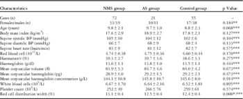
Neurally mediated syncope was diagnosed in 72 children, including 55 children with vasovagal syncope and 17 with postural orthostatic tachycardia syndrome. Arrhythmic syncope was diagnosed in 21 children, including 13 children with long QT syndrome, four children with sick sinus syndrome, and four children with catecholaminergic polymorphic ventricular tachycardia. Of the 72 patients with neurally mediated syncope, 33 were girls (females:males: 33:39). Their mean age was 9.8±2.1 years. Of the 21 patients with arrhythmic syncope, 10 were girls (females:males: 10:11). Their mean age was 9.7±3.0 years. Of the 55 control subjects, 17 were girls (females:males: 17:38). Their mean age was 8.8±2.3 years. The basal clinical characteristics did not differ among the neurally mediated syncope, arrhythmic syncope, and control groups (Table 1).
Comparison of baseline characteristics and complete blood count.

AS=arrhythmic syncope; BP=blood pressure; NMS=neurally mediated syncope
Values are expressed as mean±SD
** p values calculated by χ2
*** p values calculated by F
*p<0.05 NMS group versus arrhythmic syncope or control group
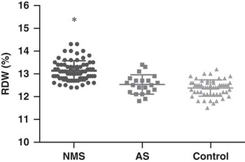
There were also no statistically significant differences in complete blood count relative parameters except for red cell distribution width among the three groups (Table 1); however, the value of red cell distribution width was obviously higher in the neurally mediated syncope group than in the arrhythmic syncope group or control group (Fig 1).
Peripheral red cell distribution width (RDW) according to causes of syncope. RDW was significantly higher in children with neurally mediated syncope (NMS) than in children of the arrhythmic syncope (AS) group or of the control group. *p<0.05 versus AS and control group.

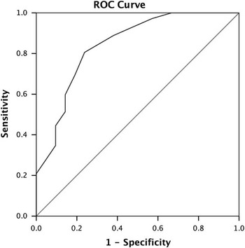
A receiver operating characteristics curve on the predictive value of red cell distribution width in differentiating neurally mediated syncope from arrhythmic syncope showed that the area under the curve was 0.841 (95% confidence interval: 0.737–0.945, p<0.05). The number of red cell distribution width at 12.8% taken as the cut-off value yielded a sensitivity of 80.6% and a specificity of 76.2% in the initial discrimination between neurally mediated syncope and arrhythmic syncope (Fig 2).
Receiver operating characteristics (ROC) curve of the predictive value of peripheral red cell distribution width for differential diagnosis between neurally mediated syncope and arrhythmic syncope in children. The y-axis represents the sensitivity to predict the diagnosis of neurally mediated syncope; the x-axis represents the false-positive rate (1-specificity).

Discussion
In this study, we report that red cell distribution width is a valuable adjunct for differentiating syncope in children and adolescents. The results indicate that children with neurally mediated syncope had a higher red cell distribution width value than those with arrhythmic syncope. The red cell distribution width cut-off value of 12.8% had a sensitivity of 80.6% and specificity of 76.2% in correctly identifying neurally mediated syncope and arrhythmic syncope in children. As red cell distribution width is a routine index involved in complete blood count, it is regularly measured and inexpensive. Therefore, the above findings of our study have important value for the initial differential diagnosis of syncope in children, based on history, physical signs, and electrocardiography review, especially for primary-care physicians.
About 15% of children would have had one syncopal episode at least before 18 years of age.Reference Friedman and Alexander 2 In determining the underlying aetiology of syncope in children, sometimes excessive or inefficient use of diagnostic tests might lead to ineffective and even harmful treatment, as well as greatly increase the diagnostic and therapeutic expense. In the past, we found that serum brain natriuretic peptide was helpful in differentiating cardiac syncope from non-cardiac syncope in children and adolescents.Reference Zhang, Jin, Qi, Yan and Du 24 Simple biomarkers to differentiate neurally mediated syncope from cardiac syncope, especially from arrhythmic syncope, would be useful adjuncts in the evaluation of syncope in the primary-care setting.
Nevertheless, in clinical practice, arrhythmic syncope is more difficult to diagnose than non-arrhythmic cardiac syncope as paediatricians can easily find some diagnostic clue in non-arrhythmic cardiac syncope by a careful history analysis and physical examination, such as the murmurs in hypertrophic cardiomyopathy and enhanced P2 in pulmonary hypertension. It is relatively difficult to acquire the abnormal electrocardiogram in arrhythmic syncope, especially during the intermittent period of syncope. Moreover, arrhythmic syncope is perhaps the most serious of all syncopal diseases, even among cardiac causes. Arrhythmias are associated with a high risk, thus requiring timely and correct diagnosis; however, serum brain natriuretic peptide could not differentiate neurally mediated syncope from arrhythmic syncope cases, which limits the value of serum brain natriuretic peptide in the clinical differential diagnosis of syncope. Therefore, looking for easy and convenient biomarkers in the initial differential diagnosis of syncopal children is particularly important, especially in the paediatric emergency department.
Previous studies have indicated the association between a nutritional deficiency – iron or vitamin B12 – and neurally mediated syncope.Reference Jarjour and Jarjour 8 – Reference Ganjehei, Massumi, Razavi and Wilson 13 In fact, patients with neurally mediated syncope are likely to have a lower levels of iron storage than patients with other types of syncope.Reference Jarjour and Jarjour 8 In addition, a therapeutic trial of iron also showed benefits in non-anaemic children with breath-holding spells, which is a form of neurally mediated syncope in early childhood.Reference Daoud, Batieha, al-Sheyyab, Abuekteish and Hijazi 12 Öner et alReference Öner, Guven, Tavli, Mese, Yilmazer and Demirpence 9 declared that there was a relative shortage of vitamin B12 in the neurally mediated syncope group as compared with the control group. Furthermore, Ganjehei et alReference Ganjehei, Massumi, Razavi and Wilson 13 suggested that orthostatic hypotension could be induced by the shortage of vitamin B12 and might be corrected with vitamin B12 supplementation. In fact, both low iron storage and vitamin B12 deficiency would cause anisocytosis with an elevated red cell distribution width number;Reference Salvagno, Sanchis-Gomar, Picanza and Lippi 14 however, other indicators such as haematocrit, mean corpuscular volume, mean corpuscular haemoglobin, and mean corpuscular haemoglobin concentration had different changes between iron deficiency and vitamin B12 deficiency, and these values were relatively unstable in neurally mediated syncope.Reference Salvagno, Sanchis-Gomar, Picanza and Lippi 14 Furthermore, red cell distribution width was reported to have a higher predictive value for iron-deficiency anaemia than the other indicators mentioned above.Reference Sazawal, Dhingra and Dhingra 25
As a result, we supposed that red cell distribution width might be a convenient and an inexpensive adjunct for the initial differential diagnosis of syncope in children on the basis of careful history analysis, systemic physical examination, electrocardiography, etc.
Nevertheless, there are still certain limitations to this study – for instance, the relatively small sample size might result in bias, and we only included long QT syndrome, sick sinus syndrome, and catecholaminergic polymorphic ventricular tachycardia cases, which could not represent all arrhythmic syncope patients. Further studies with a multicentre design to increase the case number are needed.
Conclusions
Our study suggests that red cell distribution width is a useful adjunct for differentiating neurally mediated syncope from arrhythmic syncope. These findings are of great importance for primary-care physicians to quickly determine the potential, aetiological entity of syncopal children and adolescents.
Acknowledgements
None.
Financial Support
This research received no specific grant from any funding agency or from commercial or not-for-profit sectors.
Conflicts of Interest
None.
Ethical Standards
All procedures performed in this study were in accordance with the ethical standards of the Ethics Committee of Peking University First Hospital, and with the Helsinki Declaration of 1975, as revised in 2008.