Introduction
Although the production of bovine embryos in vitro has improved remarkably over the past decades, the blastocyst rate and the pregnancy rate were still lower than the embryos developed in vivo. As culture medium is one of the most important factors that influences in vitro embryo development (Lonergan et al., Reference Lonergan, Rizos, Gutierrez-Adan, Fair and Boland2003; Krisher, Reference Krisher2004), various culture media have been developed to improve embryo development, in vitro such as mSOF (Tervit et al., Reference Tervit, Whittingham and Rowson1972), CR1aa and CR2 (Rosenkrans & First, Reference Rosenkrans and First1991), KSOM (Erbach et al., Reference Erbach, Lawitts, Papaioannou and Biggers1994), CZB (Ellington et al., Reference Ellington, Carney, Farrell, Simkin and Foote1990), G1 and G2 (Gardner, Reference Gardner1994), BECM (Lim et al., Reference Lim, Reggio, Godke and Hansel1999) and so on. Among those options, mSOF (modified synthetic oviduct fluid medium) was the most successful and widely used alternative in bovine fertilized (IVF)-derived embryo in vitro culture (Farin et al., Reference Farin, Crosier and Farin2001; Gomez et al., Reference Gomez, Rodrıguez, Munoz, Caamano, Hidalgo, Moran, Facal and Diez2008; Feugang et al., Reference Feugang, Camargo-Rodríguez and Memili2009) and it was used as a standard medium in our laboratory. G1/G2 was one of the sequential media that was developed to response to the scalable requirement of development embryos over the last few years (Bavister, Reference Bavister1995; Gardner & Lane, Reference Gardner and Lane1997; Pool, Reference Pool2004). It has been proposed for commercial use in bovine embryo culture, which was suggested to provide qualitative and quantitative bovine IVF-derived (Lane et al., Reference Lane, Gardner, Hasler and Hasler2003) and somatic cell nuclear transfer (SCNT)-derived embryos. Above all, we found that mSOF and G1.5/G2.5 were good media for the in vitro culture of bovine embryos, but as shown on bovine (Mastromonaco et al., Reference Mastromonaco, Semple, Robert, Rho, Betts and King2004) and mice (Heindryckx et al., Reference Heindryckx, Rybouchkin, Vander Elst and Dhont2001), the culture requirement between IVF- and SCNT-derived embryos was different, it would be necessary and meaningful to clarify which medium is more suitable for bovine IVF- and SCNT-derived embryos.
Due to incomplete reprogramming of gene expression, we ascertained that the media requirements of SCNT-derived embryos might be different from that of normally fertilized bovine embryos. As the culture requirements for bovine IVF- and SCNT-derived in particular are still poorly understood, further investigation was needed urgently. So far, few research groups have been successful in examining the different preferences for culture medium between bovine IVF- and SCNT-derived embryos and little comparison has been made among various embryo culture media in their ability to sustain in vitro development of these two kinds of embryos in bovine. Therefore, we investigated the effects of mSOF and G1.5/G2.5 on the developmental potential and quality of bovine embryos derived from IVF and SCNT in the present study.
The aim of the present study was to investigate the effects of two culture media, mSOF and G1.5/G2.5, on bovine IVF- and SCNT-derived embryos development and embryo quality determined by total cell number, apoptotic index and expression patterns of two qualitatively important genes related to stress (Hsp70) and apoptosis (Bax) in blastocysts.
Materials and methods
All chemicals used in this study were purchased from the Sigma Chemical Company (St. Louis, MO, USA) unless otherwise noted. Disposable, sterile plasticware was purchased from Nunclon (Roskilde, Denmark).
All procedures in this experiment were approved by the Animal Care and Use Committee of Northwest A & F University and performed in accordance with animal welfare and ethics considerations.
Oocyte collection and in vitro maturation
Bovine ovaries were obtained from local slaughter houses and transported to the laboratory within 4 h of slaughter and in sterile 0.9% NaCl saline at 25–30°C in a thermos bottle. Cumulus–oocyte complexes (COCs) were aspired from 2 mm to 8 mm antral follicles using a 12-gauge disposable needle. The COCs with several layers of cumulus cells were collected and washed first in phosphate-buffered saline (PBS) and then washed in TCM-199 (Gibco) before being placed in maturation medium (TCM-199 supplemented with 10% (v/v) fetal bovine serum (FBS), 1 μg/ml 17β-estradiol and 0.075 IU/ml human menopausal gonadotropin), and then incubated at 38.5°C in a humidified incubator of 5% CO2 in air for approximately 20 h. All matured oocytes were randomly allocated to two groups respectively for IVF and SCNT.
Donor cells preparation
Donor somatic cells were derived from a 1-week-old Holstein heifer. Briefly, ear skin was collected and minced with sterile scissors in a 35 mm Petri dish. Explants (approximately 1 mm in diameter) were cultured in Dulbecco's Modified Eagle's medium (DMEM; Gibco) that contained 10% FBS, 1 mM sodium pyruvate, 100 IU/ml penicillin and 100 mg/ml streptomycin under 5% CO2 in air at 37.5°C. Once cells had reached 90% confluency, they were trypsinized and reconstituted at a concentration of 1 × 10⁶ cells/ml. The second to fifth passages of the cell line were utilized as nuclei donors. Cells were induced in the G0/G1 phase of cell cycle by serum deprivation in culture medium (0.5% FBS, for 2 days) for subsequent use for SCNT.
Production of in vitro fertilization (IVF) presumptive zygotes
For fertilization, mature oocytes were fertilized at 20 hpm (hours post maturation) in Fert-Talp medium supplemented with 30 mg/ml heparin, 1.65 mg/ml hypotaurine, 0.27 mg/ml epinephrine, and 4.5 mg/ml penicillamine, and the frozen–thawed spermatozoa used in fertilization were from a Holstein bull separated by Percoll density gradient and further incubated for 24 h. Presumptive zygotes were denuded by treatment with 0.1% bovine testicular hyaluronidase in PBS.
Production of SCNT presumptive zygotes
After maturation, the cumulus cells were dispersed by treatment with 0.1% bovine testicular hyaluronidase in PBS. Only oocytes with an extruded first polar body were selected. The mature oocytes were transferred into droplets of PBS supplemented with 7.5 μg/ml cytochalasin B (CB) and 10% FBS. Enucleation was performed with a 20 μm (internal diameter) glass pipette by aspiration of the first polar body and a small amount of surrounding cytoplasm. Enucleation was confirmed by staining the oocytes with 10 μg/ml Hoechst 33342 for 15 min. The trypsinized donor cell was transferred to the perivitelline space of enucleated oocytes. The oocyte-cell couplets were sandwiched vertically by a pair of platinum microelectrodes connected to the micromanipulator in fusion medium (0.27 M mannitol, 0.1 mM MgSO4, 0.5 mM HEPES and 1 mg/ml bovine serum albumin (BSA)) droplet, then a double electrical pulse of 35 V for 10 μs was applied for oocyte-cell fusion (Liu et al. Reference Liu, Zhang and Zheng2007). Successfully reconstructed presumptive zygotes were kept in mSOF that contained 5 μg/ml cytochalasin B for 2 h until activation. All fused presumptive zygotes were activated further in 5 μM ionomycin for 5 min followed by 4 h exposure to 1.9 mM 6-dimethynopyridine in mSOF.
In vitro culture of presumptive zygotes
IVF-derived and activated SCNT-derived presumptive zygotes were washed three times in the culture medium and randomly allocated to two groups respectively to culture at 38.5°C in a humidified incubator containing 5% CO2 in air. Thirty presumptive zygotes were placed in 50 μl droplets of culture medium in 4-well dishes (Nunc, Roskilde, Denmark) under mineral oil. One group was transferred to G1.5 for 3 days, followed by 3–4 days’ culture in G2.5, G1.5/G2.5 sequential media were purchased from VitroLife (Gothenburg, Sweden). The other group was cultured in mSOF medium (Takahashi & First, Reference Takahashi and First1992), supplemented with 8 mg/ml fatty acid-free BSA, 1% MEM non-essential amino acid solution and 2% BME essential amino acid solution. One-half of mSOF medium was replaced at 72 h (day 3) during the culture period. Subsequent in vitro development to the 2-cell and blastocyst stages were monitored at 24 and 168 h respectively after presumptive zygotes, blastocysts were selected for further analysis.
Apoptosis detection
The numbers of apoptotic cells on day 7 blastocysts cultured in these two culture media systems were determined using a Dead End Fluorometric TUNEL System (Promega, Madison, WI, USA) according to the manufacturer's protocol. Briefly, blastocysts were fixed in 4% paraformaldehyde in PBS supplemented with 0.5% polyvinyl pyrrolidone (PVP), and permeabilized in 0.2% Triton X-100 in PBS for 5 min. Then, cells were incubated with FITC-conjugated dUTP and terminal deoxynucleotidyl transferase at 37°C for 60 min in dark (hereafter, all manipulations were done in dark). The tailing reaction was terminated in 2× SSC (SSC: 0.15 mol/l sodium chloride, 0.015 mol/l sodium citrate) for 15 min. Then cells were incubated in 25 μg/ml RNase A for 30 min and, without washing, the nuclei were counterstained with 10 μg/ml propidium iodide (PI) for 10 min. Assessment of total and TUNEL-positive cell numbers was determined by evaluation of each embryo under an epifluorescence microscope (Nikon, Japan). The apoptotic nuclei with fragmented DNA stained yellowish green, while normal nuclei appeared orange-red in colour.
Quantitative real-time PCR
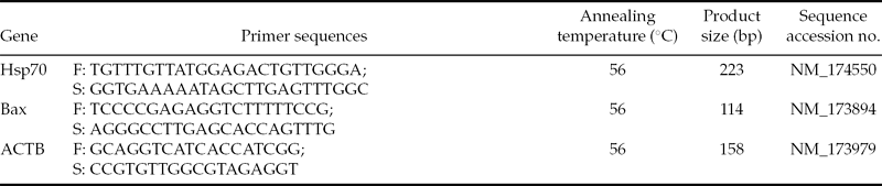
Total RNA of each culture group was isolated from 10 blastocysts on day 7 using the Cells-to-Signal™ Kit (Invitrogen, USA) according to the manufacturer's protocol. The quality and quantity of the extracted RNA were determined using an Epoch Multi-Volume Spectrophotometer System (BioTek, USA), then the total RNA samples were treated with RNA-free DNase I (Invitrogen, USA) to digest the genomic DNA in the RNA samples, and stored at –80°C. cDNA was produced and amplified using the cDNA synthesis kit (TaKaRa, Japan), according to the manufacturer's instructions. Primers for all genes were designed by Primer 5.0 software (Premier Biosoft International, Palo Alto, CA, USA) and were based on bovine RNA sequences found in the GenBank (Table 1). The specificity of all primers was tested using a BLAST analysis against the genomic NCBI database, PCR amplicons were characterized using Mfold (Zuker, Reference Zuker2003) and sequenced for verification.
Primer sequences and PCR conditions used to evaluate expression of Hsp70 and Bax in bovine blastocysts produced in vitro from IVF- or SCNT-derived presumptive zygotes cultured in mSOF or G1.5/G2.5 media

IVF, in vitro fertilized; SCNT, somatic cell nuclear transfer.
All PCR reactions were performed in triplicate in a 20-μl reaction volume on the quantitative real-time PCR CFX96 detection system (Bio-Rad, Nazareth) using the SYBR Premix Ex Taq™ (Takara, Japan), contain 10 μl 2× SYBR Green Premix, 0.8 μl of forward and reverse primers (20 pmol/ml) respectively, 2 μl embryonic cDNA and RNase-free, DNase-free water. The PCR reaction was initiated with 10 min at 95 °C to activate hot-start Taq polymerase followed by 40 cycles of denaturation at 95°C for 10 s, then 56°C for 20 s, extension at 72°C for 20 s and during which fluorescence was measured and was followed by a melting curve analysis to confirm a single gene-specific peak and to detect primer/dimer formation. A negative template control of diethyl pyrocarbonate (DEPC)-treated water was used to ensure that there was no nucleotide contamination. The relative amount of each mRNA was determined using the 2−ΔΔCt method (Rutledge & Cote, Reference Rutledge and Côté2003) and calculated by dividing the intensity of target primers by the intensity of the corresponding housekeeping gene ACTB.
Experimental design
This experiment comprised two zygote production methods (IVF and SCNT) × two culture media (mSOF and G1.5/G2.5) factorial design in which culture droplets that contained approximate 30 presumptive zygotes formed the experimental plots for the assessment of cleavage and blastocyst development. The number of plots per treatment was 20 for IVF/mSOF and G1.5/G2.5 respectively, 15 for SCNT/mSOF and G1.5/G2.5 respectively. Sub-samples of 30–41 blastocysts produced were assessed for cell number and cell apoptosis. A further 10 blastocysts per treatment combination were used for quantitative real-time PCR to evaluate the relative abundance of Hsp70 and Bax mRNA.
Statistical analysis
Each replicate of the experiments was carried out using oocytes matured on the same day, thus removing any batch effect of oocytes. All embryos were allocated randomly to each culture medium. The cleavage rate, blastocysts formation rate, cell numbers, apoptotic index and the levels of gene expression among those groups were analysed by two-way analysis of variance (ANOVA) and Tukey's least significant difference (LSD) test using SPSS 13.0 software. Differences were considered significant at P < 0.05. Data were presented as mean ± standard deviation (SD).
Results
Effect of culture media on in vitro development of IVF and SCNT-derived zygotes
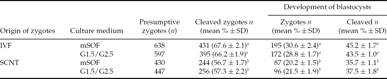
Presumptive zygotes produced by IVF were developmentally more competent than SCNT zygotes in terms of cleavage rate (66.9 vs. 57.0%; P < 0.05) and blastocyst development rates (blastocysts of presumptive zygotes 29.7 vs. 24.8%; blastocysts of cleaved zygotes 44.4 vs. 36.6%; P < 0.05). Over both zygote production systems, however, the results were similar whether culture was in mSOF or in G1.5/G2.5 media for cleavage rate (63.2 vs. 62.4 %) and blastocyst development rate (blastocysts of presumptive zygotes 26.4 vs. 25.7%; blastocysts of cleaved zygotes 41.8 vs. 41.2 %). Table 2 shows that these overall results reflect the individual treatment comparisons of culture media within zygote production system or the method of zygote production within culture medium.
Effects of culturing IVF or SCNT bovine presumptive zygotes in mSOF or G1.5/G2.5 media on cleavage rate and blastocyst production to day 7 of culture
a,bValues with different superscripts within a column are significantly different (P < 0.05). IVF, in vitro fertilized; SCNT, somatic cell nuclear transfer; SD, standard deviation.
Effect of culture media on frequency of apoptotic nuclei in IVF- and SCNT-derived blastocysts
Overall, there were no significant differences between the two methods of zygote production IVF and SCNT for the total number of cells per blastocyst (97.4 ± 3.9 vs. 96.3 ± 4.6), number of apoptotic blastomeres per blastocyst (7.1 ± 3.7 vs. 7.9 ± 4.1) or the apoptotic index (7.3 ± 2.9% vs. 8.2 ± 3.3%). Similarly, there were no significant differences between the overall means for the two culture media, mSOF and G1.5/G2.5, for the total number of cells per blastocyst (96.9 ± 2.8 vs. 97.0 ± 4.1), number of apoptotic blastomeres per blastocyst (7.7 ± 3.4 vs. 7.3 ± 2.1) or the apoptotic index (8.0 ± 3.8% vs. 7.5 ± 2.7%).
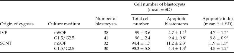
Table 3, however, shows that there was a significant interaction between the method of zygote production and culture medium for the number of apoptotic cells per blastomere and the apoptotic index. The interaction was such that IVF-produced zygotes cultured in mSOF had a lower number of apoptotic cells and apoptotic index compared with those cultured in G1.5/G2.5 whereas SCNT zygotes had a higher number of apoptotic cells and apoptotic index when cultured in mSOF compared to those cultured in G1.5/G2.5.
Effects of culturing IVF or SCNT presumptive zygotes in mSOF or G1.5/G2.5 media on the apoptotic index of bovine blastocysts at day 7 of culture
a,bValues with different superscripts within a column are significantly different (P < 0.05). IVF, in vitro fertilized; SCNT, somatic cell nuclear transfer; SD, standard deviation.
Expression level of Hsp70 and Bax in IVF- and SCNT-derived blastocysts
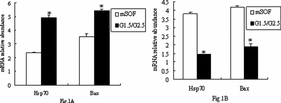
Expression levels of Hsp70 and Bax were compared between mSOF and G1.5/G2.5 group by quantitative real-time PCR (Fig. 1). Figure 1, however, shows that there was a significant interaction between the method of zygote production and culture medium for the relative transcript abundance of Hsp70 and Bax. The interaction was such that IVF-produced zygotes cultured in G1.5/G2.5 had a higher relative abundance of Hsp70 and Bax compared with those cultured in mSOF, but SCNT zygotes had a higher relative abundance of Hsp70 and Bax when cultured in mSOF compared with those cultured in G1.5/G2.5.
Relative mRNA abundance in in vitro fertilized (IVF)-derived (A) and, somatic cell nuclear transfer (SCNT)-derived (B) day 7 bovine blastocysts after in vitro culture from presumptive zygotes in either mSOF or G1.5/G2.5 culture media as assessed by quantitative real-time polymerase chain reaction (RT-PCR). *Differences between culture media within a zygote production method are significantly different.
Discussion
The present study showed that mSOF and G1.5/G2.5 media have a similar capability to support the in vitro development of preimplantation bovine IVF- and SCNT-derived embryos, but compared with G1.5/G2.5 groups, IVF embryos derived from mSOF groups had better quality, while SCNT-derived embryos that were cultured in G1.5/G2.5 medium were better than mSOF counterparts in blastocyst quality. In our experimental conditions (5% CO2 in air), these results indicated that mSOF was more suitable than G1.5/G2.5 for the in vitro culture of bovine IVF-derived embryos, while G1.5/G2.5 supported a better culture condition for bovine SCNT-derived embryos than mSOF.
In vivo, the developing embryo migrates from the oviduct to the uterine lumen where the fluid composition and gas atmosphere are likely to be different (Biggers et al., Reference Biggers, McGinnis and Summers2006; Fukui et al., Reference Fukui, Lee and Araki1996; Gandhi et al., Reference Gandhi, Lane, Gardner and Krisher2000), G1.5/G2.5 was one of the sequential media which were developed to respond to the scalable requirement of developing embryos. It was designed on the switch from pyruvate metabolite (the preferred nutrient of the early cleavage stage) to glucose during the post-compaction period, and restricted in amino acids at an earlier embryonic stage, while there was a full array of 20 amino acids after genomic activation (Lane et al., Reference Lane, Gardner, Hasler and Hasler2003; Steeves & Gardner, Reference Steeves and Gardner1999). Therefore, it was thought to be optimal for the in vitro development of embryos. This opinion was supported by some scholars. Michelle Lane (2003) demonstrated that G1.2/G2.2 could produce bovine IVF-derived embryos at an equivalent blastocysts rate and pregnancy rate to co-culture, and Anne Van Langendonckt (Reference Van Langendonckt, Demylle, Wyns, Nisolle and Donnez2001) reported that human embryos derived from G1.2/G2.2 medium had faster first cleavages and higher implantation rates. The same results were achieved in mouse (Xu et al., Reference Xu, Chan, Lee, Lee and Yeung2004) and porcine (Gandhi et al., Reference Gandhi, Lane, Gardner and Krisher2001). But different results were obtained in human (Sepulveda et al., Reference Sepulveda, Garcia, Arriaga, Diaz, Noriega-Portella and Noriega-Hoces2009), porcine (Swain et al., Reference Swain, Bormann and Krisher2001), mouse (Biggers et al., Reference Biggers, McGinnis and Lawitts2005) and sheep (Garcia-Garcia et al., Reference Garcia-Garcia, Ward, Fair, O'Meara, Wade, Duffy and Lonergan2007). They found that a single medium was as good as, or better than, a sequential media system in blastocyst rate, implantation rate and pregnancy rate, so it was thought that the sequential media systems were not necessary. In our study, we found that G1.5/G2.5 is not as good as mSOF in the culture of bovine IVF-derived embryos but for SCNT-derived embryos, it was better than mSOF. However, the compositions of mSOF and G1.5/G2.5 are very complex, such as BSA and human serum albumin (HSA), it is impossible to clarify that the capability to support development differences described in this study were an effect of a single or of a few components. Therefore, defined culture media are necessary to develop to analyse the effect of single component of culture media to the embryo development.
Studies in mice have shown that culture requirements were different between SCNT-derived and IVF-derived embryos (Chung et al., Reference Chung, Mann, Bartolomei and Latham2002; Heindryckx et al., Reference Heindryckx, Rybouchkin, Vander Elst and Dhont2001). For successful SCNT, sufficient reprogramming of the donor nucleus should occur. Incomplete or delayed reprogramming over many cleavage divisions results in abnormal production of transcripts from the donor nucleus in embryos, and altering the metabolic and physiologic behavior of embryos (Chung et al., Reference Chung, Mann, Bartolomei and Latham2002). Exposure to standard embryo culture conditions may not be sufficient for maintaining homeostasis and may inhibit essential developmental processes of SCNT embryos (Gao et al., 2003). Gao et al. (2003) showed that a significant number of blastocysts with robust morphologies, high total cell numbers and normal cell allocation were produced when cells were cultured in somatic cell culture conditions. This finding suggests that the preferences of SCNT-derived embryos for somatic cell-specific culture media were associated with the continued expression of somatic genes. Therefore, the initial physiologic state of the donor cell affects the response of SCNT-derived embryo to the culture environment (Chung et al., Reference Chung, Mann, Bartolomei and Latham2002; Gao et al., 2003), which made it important to develop media for the specific requirements of the preimplantation of SCNT-derived embryo. In the present study, we found that G1.5/G2.5 was better for the in vitro culture of bovine SCNT embryos. These findings show that G1.5/G2.5 may provide a better condition for the reprogramming than mSOF, which was helpful for the development of SCNT-derived embryo, but the concrete mechanism needs further study.
Studies in in vitro-produced bovine embryos have demonstrated that the culture medium can alter gene expression patterns (Wrenzycki et al., Reference Wrenzycki, Herrmann, Carnwath and Niemann1999; Rizos et al., Reference Rizos, Clemente, Bermejo-Alvarez, de La Fuente, Lonergan and Gutierrez-Adan2008) and bovine IVF-derived embryos generated in the mSOF medium were suggested to be more similar to their in vivo counterparts in the gene expression patterns than the embryos derived from the other culture media (Niemann & Wrenzycki, Reference Niemann and Wrenzycki2000; Wrenzycki et al., Reference Wrenzycki, Herrmann, Keskintepe, Martins, Sirisathien, Brackett and Niemann2001; Sagirkaya et al., Reference Sagirkaya, Misirlioglu, Kaya, First, Parrish and Memili2006). In the present study, we found that the apoptotic index of IVF-derived embryos in mSOF group was lower than in the G1.5/G2.5 group. Apoptosis is regulated by the expression of a cascade of pro-apoptotic and anti-apoptotic proteins. The ratios between these proteins determine whether a cell survives or undergoes apoptosis (Oltval et al., Reference Oltval, Milliman and Korsmeyer1993; Wyllie, Reference Wyllie1995). So the expression of the apoptosis-related gene Bax was detected in blastocysts cultured in different media in present study. Previous studies had found that the Bax expression level in blastocysts produced in vitro was higher than blastocysts developed in vivo (Gjorret et al., Reference Gjorret, Avery, Larsson, Schellander and Hyttel2001; Rizos et al., Reference Rizos, Lonergan, Boland, Arroyo-Garcia, Pintado, Fuente and Gutiérrez-Adán2002), showing that a lower level of Bax might indicate better blastocysts quality. Consistent with these observations, a lower incidence of apoptosis was found in our study with a relevant lower expression level of Bax in the IVF-derived blastocysts produced in mSOF than those produced in G1.5/G2.5. In addition, SCNT-derived embryos cultured in G1.5/G2.5 showed lower expression level than mSOF counterparts, indicating that G1.5/G2.5 may be more suitable for culture of bovine SCNT-derived embryos.
Hsp70 is a key gene and a useful indicator of response to biological and physicochemical stress in preimplantation embryos, and also a marker of culture conditions (Vireque et al., Reference Vireque, Camargo, Serapiao, Rosa e Silva, Watanabe, Ferreira, Navarro, Martins and Ferriani2009). Hsp70 expression is sensitive to the embryonic stress (Wrenzycki et al., Reference Wrenzycki, Herrmann, Carnwath and Niemann1999) and even minor manipulations of embryos can lead to alterations of its mRNA relative abundance (Vireque et al., Reference Vireque, Camargo, Serapiao, Rosa e Silva, Watanabe, Ferreira, Navarro, Martins and Ferriani2009). Some authors have found that there were no significant differences in the level of Hsp70 gene transcripts in different culture conditions (Lazzari et al., Reference Lazzari, Wrenzycki, Herrmann, Duchi, Kruip, Niemann and Galli2002; Vireque et al., Reference Vireque, Camargo, Serapiao, Rosa e Silva, Watanabe, Ferreira, Navarro, Martins and Ferriani2009; Wrenzycki et al., Reference Wrenzycki, Herrmann, Keskintepe, Martins, Sirisathien, Brackett and Niemann2001), while some authors reported that the expression level of Hsp70 was increased in vitro rather than in vivo, a finding that may suggest that blastocysts with good quality have lower Hsp70 levels (Bernardini et al., Reference Bernardini, Fantinati, Castellani, Forni, Zannoni, Seren and Bacci2003; Sagirkaya et al., Reference Sagirkaya, Misirlioglu, Kaya, First, Parrish and Memili2006). Our result was consistent with the latter opinion. In the present study, the expression levels of Hsp70 in IVF-derived blastocysts cultured in mSOF medium were significantly lower than that in G1.5/G2.5, which indicated that the bovine IVF-derived blastocysts cultured in mSOF might be of better quality. For SCNT-derived embryos, the expression level of Hsp70 was higher in mSOF than that of cells cultured in G1.5/G2.5, which may suggest that SCNT-derived embryos cultured in G1.5/G2.5 are of better quality than their mSOF counterparts.
Conclusion
The results of this study show that bovine IVF- and SCNT-produced presumptive zygotes have different nutrient requirements for in vitro culture to the blastocyst stage of development. IVF-derived zygotes have a preference for mSOF as the culture medium whereas the G1.5/G2.5 medium is more suitable for the culture of bovine SCNT-derived zygotes. The challenge for the future is to devise optimized, defined in vitro culture media for the production of high quality embryos from IVF- and SCNT-derived zygotes.
Acknowledgements
The authors thank Younan Wang for providing the Holstein cow ovaries used in this study. We thank Haijun Li for embryo transfer and care for recipient cows. This work was supported by grants from the National Key Project for Production of Transgenic Livestock (No.2008ZX-08007–004) and the Fundamental Research Funds for the Central Universities of Southwest University for Nationalities (No.11NZYQN33).
Conflict of interest
We declare that we have no financial and personal relationships with other people or organizations that can inappropriately influence our work, there is no professional or other personal interest of any nature or kind in any product, service and/or company that could be construed as influencing the position presented in, or the review of, the manuscript entitled Different preferences of IVF and SCNT bovine embryos for culture media.
Author contributions
Xian-rong Xiong made substantial contributions to research design and drafting the paper. Li-jun Wang made substantial contributions to drafting the paper and revising it critically. Yong-sheng Wang made substantial contributions to nuclear transfer and embryo culture. Xiang-dong Zi provided great help for analysis or interpretation of data. Song Hua provided great help for revising the paper critically. Yong Zhang has substantial contributions to research design and provides funds for this research.