A complete paroxysmal atrioventricular block is rare in paediatric patients with normal cardiac anatomyReference Blaufox 1 , Reference Park 2 and the majority of blackouts in early childhood are due to seizures or benign phenomena, such as breath-holding spells (cyanotic spells), reflex syncope (pale spells), etc. Occasionally, blackouts are cardiac in origin, resulting from arrhythmia, and are potentially lethal. We present a case with a history of atypical syncope in a previously healthy 2-year-old girl, in whom blackouts were caused by a complete paroxysmal atrioventricular block.
Case presentation
A 2-year-old girl was admitted to the local paediatric department after three episodes of syncope within a month. The observed episodes all occurred during daytime and were observed by staff in the nursery. The first episode was described as a sudden total loss of tone and consciousness lasting for a few seconds, the second as loss of tone and consciousness for 30 seconds, and the third as loss of tone without loss of consciousness. In the moments leading up to the syncope, the girl was calm and not emotionally upset in any way. All three episodes resolved spontaneously with no convulsions, post-ictal symptoms, involuntary bowel motion, or voiding.
The patient had no family history of syncope, cardiac disease, sudden unexpected death, or autoimmune disease. She was not receiving any medication.
Owing to her young age and because there were no obvious precipitating factors or events leading to the blackouts, the syncope was regarded as atypical and a thorough diagnostic workup was performed, including an electrocardiogram, a transthoracic echocardiogram, orthostatic hypotension test, routine biochemistry, Borrelia titres, and antinuclear antibodies. All tests were negative, with the echocardiogram showing situs solitus, levocardia, inferior and right superior caval veins to the right atrium, pulmonary veins to the left atrium, d-loop ventricles, atrioventricular and ventriculoarterial concordance, normal coronary anatomy, and left-sided aortic arch with normal branching.
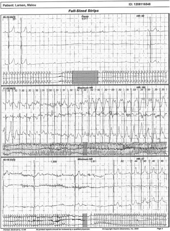
Ambulatory electrocardiographic monitoring for 48 hours showed 24 episodes of complete atrioventricular block, with RR pauses of up to 6 seconds, without any escape beats or clinical symptoms (Fig 1). Spontaneous resolution of her complete atrioventricular block was felt to be unlikely, and thus a permanent epicardial VVI pacemaker was implanted. The pacemaker programming was lead impedance 347 Ohm, R-wave sensing 5.60–8.00 mV, and threshold 1.25 V at 1.0 ms with backup of 60 bpm.
The ambulatory electrocardiographic showed 24 episodes of complete atrioventrioventricular block with RR pauses of up to 6 seconds without any escape beats or clinical symptoms.

The child developed a post-procedural haematoma in the pacemaker pocket, which was evacuated without further complication. Telemetry in the hospital showed that the patient was paced twice after the implantation because of a complete atrioventricular block of several seconds’ duration without syncope. Since discharge, the patient has not experienced any further episodes of syncope. Ambulatory device check 5 months after implantation showed a pacing rate of 0.1%.
Discussion
There are few data about paediatric patients with a structurally normal heart and complete paroxysmal atrioventricular block. To our knowledge, only two previous reports on this subject have been published.Reference Siveltti, Grutter and Di Ciommo 3 , Reference Silver, Pass and Hordof 4
Silvetti et al described 17 cases of complete atrioventricular block in young children (9.6±5.7 years) over a period of 11 years. The 12-lead electrocardiogram was normal, and therefore not useful in excluding the complete atrioventricular block, in seven out of 17 cases. Ambulatory electrocardiographic monitoring, on the other hand, documented complete atrioventricular block in 15 out of 17 patients. Only three patients (all female) did not suffer from any congenital or acquired heart disease or autoimmune diseases. Like our patient, these three girls had syncope as the only symptom with a normal 12-lead electrocardiogram and abnormal Holter monitoring.Reference Siveltti, Grutter and Di Ciommo 3
Silver et al reviewed a paediatric arrhythmia database from 1988 to 2007 and found six cases (five female and one male) of episodes of syncope associated with complete atrioventricular block in individuals with normal cardiac anatomy.Reference Park 2 All patients had syncope as the only symptom. All patients had a normal physical examination, electrocardiogram, and echocardiogram and did not take any kind of medicine known to interfere with atrioventricular nodal function. Laboratory tests were negative, including antinuclear antibody and Borrelia titres. Syncope in five of the six patients was atypical and all six patients had documented complete atrioventricular block upon ambulatory electrocardiography monitoring, or event recorder. It was concluded that paroxysmal atrioventricular block should be considered in patients with recurrent episodes atypical of vasovagal syncope and that the diagnosis can be made with the use of electrocardiogram monitoring/event monitor or loop recorder.Reference Silver, Pass and Hordof 4
Bringnole et al conducted a follow-up study on 18 adult patients with a similar clinical presentation with an unexplained syncope, normal baseline electrocardiogram, absence of structural heart disease, and documented paroxysmal third-degree atrioventricular block. They found that adenosine with low-to-normal values in an adenosine test was sensitive in identifying patients with idiopathic atrioventricular block when patients with a typical reflex syncope had high adenosine levels.Reference Brignole, Deharo and Roy 5
Our case describes a rare and potentially life-threatening condition that should not be overlooked when a child presents with atypical syncope. The clinical history in combination with normal first-line investigations led to ambulatory cardiac Holter monitoring of the patient when a complete atrioventricular block was diagnosed.
In children presenting with syncope, we encourage taking a thorough clinical history including a description of the episodes, any precipitating factors such as physical activity, stress, anger, or fasting, and any associated symptoms including palpitations, cyanosis, premonitory signs, seizures, pain, discomfort, vomiting, or post-ictal symptoms. Neurological disease, infection, and autoimmune disease should be excluded by clinical examination and relevant investigations. It is important to ask about a family history of arrhythmia, because some genetic causes such as LMNA and SCN5A can cause an atrioventricular block.Reference Fatkin, MacRae and Saski 6 , Reference McNair, Ku and Taylor 7 A normal electrocardiogram does not rule out a complete atrioventricular block and ambulatory electrocardiogram monitoring or, if necessary, an implantable loop recorder is the investigation of choice to detect a complete atrioventricular block. It is important to stress that short pauses can be asymptomatic; this does not exclude longer pauses causing symptoms.
In conclusion, a complete atrioventricular block is a rare but potentially lethal cause of syncope in paediatric patients. We stress the importance of a thorough clinical history and diagnostic workup in patients with unexplained syncope with no precipitating events.
Acknowledgements
We acknowledge James Dodd, MD, Klaus Juul, MD and Henrik Ørbæk Andersen, MD, for their help with this paper.
Financial Support
None.
Conflicts of Interest
None.
Ethical Standards
There was no experimentation on humans or animals in this case report.