CASE REPORT
A 50-year-old man was referred for a dental assessment before receiving adjuvant radiotherapy following right partial glossectomy and ipsilateral supraomohyoid neck dissection for a T1N2b squamous cell carcinoma. The planned radiotherapy dose to the right neck was 50 Gy with a 10 Gy boost. His medical history was essentially unremarkable, with no known allergies and no current medications. He reported a longstanding history of smoking (30 cigarettes/day for 35 years) and alcohol intake (40 g/day). Extra-oral examination revealed a slight palsy of the right lower lip, as well as a dysaesthesia affecting the right border of the mandible and submandibular area, present post-surgery. Of significance, no regional lymphadenopathy was noted.
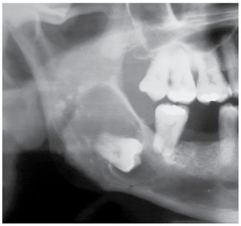
Intra-orally, soft tissue examination showed the healing surgical site on the right posterior third of the tongue. The patient was noted to be partially dentate, with several teeth lying within the proposed field of radiotherapy. In addition, several retained roots and carious teeth were noted. Routine radiographic examination, in the form of an orthopantomogram (OPG), revealed a 20 × 40 mm unilocular radiolucent lesion associated with the crown of the horizontally impacted unerupted right mandibular third molar, and extending superiorly to involve the ascending ramus of the mandible (Figure 1). There was downwards displacement of the inferior alveolar nerve canal. No radiographic evidence of resorption of the adjacent right mandibular second molar was noted. No clinical evidence of buccal and lingual expansion of this area of the mandible was noted. This lesion was consistent, radiographically, with a dentigerous cyst. The differential diagnoses were unicystic ameloblastoma and a keratocystic odontogenic tumour (formerly known as odontogenic keratocyst)
Orthopantomogram revealing unilocular radiolucent lesion associated with the crown of the horizontally impacted unerupted right mandibular third molar.

Continuation with the pre-radiotherapy dental “work up” involved an in-depth discussion with the patient regarding the complications of radiotherapy on the oral cavity, in particular, the resultant salivary gland hypofunction and the subsequent increased risk of dental caries, along with the inherent risk of developing osteoradionecrosis following future extraction of teeth that were in the field of radiotherapy. In light of this, it was recommended to extract all the posterior teeth within the field, in addition to teeth with extensive caries deemed to have a poor prognosis that did not lie in the radiotherapy field. It was advised that these extractions be performed at the time of surgical removal of the unerupted right mandibular third molar and associated cyst, essential for histopathological examination before commencement of radiotherapy, whilst still allowing an adequate healing period.
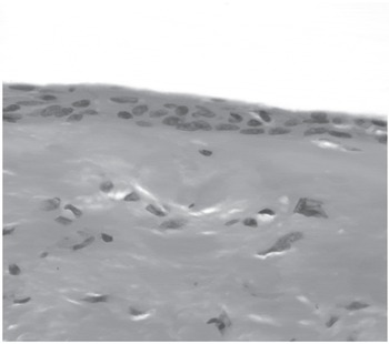
Following a case discussion with the treating Radiation Oncologist, only the right mandibular second molar, the unerupted right mandibular third molar and associated cyst were surgically removed under general anaesthesia. The remaining teeth were spared as the decision whether or not to proceed with radiotherapy was unclear at this stage due to the risks of pathological fracture of the mandible and osteoradionecrosis in this area, as well as the patient’s reluctance to proceed with radiotherapy. The patient was reviewed 2 weeks following surgery, and the area was noted to be healing well. Histological examination of the lesion from the right angle of the mandible revealed a cyst lined by a thin regular stratified squamous epithelium (Figure 2) with foci of mucous metaplasia. These features were consistent with a dentigerous cyst. Repeat radiographic examination 4 weeks post-surgery showed that the area was healing well.
Cyst lined by a thin regular stratified squamous epithelium with minimal inflammation of cyst wall. Haematoxylin and eosin stain.

A positron emission tomography (PET) scan was performed to reassess the need for adjuvant radiotherapy, given the additional risks involved in irradiating this area of the mandible. Unfortunately, there was a focus uptake in the right upper neck, which was very suggestive of residual nodal carcinoma, indicating a clear role for adjuvant treatment. The patient subsequently underwent external beam radiotherapy. In light of the odontogenic cyst and residual healing defect, the total dose to the right angle of the mandible was limited to receive 35 Gy. No complications to this area were noted to develop post-radiotherapy.
The patient has been reviewed at regular intervals since the completion of treatment. Clinical examination remains unremarkable and radiographs show a degree of bony infill in the right angle of the mandible. At 2 years review, the patient remains free of primary disease.
DISCUSSION
The oral complications of radiotherapy administered to the head and neck region are well recognised and include mucositis, opportunistic infections, xerostomia with resultant increased caries, loss of taste, trismus and the risk of osteoradionecrosis. Protocols have been developed to minimise or manage the early and late oral sequelae of radiotherapy of the head and neck region,Reference Jansma, Vissink and Spijkervet1 however, despite advances in medical therapy, such as granulocyte–macrophage colony stimulating factor,Reference Ryu, Swann and LeVeque2 these sequelae are still difficult to manage. The most successful approach to managing these complications is prevention, patient education and early consultation by involvement of a dentist or dental specialist. The advantages of patient care being co-ordinated by Multidisciplinary Clinics are well established and often include a dentist or dental specialist as part of the team. The role of the dentist with patients undergoing radiotherapy to the head and neck region can be considered in three phases, before commencement of radiotherapy, during and following completion of therapy. A particular benefit of the dentist being present during the Multidisciplinary Clinics is, with early involvement, potential oral problems are identified and managed in an appropriate manner and timing. It has been demonstrated that panoramic radiographs, such as OPGs, have a limited diagnostic yield when compared to selective intra-oral radiographsReference Rushton, Horner and Worthington3 and are not generally recommended as a screening tool. Patients receiving head and neck radiotherapy are often first seen in a Multidisciplinary Clinic set within a hospital and radiology services may be limited in respect to intra oral films.
This case report exemplifies the value of a screening panoramic film in the setting of a pre-radiotherapy dental workup. It facilitates discussion between team members to accurately identify structures lying within the radiotherapy field and be able to explain this clearly to the patient. A general dental assessment can be ascertained quickly by the dentist including an assessment of the extent of work required prior to commencement of radiotherapy. As in our case, presence of any underlying pathology can also be determined.
Dentigerous cysts are developmental odontogenic cysts and are the most common developmental jaw cyst.Reference Jones, Craig and Franklin4 Prompt histological diagnosis was indicated in our case as the differential diagnosis included other lesions, such as unicystic ameloblastoma and keratocystic odontogenic tumour (previously known as odontogenic keratocyst), which have a more locally aggressive nature and propensity for recurrence. The effects of radiotherapy on dentigerous cysts are not known; however, radiotherapy involving the jaws is not advised with individuals with Gorlin Goltz syndrome due to the presence of multiple keratocystic odontogenic tumours.
The patient was not keen to proceed with radiotherapy particularly after the identification of a large cystic lesion involving the field of radiotherapy and the inherent risk of osteoradionecrosis. Utilisation of a PET scan was most valuable in the decision-making process as it identified a focus uptake suggestive of residual nodal carcinoma. The Multidisciplinary setting provided a forum for team members to discuss this interesting case and determine a strategy to achieve curative intent with minimal adverse effect of the mandible. The patient is free of disease at 2 year follow-up and the right angle of the mandible demonstrates a degree of bony infill, but more importantly no evidence of osteoradionecrosis. This case highlights the role of the dentist in a multidisciplinary team for patients undergoing head and neck radiotherapy and the value of screening panoramic radiographs in this setting.
Acknowledgements
No financial grants are associated with this case report.