INTRODUCTION
Infections with parasitic helminths continue to cause considerable morbidity and mortality in humans and their domestic livestock, translating into substantial socioeconomic losses. For instance, morbidity associated with Schistosoma mansoni or Echinococcus granulosus infections is debilitating and sometimes fatal (Carod-Artal, Reference Carod-Artal2008; King and Dangerfield-Cha, Reference King and Dangerfield-Cha2008), and infections with hookworms retards the growth of children and can impair their cognitive functions (Oberhelman et al. Reference Oberhelman, Guerrero, Fernandez, Silio, Mercado, Comiskey, Ihenacho and Mera1998). The magnitude of the problems is illustrated by estimates from the the World Health Organization (WHO, 1999) that, even in the midst of successful anthelmintic programs, 30% of the world's population harbours at least 1 species of helminth parasite; a staggering 2 billion people are affected! However, numerous studies have shown that modulation of the immune response of the host by parasitic helminths can have a concomitant health benefit (reviewed by McKay, Reference McKay2006). In addition to infections with live organisms, helminth-derived molecules are potent immunomodulators and can serve as templates for the design of novel anti-inflammatory drugs, and may also be candidates for vaccine development (Harnett and Harnett, Reference Harnett and Harnett2008; Perrigoue et al. Reference Perrigoue, Marshall and Artis2008). The former aspect of the host-parasite interaction is the topic of this review.
HELMINTH THERAPY: THE GOOD, THE BAD AND THE WRIGGLY
Parasitic helminths have been humans' long-time companion and have evolved as ‘master regulators’ of host immune responses (Maizels et al. Reference Maizels, Balic, Gomez-Escobar, Nair, Taylor and Allen2004). Consequently, knowledge of how the parasite modifies host immunity can be used to develop therapies for other diseases. In considering ‘helminth therapy’, one can either use viable infections or immunomodulatory molecules from the parasite; both strategies have been used to treat diseases in animal models. Infection with a variety of species of parasitic helminths has been shown to reduce the severity of airway, cerebral and intestinal inflammation in murine models of human disease (McKay, Reference McKay2006), and a number of investigators are pursuing the mechanism(s) underlying this health benefit (Weinstock et al. Reference Weinstock, Summers and Elliott2005). Moreover, Weinstock and colleagues have shown that the ingestion of viable eggs of Trichuris suis by patients with inflammatory bowel disease (Crohn's disease and ulcerative colitis) led to a reduction in disease activity, and adverse side-effects were not apparent (Summers et al. Reference Summers, Elliott, Qadir, Urban, Thompson and Weinstock2003, Reference Summers, Elliott, Urban, Thompson and Weinstock2005). However, there can be a number of concerns regarding the use of an infection as a therapy. First, a parasite that has limited impact on the host must be used, which prohibits the use of particular species, such as S. mansoni, despite its potent stimulation of host immunity. Second, there are patient groups for which this approach would not be advocated, typically children, the elderly, immunocompromised individuals and pregnant women (Farah et al. Reference Farah, Langoi, Nyaundi and Hau2007). Third, there are examples of helminth infections worsening disease evoked by bacteria or protozoa (Chen et al. Reference Chen, Louie, McCormick, Walker and Shi2005; Marshall et al. Reference Marshall, Brunet, van Dessel, Alcarez, Bliss, Pearce and Denkers1999) and, in at least one case, chemically-induced colitis (Hunter et al. Reference Hunter, Wang and McKay2007). Finally, when introducing a species to a novel environment, there is the potential for maladaptive responses and unpredicted pathological effects/changes in remote tissues. Moreover, the possibility exists for parasite establishment in, rather than expulsion from the host (Kradin et al. Reference Kradin, Badizadegan, Auluck, Korzenik and Lauwers2006). Despite these issues, the data on ‘helminth therapy’ are encouraging, and animal models can be used to identify novel anti-inflammatory mechanisms for translation into medical treatments, negating the need for actual infection.
The use of helminth-derived molecules as drugs overcomes the concerns with the use of live parasites. However, the issues here are often technical and revolve around a single basic question: how can pure molecules be isolated from miniscule amounts of parasite material to allow testing in biological systems and to serve as blueprints for drug development?
IMMUNE RESPONSES TO HELMINTH PARASITES: THE BASICS
A number of authorative reviews of the mammalian immune system and the responses to infection with helminths are available (Hayes et al. Reference Hayes, Bancroft and Grencis2004; Maizels et al. Reference Maizels, Balic, Gomez-Escobar, Nair, Taylor and Allen2004; Antony et al. Reference Antony, Rutitzky, Urban, Stadecker and Gause2007; Khan, Reference Khan2008; Perrigoue et al. Reference Perrigoue, Marshall and Artis2008). As metazoans, helminths cannot be ingested by macrophages and other phagocytes, so that a variety of effector mechanisms have evolved to combat helminths. These involve the mobilization and activation of T and B lymphocytes, mast cells, eosinophils, natural killer (NK) cells, and, if the infection is at a mucosal surface, mucus-producing goblet cells. Briefly, while there is likely to be an innate immune response to helminth-derived products by macrophages and natural killer cells, the current view is that the predominant anti-worm response is the remit of an adaptive immune response. Thus, following antigen processing and presentation by professional antigen presenting cells (e.g., dendritic cells (DCΦs)), humans and mice display a stereotypic T cell response which is characterized by the development of CD4+ T helper (TH)-2 cells that produce interleukin (IL)-4, -5, -13 and others. These chemical signals mobilize other cells; for example, they direct isotype switching, so that high affinity immunoglobulin (Ig) G, E and A can be produced. These antibodies then bind to receptors on effector cells, allowing for their activation by worm antigen and the release of molecules (e.g., mast-derived histamine and trypases, eosinophil-derived cationic protein) that either disrupt the surface of the helminth and/or alter the environment, making it less hospitable to the helminth. In addition, IgG can bind directly to the surface of the helminth (opsonization), interfering with worm behaviour and targeting the worm for attack by complement and polymorphonuclear cells. Thus, the host response to infection with parasitic helminths is multi-cellular and is orchestrated by Th2 cells/cytokines. Recent studies are elucidating a role for additional cytokines such as IL-21 and IL-33 (Humphreys et al. Reference Humphreys, Xu, Hepworth, Liew and Grencis2008) and regulator cells, including the CD4+/Foxp3+ natural regulatory T cells and, alternatively activated macrophages (AAM) (Baumgart et al. Reference Baumgart, Tomkins, Leng and Hess2006; Taylor et al. Reference Taylor, Mohrs and Pearce2006; Persaud et al. Reference Persuad, Wang, Reardon and McKay2007). While all immune cells can be considered as immunomodulatory (i.e., they affect other immune and stromal cells in their vicinity), regulatory T cells and AAMs (of which there are multiple types for each; Belkaid and Oldenhove, Reference Belkaid and Oldenhove2008; Reyes and Terrazas, Reference Reyes and Terrazas2007) have emerged as prototypic immunomodulators whose primary function is to switch-off or dampen immune reactions, or engage reactions that promote tissue recovery and restitution. Much research remains to be done to precisely define the biology of regulatory T cells and AAMs; however, the mobilization of both classes of cells following an infection with helminths would generate an immunoregulatory/immunosuppressive environment in which a spectrum of immune events would be inhibited. Thus, by extrapolation, the ability of products from the helminth to elicit regulatory T cell or AAM activity could contribute to worm infectivity and survival.
HELMINTH-DERIVED IMMUNOMODULATORS
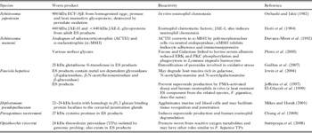
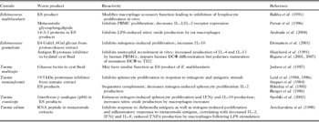
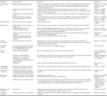
An analysis of the biology of a parasite should include studying the mechanisms of the interaction of the parasite with the host immune system, so that potential parasite-derived immunomodulators (used to develop drugs to treat immuno-pathological changes/disease) can be identified. During the last 40 years, a staggering number of helminth-derived immunomodulators have been partially characterized, both in terms of structure and bioactivity (we define an ‘immunomodulator’ as any molecule that regulates host immunity, but not antigens recognized by the host that are used to generate immune responses aimed at the destruction or elimination of the parasite from the host). Here, we focus on selected examples from the 3 main groups of parasitic helminths and draw the reader's attention to the fact that a literature search will reveal many other examples, all worthy of discussion, and excluded only on the basis of space restrictions (the examples given below are supplemented by additional material in Tables 1–3).
Selected examples of immunomodulatory molecules from trematodes

Selected examples of immunomodulatory molecules from cestodes

Selected examples of immunomodulatory molecules from nematodes

Trematodes
Schistosoma spp. Schistosomes, because of their impact on human health (King and Dangerfield-Cha, Reference King and Dangerfield-Cha2008), have been the focus of intense research efforts, and a variety of glycoconjugates have been extracted from the eggs and adult tegument (Bennett, Reference Bennett1963). Soluble egg antigen (SEA) from S. mansoni has been implicated in the host-response to infection, and has been shown consistently to have immunomodulatory properties. For example, SEA appears to be responsible for the reduction in CD4+ lymphocytes 6 weeks following infection, an event that was attributed to enhanced Fas-FasL-induced apoptosis involving CD19+ B cells (Lundy et al. Reference Lundy, Lerman and Boros2001). Excretory/secretory (ES) products from schistosomula contain a 23 kDa molecule that results in T cell depletion in vivo via Fas-FasL killing. Exposure to SEA results in reduced levels of nitric oxide (NO) and T helper cell-type 1 (TH1) chemokines (CCL3) and cytokines (tumour necrosis factor-α (TNFα), IL-12) and an increased expression of markers indicative of AAMs. These events are all compatible with the mobilization of TH2-type cytokines (e.g. IL-4, IL-13) which is stereotypic of the mammalian immune response to infection by helminth parasites. The induction of AAMs is of particular note, as these cells exert anti-inflammatory effects and are active in tissue restitution after injury (Gordon and Taylor, Reference Gordon and Taylor2005). Thus, the ability to use a consitutent of SEA to induce this cellular response pharmacologically could be of direct health benefit.
Mice treated with SEA display endothelial cell proliferation and capillary formation which appear to be driven by a heat- and protease-insensitive molecule, indicative of a glycan (Kanse et al. Reference Kanse, Liang, Schubert, Haas, Preissner, Doenhoff and Dennis2005). These findings complement the work of van de Vijver et al. (Reference van de Vijver, Deelder, Jacobs, van Marck and Hokke2006) that detected LDN (GalNAcβ1,4GlcNAc) and LN (Galβ1,4GlcNAc) modified glycans in SEA-stimulated granulomata. SEA has been shown to induce vascular endothelial growth factor (VEGF) secretion (Loeffler et al. Reference Loeffler, Lunfy, Singh, Gerard, Hudson and Boros2002) and fibroblast-stimulating factor (FSF-1) (Greenwel et al. Reference Greenwel, Wyler, Rojkind and Prakash1993), which could contribute to the formation of the fibrous capsule around the granulomata, although it is unclear whether the stimulation of the production of either growth factor was due to a glycan component of the SEA. Nevertheless, one could speculate that SEA evoked growth factor release from host cells to stimulate angiogenesis could be used to enhance the recovery from tissue damage, although the potential to elicit metastatic events should not be overlooked.
In addition, while the exposure to SEA decreased the expression of Toll-like receptors (TLR) on macrophages, the responses of these cells to TLR agonists (e.g., lipopolysaccharide (LPS)) were not appreciably retarded (Joshi et al. Reference Joshi, Raymond, Coelho, Kunkel and Hogaboam2008). These studies are intriguing, as they demonstrate a cross-talk between infection with parasitic helminths and responsiveness to bacteria and may illuminate a way in which one could exploit one type of infection to treat another. In addition, the complexity of the host response to infection, which is linked temporally or spatially and/or parasite-specific, is aptly illustrated by these studies.
The identification of a >100 kDa heat-insensitive, carbohydrate-positive fraction from S. mansoni (see Hayunga et al. Reference Hayunga, Murrell, Taylor and Vannier1979) was followed by studies characterizing the glycoproteins of schistosomes and notably led to the elucidation of lacto-N-fucopentose (LNFPIII) (carbohydrate containing the mammalian LewisX trisaccharide [Galβ1,4[Fucα1,3]GlcNAc]) and lacto-N-neotetrose (LNT) (Ko et al. Reference Ko, Drager and Harn1990). Analysis of LNT revealed that its injection into mice caused an expansion of a Gr1+ cell population which suppressed CD4+ T cell proliferation and cytokine production via an apoptosis-independent but cell contact-dependent mechanism (Terrazas et al. Reference Terrazas, Walsh, Piskorska, McGuire and Harn2001). The authors postulated that this treatment ‘imprinted’ the Gr1+ cells with a TH2-type phenotype.
Since the LewisX antigen is present in the lymphocyte function-associated molecule-1 (LFA-1), the role of LNFPIII in lymphocyte proliferation was assessed and shown to stimulate IL-10 and prostaglandin E2 (PGE2) production from B-cells of infected mice without hampering antigen presentation (Velupillai and Harn, Reference Velupillai and Harn1994). Also, the co-culture of LNFPIII-treated macrophages and NK cells enhanced NK cell activation as gauged by the expression of CD69 and interferon-γ (IFNγ) production (Atochina and Harn, Reference Atochina and Harn2005). Lacto-N-fucopentose III also generates anti-inflammatory responses by signalling through Toll-like receptor 4 (TLR4) to induce the phosphorylation and activation of the mitogen-activated protein kinase (MAPK), ERK (Thomas et al. Reference Thomas, Carter, Atochina, Da'Dara, Piskorska, McGuire and Harn2003).
The presence of fucose in the LewisX moiety of LNFPIII is essential for bioactivity (Okano et al. Reference Okano, Satoskar, Nishizaki, Abe and Harn1999), since lacto-N-neotetrose lacking LewisX had no effect on DCΦ differentiation to an IL-4 producing phenotype in response to LPS. Importantly, the induction of accessory molecules is required to increase the strength of an immune response; DC-SIGN has strong affinity for intercellular adhesion molecule-3 (ICAM3) via the LewisX structure (Bogoevska et al. Reference Bogoevska, Nallau, Lucka, Grunow, Klampe, Uotila, Samsen, Gahmberg and Wagener2007), suggesting a possible mechanism used by LNFPIII to bind to DCΦ and interfere with cell-to-cell communication with other immune cells (Appelmelk et al. Reference Appelmelk, Van Die, Van Vleit, Vandenbroucke-Grauls, Geijtenbeek and Van Kooyk2003). The multivalency of LNFPIII is important for its bioactivity, since monovalent LNFPIII was shown to be incapable of inhibiting antigen-induced peripheral blood mononuclear cell (PBMC) activation (Velupillai et al. Reference Velupillai, dos Reis, dos Reis and Harn2000). At the signalling level, LNFPIII increases nuclear factor (NF)-κB activity in DCΦ to a level similar to LPS; however, a second phase (prolonged NF-κB activation associated with IκBβ degradation) was not observed. Use of the nuclear import inhibitor, SN50, and NFκB-deficient mice revealed that LNFPIII effects on DCΦ were indeed NFκB-dependent (Thomas et al. Reference Thomas, Carter, Da'Dara, DeSimone and Harn2005). Collectively, these studies suggest that LNFPIII (or possibly fragments thereof) could be used to extrinsically manipulate DCΦ function as a way of limiting immunopathological reactions/changes or controlling immune cell responsiveness.
The stimulation of IL-10 and IL-12p40 from murine peritoneal macrophages by cercarial ES products required the presence of TLR4, whereas the production of IL-6 was TLR4 independent (Jenkins et al. Reference Jenkins, Hewitson, Ferret-Bernard and Mountford2005). Furthermore, IL-6 release was reduced following protease treatment of the ES products suggesting a proteinaceous ligand, and sodium periodate ablated activity of the cercarial extract, indicating the presence of glycans. It is plausible that cercarial products contain proteins/glycoprotiens that can act via TLR4, highlighting the promiscuity of TLR4 as more than an LPS receptor. Other TLRs have been implicated as receptors for schistosome ligands. Lyso-phosphatidlyserine from S. mansoni induces DCΦ maturation and decreases IL-12p70 production via TLR2. The activity is abrogated by TLR2-blocking antibodies (van der Kleij et al. Reference van der Kleij, Latz, Brouwers, Kruize, Schmitz, Kurt-Jones, Espevik, de Jong, Kaspenberg, Golenbock, Tielens and Yazdanbakhsh2002), whereas host phosphatidylserine neither activates TLR2 nor induces DCΦ maturation. Additionally, phosphoserine alone is not effective in this system, indicating that the acyl and phosphoserine groups of S. mansoni lysophosphatidylserine are required for the effect. Inhibition of lysophosphatidylserine acyltransferase (LPC-AT) did not affect TLR4 expression or the binding of LPS, but it did prevent TLR4 translocation to lipid rafts and thus blocked the formation of the TLR4 complex with a resultant decrease in TNFα and IL-6 production (Jackson et al. Reference Jackson, Abate, Parton, Jones and Harwood2008). It is possible that S. mansoni lysophosphatidylserine also signals through TLR4 in a similar manner. Studies such as these illustrate an emergent theme, in which reponses to helminth-derived molecules occur via receptors that have been defined on the basis of their ability to recognize conserved microbe-derived products; however, the data must be interpreted with caution, since the possibility of bacterial contamination of the helminth extracts is omnipresent.
Fasciola hepatica
ES products from adult F. hepatica (FhES) contain a glycan that is cytotoxic to eosinophils and acts via tyrosine kinase and caspase-dependent mechanisms (Serradell et al. Reference Serradell, Guasconi, Cervi, Chiapello and Masih2007). Induction of AAMs often accompanies an infection with helminths, and it has been reported that F. hepatica thioredoxin peroxidase elicits AAMs production (Donnelly et al. Reference Donnelly, O'Neill, Sekiya, Mulcahy and Dalton2005). Numerous studies have shown that AAMs are induced by IL-4/IL-13 (Stein et al. Reference Stein, Keshav, Harris and Gordon1992); however, in the case of F. hepatica thioredoxin peroxidase, this conversion appeared to be IL-4-independent (Donnelly et al. Reference Donnelly, O'Neill, Sekiya, Mulcahy and Dalton2005). The peroxidase also increased transforming growth factor-β (TGFβ) and PGE2 while decreasing IL-10, IL-12 and IL-18 from stimulated macrophages – such events in vivo would skew the immune reponse in favour of immunoregulation/immunosuppression and away from TH1-dominated pro-inflammatory reactions. Complementing these findings, the in vitro treatment of bovine macrophages with FhES products inhibited IFNγ and NO production in response to bacterial products and skewed the macrophage population towards an AAM phenotype; the latter event was lost by heat treatment of the ES products (Flynn and Mulcahy, Reference Flynn and Mulcahy2008). This emphasizes that we must constantly be vigilant of the possibility that parasite-derived molecules, as well as those produced by the host (e.g., cytokines), can modify the host's immunity. Finally, cathepsin L appears to be a dominant molecule in FhES and may aid F. hepatica by degrading host tissue to assist parasite entry/migration, and interfering with the host's immune response by, for example, the degradation of IgG (Smith et al. Reference Smith, Dowd, Heffernan, Robertson and Dalton1993; O'Neill et al. Reference O'Neill, Mills and Dalton2001).
Cestodes
Pseudophyllidean and cyclophyllidean tapeworms usually elicit limited damage in the alimentary tract of their definitive host. However, they are recognized by their host (i.e., anti-worm antibodies are generated) so, the ability to suppress or negate host immunity would remain a priority for the helminth (see Table 2).
Taenia crassiceps
T cells from T. crassiceps-infected mice are hypo-proliferative and, in the presence of excretory/secretory (TcES) products, show diminished responses to mitogen stimulation in vitro (Spolski et al. Reference Spolski, Corson, Thomas and Kuhn2000, Scuitto et al. Reference Scuitto, Fragoso, Baca, de la Cruz, Lemus and Lamoyi1995). Administration of TcES products to mice caused expansion of F4/80+ Gr1+ cells, and these cells were capable of blocking T cell activation in vivo. Glycans in the TcES were essential for this response, since the treatment with sodium periodate significantly reduced F4/80+ Gr1+ cells in the peritoneal cavity of infected mice and the production of cytokines and AAM (Gomez-Garcia et al. Reference Gomez-Garcia, Lopez-Marin, Saaverdra, Reyes, Rodriguez-Sosa and Terrazas2005). Extracts from the adult worm also directed a strong TH2 polarization (IL-4 production) and enhanced antibody production (IgG1 and IgE) in mice co-treated with ovalbumin (Gomez-Garcia et al. Reference Gomez-Garcia, Rivera-Montoya, Rodriguez-Sosa and Terrazas2006). Concomitant with these events was an increased expression of markers indicative of AAMs, a predicted decrease in IFNγ and iNOS expression and increased synthesis of IL-10 and TGFβ. Like the ES products, the glycan component was critical for these effects of a crude antigen preparation from the adult worm, because all were abolished or significantly inhibited by sodium periodate treatment of the extract.
Extracts from the T. crassiceps metacestodes are highly mannosylated and contain both non-fucosylated and fucosylated structures, including the LewisX antigen (Dissanayake et al. Reference Dissanayake, Amith and Shahin2004). In contrast to the adult worm antigen, the effect of which was unaltered by the knock-out of TLR4 (Gomez-Garcia et al. Reference Gomez-Garcia, Lopez-Marin, Saaverdra, Reyes, Rodriguez-Sosa and Terrazas2005), the ability of the glycans in the metacestode extract to elicit IL-6 production from murine peritoneal macrophages was reduced in cells lacking TLR2 and TLR4 (Dissanayake et al. Reference Dissanayake, Amith and Shahin2004). The same researchers recently found that the terminal beta-(1-4)-galactose residue in the LewisX analogue can evoke IFNγ production by murine splenocytes and activate the NF-κB pathway. The IFNγ production was reduced by inclusion of anti-TLR2, -4 and -6 antibodies in the assay (Dissanayake and Shahin, Reference Dissanayake and Shahin2007), again reiteriating the notion that helminth-derived products can exert their effects via TLRs.
Spirometra erinaceieuropaei
Excretory/secretoy (SeES) products contain immunoglobulinases and a heat-sensitive factor that inhibits LPS- and lipoteichoic acid (LTA)-induced TNFα and IL-1β production from murine macrophages (Diragahayu et al. Reference Diragahayu, Fukumoto, Tademoto, Kina and Hirai2004). The inhibitory effect of the SeES products was not due to the stimulation of PGE2, IL-10 or TGFβ in the target cells (Fukushima et al. Reference Fukushima, Isobe, Hojo, Shiwaku, Yamane and Torii1993). The inhibition of macrophage production of TNFα, IL-β and NO was also shown in a study by Kina et al. (Reference Kina, Fukumoto, Miura, Tademoto, Nunomura, Diraghayu and Hirai2005), in which a 90 kDa trypsin-sensitive glycoprotein was identified as the active component in the SeES products. Similarly, a 94 kDa component of SeES products could block LPS-induced MAPK (i.e., ERK1, ERK2 and p38MAPK) phosphorylation and the accompanying synthesis of TNFα (Dirgahayu et al. Reference Dirgahayu, Fukumoto, Miura and Hirai2002).
Hymenolepis diminuta
Extracts from adult H. diminuta reduced ConA-induced proliferation, IL-2 and IL-4 synthesis by murine spleen cells and human PBMC cells in a concentration-dependent manner (Wang and McKay, Reference Wang and McKay2005). These events were mirrored, to a large extent, by use of ES products from the adult worm. A crude phosphate-buffered saline (PBS)-solube extract of the adult worm stimulated IL-10 release from murine splenocytes and also contained a component with some structural similarity to the p40 chain of IL-12, as detected by enzyme-linked immunosorbent assay (ELISA) and immunoblotting. The bioactivity in both the worm extract and the ES products was within a >50 kDa fraction and was partially sensitive to boiling (Wang and McKay, Reference Wang and McKay2005).
Nematodes
Parasitic nematodes have been the focus of extensive research efforts because of their major socio-economic impacts globally (Hall et al. Reference Hall, Hewitt, Tuffrey and de Silva2008). Therefore, a number of immunomodulatory molecules have been identified from this phylum (Table 3). We focus this discussion by highlighting known molecules from Nippostrongylus brasiliensis, Trichinella spiralis, Necator americanus, Trichuris suis, Ascaris suum and Acanthocheilonema viteae.
Nippostrongylus brasiliensis
ES products from this parasite direct a range of immunomodulatory reactions. As examples, (i) they reduce LPS-induced IL-12p40 production and expression of a panel of cytokine/chemokine genes in murine bone-marrow-derived DCΦ, (ii) an undefined protein component induces IL-4, IL-10 and IgE production, with the IL-4 response requiring the presence of major histocompatibility class II (MHC II) antigens and so presumably MHC II-T cell restricted events, and (iii) suppress TH1-events via an IL-10-independent mechanism (Holland et al. Reference Holland, Harcus, Riches and Maizels2000, Reference Holland, Harcus, Balic and Maizels2005; Balic et al. Reference Balic, Harcus, Holland and Maizels2004). The characterization of the ES products revealed the presence of a serine acetylhydrolase capable of reducing intestinal anaphylactic responses by inactivating platelet activating factor and blocking neutrophil and eosinophil aggregation (Blackburn and Selkirk, Reference Blackburn and Selkirk1992). The value of inhibiting eosinophil activity to the helminth is intuitive, and the use of this acetylhydrolase would be beneficial in limiting the pro-inflammatory effects of granulated phagocytes in other diseases. Furthermore, NbES blocked goblet cell hyperplasia, eosinophilia, neutrophilia and airway hyper-responsiveness in murine models of asthma (Keir et al. Reference Keir, Brown, Clouter-Baker, Harcus and Proudfoot2004; Trujillo-Vargas et al. Reference Trujillo-Vargas, Werner-Klein, Wohlleben, Polte, Hansen, Ehlers and Erb2007). The active component/s was/were heat and protease insensitive, suggesting a glycan-type molecule. When NbES was administered intraperitoneally to ovalbumin-sensitized mice, they developed less pathology on ovalbumin re-challenge than untreated controls; in contrast, intranasal NbES induced significant lung inflammation (Marsland et al. Reference Marsland, Camberis and Le Gros2005). Finally, and intriguingly, N. brasiliensis may be worthy of consideration in cancer therapy. When carcinogenic cells were injected with N. brasiliensis extracts, eosinophils were recruited and attacked the cells (i.e., as if they were a helminth), resulting in reduced size of the carcinoma (Frenoy, Reference Frenoy2005).
Trichinella spiralis
Structural and biochemical analysis of T. spiralis ES products (TsES) from larvae or adult worms revealed the presence of a nucleoside 5′-diphosphate phosphohydrolase (apyrase, 17 kDa), a 5′nucleotidase that preferentially targets AMP to generate adenosine and an adenosine deaminase (Gounaris, Reference Gounaris2002; Gounaris et al. Reference Gounaris, Selkirk and Sadeghi2004). The ability to regulate AMP metabolism has a number of potential immunomodulatory effects, since adenosine is known to inhibit platelet aggregation, monocytes/macrophage TH1 cytokine production, and neutrophil synthesis of reactive oxygen species (Liaudet et al. Reference Liaudet, Mabley, Pacher, Virag, Soriano, Marton, Hasko, Deitch and Szabo2002; Antonioli et al. Reference Antonioli, Fornai, Colucci, Ghisu, Tuccori, Del Tacca and Blandizzi2008), and is protective in a murine model of colitis (Mabley et al. Reference Mabley, Pacher, Liaudet, Soriano, Hasko, Marton, Szabo and Salzman2003). Also, adenosine stimulates epithelial electrogenic Cl− secretion, which creates a driving force for water to move into the gut (Kottgen et al. Reference Kottgen, Loffler, Jacobi, Nitchke, Pavenstadt, Schreiber, Frische, Nielsen and Leipziger2003) and could act to flush Trichinella larvae as well as intestinal bacteria and protozoan pathogens from the host.
Necator americanus
Nematode-derived proteases serve a number of vital roles in larval exsheathment, the digestion of host tissue (facilitating invasion and migration) and in immune evasion (Gamble et al. Reference Gamble, Purcell and Fetterer1989, McKerrow et al. Reference McKerrow, Brindley, Brown, Gam, Staunton and Neva1990). Necator americanus ES (NaES) contains at least 7 cysteine proteases, 1 serine protease and various metalloproteases, some of which act as immunoglobulinases (Kumar and Pritchard, Reference Kumar and Pritchard1992). These enzymes exert a range of bioactivities, the majority of which are unknown. However, one can speculate that the cleavage of immunoglobulins (Igs) could result in reduced complement activation and that the activity of cells that bind Igs and are activated by antibody-crosslinkage by antigen (e.g., mast cells and eosinophils) would be reduced. Eosinophil recruitment in vivo could also be perturbed by, for example, the degradation of the chemokine, eotaxin-1 (Culley et al. Reference Culley, Brown, Conroy, Sabroe, Pritchard and Williams2000). Additionally, a Necator americanus protein was shown to evoke release of IFNγ from murine and human NK cells but not NK T cells (Hsieh et al. 2004).
Trichuris suis and Ascaris suum
Trichuris suis ES (TrsES) products contain heat-stable and protease-insensitive components which reduce the growth of the bacteria Campylobacter jejuni, Escherichia coli and Staphylococcus aureus (see Abner et al. Reference Abner, Parthasarathy, Hill and Mansfield2001). High performance liquid chromatography (HPLC) did not reveal the presence of antibiotics (i.e., penicillin, streptomycin and amphotericin) in the TrsES. The active component was <10 kDa in size. A. suum pseudocoelomic fluid (PCF) also contained a 6–14 kDa heat-stable, trypsin-sensitive molecule which could kill Gram-positive bacteria (e.g., S. aureus) (Wardlaw et al. Reference Wardlaw, Forsyth and Crompton1994). The molecule was designated ASABF (Ascaris suum antibacterial factor) (Kato and Komatsu, Reference Kato and Komatsu1996) and is similar to insect defensins which penetrate Gram-positive bacterial membranes. Genes encoding antibacterial cecropins had been characterized in A. suum, and chemically synthesized versions are active against numerous species of bacteria (Pillai et al. Reference Pillai, Ueno, Zhang, Lee and Kato2005). With increasing incidence of antibiotic-resistant bacteria, the putative usefulness of these nematode-derived molecules is apparent.
Acanthocheilonema viteae
Nematode-derived cystatins (cysteine protease inhibitors) have been repeatedly shown to regulate immune cell activity. Cystatins from A. viteae (as well as those of other filarioids, including Onchocerca volvulus and Brugia pahangi) inhibit T cell proliferation, interfere with antigen processing and presentation, and enhance IL-10 production from stimulated murine spleen cells (Osborne and Devaney, Reference Osborne and Devaney1999; reviewed by Hartmann and Lucius, Reference Hartmann and Lucius2003). Recently an A. viteae-derived cystatin has been demonstrated to reduce inflammation in a murine model of airway allergy (Schnoeller et al. Reference Schnoeller, Rausch, Pillai, Avagyan, Wittig, Loddenkemper, Hamann, Hamelmann, Lucius and Hartmann2008). Onchocystatin (Ov17) from O. volvulus was specifically shown to suppress PBMC proliferation by decreasing expression of the co-stimulatory molecule B7.2 (CD86) (Schonemeyer et al. Reference Schonemeyer, Lucius, Sonnenburg, Brattig, Sabat, Schilling, Bradley and Hartmann2001), and infection with O. volvulus resulted in IL-10/TGFβ-mediated immune hypo-responsiveness (Doetze et al. Reference Doetze, Satoguina, Buchard, Rau, Loliger, Flesicher and Horeauf2000). In that study, the source of the IL-10 and TGFβ was most likely host cells, but the possibility of helminth-derived analogues of these cytokines should not be overlooked. For example, a TGFβ-like molecule has been isolated from the filarial nematodes Brugia malayi and Brugia pahangi (Gomez-Escobar et al. Reference Gomez-Escobar, Gregory and Maizels2000).
The best described helminth immunomodulator is an A. viteae ES 62 kDa glycoprotein (ES-62) product. The bioactivity of ES-62 is critically dependent on phosphorylcholine (PC) attached to an N-glycan (likely GlcNAc) via acetlyglucosaminyltransferase I, such that recombinant ES-62 lacking PC is generally non-functional (Goodridge et al. Reference Goodridge, Marshall, Wilson, Houston, Liew, Harnett and Harnett2004; Houston and Harnett, Reference Houston and Harnett2004; Houston et al. Reference Houston, Sutharsan, Steiger, Scachter and Harnett2008). Homologues of ES-62 exist in related nematodes (Stepek et al. Reference Stepek, Auchie, Tate, Watson, Russell, Devaney and Harnett2002, Reference Stepek, Houston, Goodridge, Devaney and Harnett2004; Hewitson et al. Reference Hewitson, Harcus, Curwen, Dowle, Atmadja, Ashton, Wilson and Maizels2008). Harnett et al. (Reference Harnett, McInnes and Harnett2004) continue to explore the bioactivity of ES-62 in cell culture systems and murine models. Thus, ES-62 has been shown to enhance the production of IL-4 by murine splenocytes (Harnett et al. Reference Harnett, Deehan, Houston and Harnett1999) and decrease IFNγ, TNFα, IL-1β, IL-12 and IL-18 from DCΦ stimulated with TLR ligands (Whelan et al. Reference Whelan, Harnett, Houston, Patel, Harnett and Rigley2000; Goodridge et al. Reference Goodridge, Wilson, Harnett, Campbell, Harnett and Liew2001, Reference Goodridge, Marshall, Wilson, Houston, Liew, Harnett and Harnett2004). Complementing the latter, macrophages from mice implanted with mini-osmotic pumps containing ES-62 were hypo-responsive to bacterial stimuli. ES-62 treatment in a murine model of arthritis resulted in significantly less inflammatory disease, which was accompanied by reduced IFNγ, TNFα and IL-6, and increased IL-10 synthesis by stimulated lymphocytes (McInnes et al. Reference McInnes, Leung, Harnett, Gracie, Liew and Harnett2003). Significantly, IFNγ and T cell proliferation remained suppressed for up to 50 days after treatment whereas TNFα and IL-6 suppression required ongoing exposure, as did the inhibition of the production of collagen-specific antibodies. ES-62 also prevented B lymphocyte proliferation, and less IgG2α would contribute to reduced auto-immune driven arthritis (Wilson et al. Reference Wilson, Deehan, Katz, Brown, Houston, O'Grady, Harnett and Harnett2003).
ES-62 inhibits phosphorylation of immunoreceptor tyrosine-based activation motifs (ITAM) following B cell receptor ligation via the induction of the Src homology protein (SHP-1) tyrosine phosphatase. This effectively prevents the recruitment of crucial signalling molecules, such as protein kinase C (PKC) (Goodridge et al. Reference Goodridge, Deehan, Harnett and Harnett2005a). A similar inhibition of PKC activity may relate to the ability of ES-62 to inhibit TH1 cytokine production (Deehan et al. Reference Deehan, Harnett and Harnett1997). In contrast, ES-62 inhibition of mast cell mediator release following the cross-linking of FcεRI did not require tyrosine phosphatase activity (Melendez et al. Reference Melendez, Harnett, Pushparaj, Wong, Tay, McSharry and Harnett2007). Instead, the increase in intracellular Ca2+ elicited by receptor cross-linking was affected and mobilization of phospholipase D and sphingosine kinase was disrupted (Goodridge et al. Reference Goodridge, Deehan, Harnett and Harnett2005a). FcεRI-mediated NF-κB activation and the consequent synthesis of pro-inflammatory cytokines were abrogated by ES-62 through the disruption of the p50 and p65 subunits of NF-κB (Melendez et al. Reference Melendez, Harnett, Pushparaj, Wong, Tay, McSharry and Harnett2007). Given the pivotal role that mast cells play in immediate hypersensitivity and allergic reactions, a molecule, such as ES-62, could have many therapeutic applications.
The effect of ES-62 on mast cells appears to require TLR4 (Melendez et al. Reference Melendez, Harnett, Pushparaj, Wong, Tay, McSharry and Harnett2007), and this is in agreement with the examination of the effects of ES-62 on murine macrophages and DCΦ. This is not entirely unexpected, since PC can be a component on pathogen-associated molecular patterns (Goodridge et al. Reference Goodridge, Marshall, Else, Houston, Egan, Al-Riyami, Liew, Harnett and Harnett2005b). Thus, cells deficient in TLR4 or MyD88 (a key intracellular adaptor molecule) were unaffected by ES-62. The utilization of MyD88 may be critical, since cytokine-inducing stimuli that do not require this molecule, for example, the ligation of TLR3 or the accessory molecule CD40 in macrophages, are not affected by ES-62 treatment (Goodridge et al. Reference Goodridge, Marshall, Else, Houston, Egan, Al-Riyami, Liew, Harnett and Harnett2005b). Moreover, a natural mutation in the TIR domain in the TLR4 gene of C3H/HeJ mice renders these animals unresponsive to LPS, yet the ES-62 response remains, indicating that the helminth-derived molecule uses TLR4 in an unorthodox manner. It should be noted that the inhibitory effect of ES-62 can be overridden, as observed in Toxoplasma gondii-infected mice, in which immunity relies on TH1 cytokines from a variety of cell types (Couper et al. Reference Couper, Chen, Houston, Harnett and Johnson2005), or when T or B cells are challenged with two TH1-inducing antigens from O. volvulus (see Al-Riyami et al. Reference Al-Riyami, Egan, Bradley, Lustigman and Harnett2008).
While the goal of this commentary is to illustrate the immunomodulatory effects of helminth-derived molecules and the potential of these molecules to serve as drugs or templates for drug design, it would be naïve to think that all parasite products would be beneficial. This point is intuitive and we will not belabour it here, but for the sake of balance in this discussion, we draw the reader's attention to a few of many examples in which helminth-derived products elicit pathological reactions (Hill et al. Reference Hill, Gamble, Rhoads, Fetterer and Urban1993; Lee et al. Reference Lee, Cho, Kwon, Shin, Lim and Kim2006; Kim et al. Reference Kim, Choi, Hong and Bae2008).
We have presented selected examples of immunomodulation by parasitic helminth-derived molecules. Since the essence of any host-parasite relationship is specificity, it follows that nuances of immunomodulation will be parasite-, and indeed life-cycle stage-specific. However, some commonality emerges from these studies and, as generalizations, helminth-derived products mobilize a TH2 cytokine response (and ablate TH-1 events), stimulate the synthesis of immunomodulatory/immunosuppressive molecules (e.g., IL-10, TGFβ, PGE2), and some evoke eosinophil (and neutrophil) apoptosis. All of these events would ameliorate inflammatory disease and, as a pharmacopoeia for new drugs, the potential of the parasitic helminths should not be underplayed. Yet, much remains to be explored, to move the field from a series of intriguing observations in vitro and in rodents toward the development of drugs with a safety profile compatible with testing in clinical trials. At such a juncture, addressing issues relating to the in vivo stability and pharmacodynamics, and even the delivery methods of the helminth-derived molecule or drug, will be critical if new therapeutic modalities are to be achieved.
HELMINTH GLYCOBIOLOGY
Parasitic helminths as a group express or secrete/excrete a variety of molecules that have immunomodulatory properties. These molecules can be classical proteins or lipid-based, and many others are glycoproteins. We have opted to focus on parasite glycoproteins as molecules that could serve as models for the development of anti-inflammatory or immunoregulatory drugs, and will describe how such molecules can be isolated, characterized and might interact directly with mammalian cells.
Glycan types
An important type of protein post-translational modification is glycosylation, in which sugar residues are added on to a core protein/sugar sequence, resulting in antennary structures of differing complexity in terms of carbohydrate variety, linkage, presence of sulphated groups and antennae numbers. N-linked glycans are attached to asparagine (Asn) via the tripeptide sequence Asn-X-Ser/Thr, where X represents any amino acid except proline. N-glycans containing LDN (GalNAcβ1,3GlcNAc) in antennary regions are considered ‘complex’ and are often capped by sialic acid or fucose via sialyl- and fucotransferases. O-linked glycans are attached to serine (Ser) or threonine (Thr) and, unlike N-glycans, do not share a common core sugar. O-glycan diversity is due to the presence of 8 possible core sugars. ‘Hybrids’ contain mannose and N-acetyllactosamine attached to a Man3GlcNAc2 chitobiose core.
Glycan isolation
Technological advances in the last 30 years have revolutionized glycoconjugate analysis, and considerable data can now be gleaned from minute amounts of material. Thus, metabolic radio-isotope labelling of carbohydrates using [3H], [125I] and [14C], HPLC and fast performance liquid chromatography (FPLC), lectin affinity chromatography, followed by borate elution to increase the yield of glycans, coupled with more traditional approaches (e.g., high temperature [95°C] hydrazinolysis, enzymatic cleavage and the use of periodate/borohydride to linearize sugar rings and remove reactive aldehydes) can yield considerable data on the nature of complex glycoproteins. Thus, mannose and fucose residues can be labelled using [2-3H]-mannose, sialic acid, GalNAc and GlcNAc with [6-3H]-glucosamine, whereas [6-3H]-galactose labels Gal and Glc. Further fractionation of labelled glycans can be accomplished via chromatography using agarose saturated with a single lectin type that reacts with carbohydrates in specific linkages (e.g., Phaseolus vulgaris agglutinin-agarose for tri-and tetra-antennary N-glycans possessing α-mannose substituted at C-2 and C-6 with Galβ1,4GlcNAc) (Morelle and Michaelski, Reference Morelle and Michaelski2004). O-linked glycans have been analysed following β-elimination/release by mild alkaline and sodium borohydride treatment. However, this procedure releases some N-linked glycans and destroys O-acetyl groups, both of which can be avoided without lowering the yield of released glycans by using ammonia-borane β-elimination, with the added benefit that this treatment improves analysis via MALDI-MS (matrix assisted laser desorption/ionization time of flight) or CE-ESI-MS (capillary electrophoresis coupled to electrospray ionization mass spectrometry) (Huang et al. Reference Huang, Konse, Mechref and Novotny2002).
All of these methodologies have been used to characterize helminth-derived glycans (Kang et al. Reference Kang, Cummings and McCall1993; Schallig and van Leeuwen, Reference Schallig and van Leeuwen1996; Casaravilla et al. Reference Casaravilla, Friere, Malgor, Medeiros, Osinaga and Carmona2003). For example, PNGase F and A treatment or methanolysis-evoked release of radio-isotope labelled N-glycans revealed fucosylated structures in the tegument of adult S. mansoni (see Nyame et al. Reference Nyame, Smith, Damian and Cummings1989) and in H11 from Haemonchus contortus (see Haslam et al. Reference Haslam, Coles, Munn, Smith, Smith, Morris and Dell1996), while Tetragonolobus purpureas agglutinin-agarose electrophoresis and immunoblotting of metabolically labelled fractions with immune antisera demonstrated the LewisX antigen in schistosome glycoproteins (Srivatsan et al. Reference Srivatsan, Smith and Cummings1992).
Other exquisitely sensitive techniques, such as fast atom bombardment (FAB)-MS, have provided structural information on glycans from Dictyocaulus viviparus, H. contortus, T. spiralis, S. mansoni and O. volvulus (see Haslam et al. Reference Haslam, Coles, Munn, Smith, Smith, Morris and Dell1996, Reference Haslam, Houston, Harnett, Reason, Morris and Dell1999, Reference Haslam, Coles, Morris and Dell2000) as well as demonstrating O-glycan abundance in Toxocara canis ES products and in the glycocalyx of schistosome cercariae (Khoo et al. Reference Khoo, Sarda, Xu, Caulfield, McNeil, Homans, Morris and Dell1995). FAB-MS also demonstrated tyvelose (3,6 dideoxy-D-arabinohexose) on tri- and tetra-antennary glycans of T. spiralis, a modification previously regarded as unique to Gram-negative bacteria (Wisnewski et al. Reference Wisnewski, McNeil, Greive and Wassom1993). Although FAB-MS provides high sensitivity, MALDI-TOF mass spectrometry is ∼10–100 times more sensitive (Huberty et al. Reference Huberty, Vath, Yu and Martin1993) and allowed the structure of the IL-4-inducing ES glycoprotein of S. mansoni, IPSEα1 to be characterized (Wuhrer et al. Reference Wuhrer, Balog, Catalina, Jones, Schramm, Haas, Doenhoff, Dunne, Deelder and Hokke2006).
Peptide mass finger-printing (PMF) determines protein identity following resolution by polyacrylamide electrophoresis. Resolved proteins are excised, cleaved by proteolysis, their masses determined by MALDI-TOF-MS and compared against a proteome database. When combined with lectin chromatography, mass spectrometry revealed the LewisY antigen (Fucα1,2Galβ1,4(Fucα1,3)GlcNAcβ1) as a modification of tegumental, but not ES glycoproteins from S. bovis (see Ramajo-Hernandez et al. Reference Ramajo-Hernandez, Olega, Ramajo-Martin and Perez-Sanchez2007). Analysis by nanoESI (nano ElectroSpray Ionization) mass spectrometry enabled the characterization of the structures of immunogenic glycosphingolipids from S. mansoni (see Wuhrer et al. Reference Wuhrer, Kantelhardt, Dennis, Doenhoff, Lochnit and Geyer2002, Reference Wuhrer, Grimm, Dennis, Idris and Geyer2004). When nanoESI is combined with fourier transform ion cyclotron resonance (FTICR), analysis of intact, heavily sialyated glycoproteins can proceed without the need for chromatography, enzymatic treatment or derivitization (Nagy et al. Reference Nagy, Vekey, Imre, Ludanyi, Barrow and Derrick2004). FTICR can include fragmentation by electron capture dissociation (ECD), which cleaves protein backbones of glycoproteins without removing glycosylated regions, allowing glycosylation site determination. Combining ECD with external accumulation (XA) reduces ‘background’ while increasing resolution of unstable products, and was used to analyse <10 kDa glycoproteins containing sialic acids O-linked to GalNAc (Haselmann et al. Reference Haselmann, Budnik, Olsen, Nielsen, Reis, Clausen, Johnsen and Zubarev2001). Recently, quadrupole ion trap (QIT) technology has been coupled to ESI and MALDI-TOF MS for glycoprotein analysis, in order to facilitate further molecular resolution of the isolated molecules (Demelbauer et al. Reference Demelbauer, Zehl, Plematl, Allmaier and Rizzi2004). When coupled to MALDI-TOF, nuclear magnetic resonance (NMR) radio-isotope labelling and glycosidase treatment, the structures of glycosphingolipids from A. suum were obtained (Friedl et al. Reference Friedl, Lochnit, Zahringer, Bahr and Geyer2003). The technology has culminated in quadrupole orthogonal acceleration time of flight mass spectrometry (Q-TOF) (Morris et al. Reference Morris, Paxton, Panico, McDowell and Dell1997), which may be the most sensitive means for glycoprotein analysis.
These techniques can be applied to pure compounds, and many are suitable for analysis of mixtures of molecules. For example, CE-ESI-MS is superior to both CE and HPLC, and provides enhanced resolving power and separation of molecules based on electrophoretic mobility. Sheathless CE-ESI avoids sample dilution while increasing ionization and resolution efficacy. These qualities make CE-ESI-MS analysis attractive for glycoprotein investigations (Bateman et al. Reference Bateman, White and Thibault1998). However, mass spectrometry has proven to be indispensable for biological analysis and has enabled comparisons of glycans among helminths (see Morelle and Michaelski, Reference Morelle and Michaelski2004). From this brief account, it is clear that the combination of these techniques with genomics, proteomics and glycomics have brought us to a point where accelerated progress in the elucidation of helminth-derived molecules (e.g., glycans) would be anticipated.
Are Siglecs receptors for helminth-derived molecules?
Some glycans from parasitic helminths appear to signal through TLRs. However, there are additional mammalian receptors which primarily recognize carbohydrates that are involved in immunomodulation, and these may have been ‘co-opted’ into the parasites strategy to overcome or skew host immunity. Siglec (sialic acid immunoglobulin-like lectin) receptors are expressed by leukocytes and recognize sialic acids; for example, Siglecs 1,3 and 4 recognize terminal α 2,3 sialic acid (Powell and Varki, Reference Powell and Varki1994; Freeman et al. Reference Freeman, Kelm, Barber and Crocker1995; Collins et al. Reference Collins, Kiso, Hasegawa, Tropak, Roder, Crocker and Schnaar1997); Siglec-2 binds α 2,6 sialic acid (Kelm et al. Reference Kelm, Pelz, Schauer, Filbin, Tang, DeBellard, Schnaar, Mahoney, Hartnell, Bradfield and Crocker1994); and, Siglecs 5, 7 and 9 recognize both sugar moieties (Cornish et al. Reference Cornish, Freeman, Forbes, Ni, Zhang, Cepeda, Gentz, Augustus, Carter and Crocker1998; Zhang et al. Reference Zhang, Nicoll, Jones and Crocker2000). The cytoplasmic tails of Siglecs possess immunoreceptor tyrosine-based inhibitory motifs (ITIMs) and, so, Siglec activation is often associated with immunosuppression. For instance, the ligation of Siglec 9, and to a lesser extent Siglec 5, reduced TNFα production by macrophages while increasing IL-10 synthesis (Ando et al. Reference Ando, Tu, Nishijima and Iijima2008). Also, the repression of Siglec 3 (or CD33) expression with siRNA increased the spontaneous release of IL-1β from monocytes (Lajaunias et al. Reference Lajaunias, Dayer and Chizzolini2005). These findings, and others (see Ikehara et al. Reference Ikehara, Ikehara and Paulson2004), lend credence to the hypothesis that Siglec-mediated recognition of ‘self’ sialic acid by leukocytes may serve to generally dampen immune responses and perhaps promote tolerogenic events.
The ligation of Siglec 8 on eosinophils (and Siglec 9 on neutrophils; von Gunten et al. Reference von Gunten, Yousefi, Seitz, Jakob, Schaffner, Seger, Takala, Villiger and Simon2005) induces apoptosis (Nutku et al. Reference Nutku, Aizawa, Hudson and Bochner2003), and the ability of helminths to trigger such a response would be to their benefit. The published literature on parasitic helminths possessing sialic acid-based glycoproteins is conflicting, with many studies suggesting that helminths do not synthesize them, and there is an equally convincing body of evidence demonstrating that sialic acid reactivity can be detected in worm extracts or ES products from many helminth species (McDiarmid and Podesta, Reference McDiarmid and Podesta1984; Apinhasmit et al. Reference Apinhasmit, Sobhon, Tarasub, Mothong, Saitongdee, Sretarugsa, Wanichanon and Upatham2000; Elayoubi and Craig, Reference Elayoubi and Craig2004; Johnston, unpublished observations). We feel that this issue is deserving of clarity that will only come with additional well-controlled experimentation. Currently, there are no data on helminth-derived molecules that can bind Siglecs, and thus we can only speculate on the possibility of helminth-derived glycans-Siglec receptor interactions as a mechanism for helminth parasites to elicit a general immunosuppression in their hosts.
Are galectins receptors for helminth-derived molecules?
Beta-galactose binding lectins (galectins) are secreted carbohydrates that bind cell surface and matrix-associated glycans. Fifteen galectins have been identified in mammals and each possesses a conserved carbohydrate recognition domain (CRD) which binds N-acetyllactosamine (LN), Galβ1, 4GlcNAc or Galβ1,3GlcNAc on N- and O-linked glycans (Milos and Zaliik, Reference Milos and Zalik1983). Galectins can be monovalent (one CRD), bivalent or chimeric (one CRD plus a unique amino terminal sequence). There are many examples of galectin modulation of immune activity, such as the induction of apoptosis, enhancement of macrophage phagocytosis, TH2-TH1 skewing and the induction of regulatory T cells (Rabinovich et al. Reference Rabinovich, Liu, Hirashima and Anderson2007). Current data suggest that the helminth-derived glycans can interfere with galectin-driven events. Galectin-1 ligation by helminth galactose can potentially decrease TNFα and IFNγ and prevent mast cell degranulation and neutrophil activity (Rabinovich et al. Reference Rabinovich, Baum, Tinari, Paganelli, Natoli, Liu and Iacobelli2002). The galectin-3 receptor recognizes lactose-containing glycans (LN and LDN) from S. mansoni. This interaction has been implicated in granuloma formation (van den Berg et al. Reference van den Berg, Honing, Franke, van Remoortere, Shiphorst, Liu, Deedler, Cummings, Hokke and van Die2004) (recently disputed by Bickle and Helmby, Reference Bickle and Helmby2007) and in eosinophil-mediated cytotoxicity (Truong et al. Reference Truong, Gruart, Liu, Prin, Capron and Capron1993), although the effect on eosinophils could be reduced by the galectin-3 inhibition of IL-5 production (Cortegano et al. Reference Corteganao, del Pozo, Cardaba, de Andres, Gallardo, del Amo, Arrieta, Jurado, Palomino, Liu and Lahoz1998). In addition, mice in which galectin-3 expression is reduced (via genetic manipulation) display reduced numbers of splenic T and B cells, less liver pathology and increased levels of TH1-type cytokines following S. mansoni infection (Breuilh et al. Reference Breuilh, Vanhoutte, Fontaine, van Stijn, Tillie-LeBlond, Capron, Faveeuw, Jouault, van Die, Gosset and Trottein2007). Similarly, galectin-3 appears to provide a degree of protection against Clonorchis sinensis-induced cholangiocarcinoma (see Junking et al. Reference Junking, Wongkham, Sripa, Sawanyawisuth, Araki and Wongkham2008). With the exception of galectin-3 (Yang et al. Reference Yang, Hsu and Liu1996), galectin activation of a cell often induces apoptosis (reviewed by Hernandez and Baum, Reference Hernandez and Baum2002), an event that can be inhibited by α 2,6 sialic acid (Leffler and Barondes, Reference Leffler and Barondes1986). Since many helminths produce sialic acids, the release of these glycans from the worm could increase the life-span of immune cells.
Many helminths also secrete galectins (Turner et al. Reference Turner, Wildblood, Inglis and Jones2008). For example, a 34 kDa galectin in O. volvulus is temporally expressed by third-stage larvae (L3) and adults (Joesph et al. Reference Joesph, Huima, Lion and Lustigman2000), shares 40% homology to a Teladorsagia circumcincta- and H. contortus-derived galectin (Greenhalgh et al. Reference Greenhalgh, Beckham and Newton1999), and is recognized by sera from infected individuals. The related galectin from O. volvulus, OvGalBP, binds IgE and may protect against eosinophil and neutrophil-mediated damage of the helminth (Klion et al. Reference Klion and Donelson1994). Galectin-1 administration is therapeutic in a plethora of murine models of autoimmune disease, where it is associated with reduced TH1 and TH17 events and increased CD4+ T cell apoptosis (Salatino et al. Reference Salatino, Croci, Bianco, Illarregui, Toscano and Rabinovich2008). A galectin-1 like molecule has been identified in nematodes (Table 3). Moreover, chitin (a GlcNAc polymer) is abundant in helminths and profoundly affects immunity by mobilizing AAMs, eosinophils and basophils in vivo (Reese et al. Reference Reese, Liang, Tager, Luster, van Rooijen, Voehringer and Locksley2007).
Are C-type lectins receptors for helminth-derived molecules?
C-type lectins (CTLs) are Ca2+-dependent carbohydrate receptors involved in signalling, antigen presentation and cell-cell adhesions (Pyz et al. Reference Pyz, Marshall, Gordon and Brown2006). CTLs include the collectins, which bind mannan proteins, the selectins, which bind sialyated/sulphated glycans, and the macrophage mannose receptor (MMR). A few studies have shown that parasitic helminths produce lectin-type molecules and molecules that bind host lectins, thus imparting the potential to affect processes as diverse as macrophage activation, immune cell trafficking and dendritic cell activity. For instance, mannan-binding lectin (MBL) interacts with glycans on S. mansoni and T. spiralis, resulting in complement fixation (Gruden-Movsesijan et al. Reference Gruden-Movsesijan, Petrovic and Sofronic-Milosavljevic2003). Similarly, the macrophage galactose-type lectin (MGL), which is also expressed on DCΦ, binds the LewisX antigen and α/β GalNAc, both of which occur in S. mansoni LDN and LDNF (van Vliet et al. Reference van Vliet, van Liempt, Saeland, Aarnoudse, Applemelk, Irimura, Geijtenbeek, Blixt, Alvarez, van Die and van Kooyk2005). Lectin-targeting of the worm surface would be aimed at worm destruction, although the shedding of the glycan from the parasite could be a component of an immune evasion strategy. Parasite-secreted CTL-like products have been identified and include ES products of ∼32 and 70 kDa from Toxocara canis which resemble the MMR (CD206) and the low affinity IgE receptor (CD23), respectively (Loukas et al. Reference Loukas, Mullin, Tetteh, Moens and Maizels1999, Reference Loukas, Doedens, Hintz and Maizels2000a). Lectins encoded by ESTs in Ancylostoma ceylanicum and Ascaris suum exist in the GenBank database and a Necator americanus lectin is predicted from transcriptome analysis (Daub et al. Reference Daub, Loukas, Pritchard and Blaxter2000). Exactly how helminth-derived glycans or lectin-like molecules modulate mammalian immune responses is not known; however, there is potential for pure forms of these molecules to be used as potent drugs. For example, glycans and SEA from S. mansoni can bind to DC-SIGN (Meyer et al. Reference Meyer, van Liempt, Imberty, van Kooyk, Geyer, Geyer and van Die2005; van Liempt et al. Reference van Liempt, van Vliet, Engering, Vallejo, Bank, Sanchez-Hernandez, van Kooyk and van Die2007). DC-SIGN is important in recognizing mannose-containing carbohydrates and is also an activation signal that helps direct DCΦ migration from tissues to lymph nodes to initiate primary adaptive immune responses.
CONCLUSIONS
The concept of helminth-derived molecules as a source of immunomodulatory agents is fully established (Hartnett and Harnett, Reference Harnett and Harnett2008), and the literature is flush with examples of how crude extracts and partially characterized molecules from many species of parasitic helminth can affect virtually all aspects of innate and adaptive immune responses. To date, this wealth of data has not been converted into a slew of new drugs. While the promise of parasitic helminths as a pharmacopeia has yet to be fulfilled, this should not deter future research in this area, as the potential pay-back in terms of new drugs is immense. Indeed, one could foresee that a number of novel drug candidates will be forthcoming in the next 5–10 years, given the advent of technology for the purification and characterization of molecules from small amounts of tissues and the synthesis of agents for testing in high throughput immunological assays. We subscribe to, and will leave the reader with the view presented by Falcone and Pritchard (Reference Falcone and Pritchard2005) : “The fact that the compounds from parasites might have evolved to unlock the human immune system selectively will also result in less attrition when compounds are tested for safety and adverse activity profiles”.