INTRODUCTION
Many ectoparasitic monogeneans exude secretions from the anterior adhesive apparatus (or head organs as they are known in monogenean systematics) for temporary attachment during movement over wet host tissues, particularly when their posterior attachment organs are detached. The assemblage of organs involved in the production and release of these secretions includes the gland cells located on either side of the pharynx, together with their associated gland ducts, microvillous pits and associated sense organs (Kritsky, 1978; El-Naggar and Kearn, 1980; see later). In ultrastructural studies each microvillous pit with its associated glands and ducts was referred to by Kritsky (1978) as a ‘head organ’, while El-Naggar and Kearn (1980) used the term ‘anterior adhesive sac’ for each pit and the term ‘anterior adhesive apparatus’ was implied as a collective term for the whole assemblage of pits, ducts and glands. Although the term ‘head organs’ is widely employed in taxonomic descriptions of monogeneans to refer to the whole assemblage of 6 pits and associated ducts and glands (see Bychowsky, 1957; Yamaguti, 1963; Gussev, 1976; Lim, 1995), in this paper the term ‘anterior adhesive apparatus’ will be used instead of ‘head organs’ to dispel possible misunderstanding arising from the use of the term ‘head organs’.
To date, the ultrastructure of the anterior adhesive apparatus (head organs) has only been described for 20 monogenean species of the subclass Polyonchoinea Bychowsky, 1937 (see Bychowsky, 1957) from 7 families and the information is summarized in Table 1. Previous ultrastructural studies on the anterior adhesive apparatus have revealed 3 types of membrane-bound secretions: rod-like bodies (S1) and 2 types of spherical bodies (S2 and S3) (see Table 1; Whittington and Cribb, 2001). The third secretion (S3) is less common and only reported from 6 monogenean species (Table 1). However, Yuan and Long (1996) and Yuan and Lang (1997) described the secretions from the anterior adhesive apparatus of Bychowskyella pseudobagri, Dactylogyrus aristichthys, Gyrodactylus sprostonae and Silurodiscoides sp. (Silurodiscoides is now a synonym of Thaparocleidus) (Lim, 1996) as elliptical bodies. After examining the micrographs of these species, we concur with the opinion of Cribb, Gould and Whittington (2001) that they are rod-like rather than elliptical.
Table 1. Summary of the general features of the anterior adhesive apparatus (head organs) of monogenean species of the subclass Polyonchoinea Bychowsky, 1937 (based on SEM and TEM) (N.R., not reported; S1, rod-like body; S2, spherical electron-dense body; S3, spherical electron-lucent body; 1 electron-dense bodies are reported.)

In the present paper, we present an ultrastructural study of the anterior adhesive apparatus and associated structures of Bravohollisia gussevi Lim, 1995 and discuss the probable mechanisms involved in the production and exudation of the secretory adhesive products.
MATERIALS AND METHODS
Adult specimens of Bravohollisia gussevi were collected from freshly excised gills of Pomadasys hasta (Bloch) obtained from a marine fish farm in Klang Straits, Malaysia. Live specimens (n=10) were studied under a stereomicroscope at 40× magnification to observe attachment and locomotion processes on gill filaments as well as on glass slides. A number of worms (n=10) were flattened with a cover-slip and fixed in ammonium picrate glycerine for examination by phase-contrast microscopy to determine the general morphology of the anterior adhesive apparatus and also to locate the positions of the associated gland cells. For scanning electron microscopy (SEM), specimens (n=5) were fixed in a combined-fixative containing glutaraldehyde and osmium tetroxide (3[ratio ]1) for 6 h at 4 °C, dehydrated through a graded series of ethanol, dried in hexamethyldisilazane, mounted on aluminium stubs, sputter-coated with gold and examined using a JEOL, JSM-6400 scanning electron microscope operating at 15 kV. For transmission electron microscopy (TEM), specimens (n=5) were fixed in 4% double-distilled glutaraldehyde, buffered to pH 7·2 with 0·1 M sodium cacodylate, containing 3% sucrose and 0·5% NaCl for 6 h at 4 °C. Following several washes in 0·1 M sodium cacodylate buffer, the specimens were post-fixed in 1% osmium tetroxide for 2 h, washed in buffer, dehydrated through an ascending series of ethanol, infiltrated and embedded in Agar 100 resin and polymerized for 48 h at 60 °C. Ultrathin sections 60–70 nm in thickness, were then cut on a Reichert Ultracut E ultramicrotome, collected on bare 200-mesh copper grids, double-stained with uranyl acetate (8 min) and lead citrate (5 min) and examined in a Philips CM100 transmission electron microscope at 100 kV.
RESULTS
Light microscopy
On freshly excised gill filaments, live B. gussevi move in a leech-like ‘looping’ manner, using the anterior adhesive apparatus (head organs) for temporary attachment. However, because the parasite is small and moves rapidly, accurate details of its locomotion could not be ascertained. It is difficult to dislodge the worm once it has attached itself by its anterior end on the gill filament or on a glass slide. However, the worm was able to release itself from the substrate readily. When worms are dislodged from the surface of a glass slide with a fine needle, ‘droplets’ or ‘prints’ of sticky secretions are observed on the glass surface at the site of adhesion.
Bright-field and phase-contrast microscopical investigations of the anterior region of B. gussevi revealed that the anterior adhesive apparatus consists of 6 anterior openings, each in the form of a pit in the antero-lateral region (3 on each side of the anterior region), 2 sets of gland cells situated on either side of the pharynx with fine ducts extending anteriorly and converging at the base of the pits (Fig. 1A).

Fig. 1. Composite diagrams of the anterior adhesive apparatus of adult Bravohollisia gussevi. (A) Three pairs of pits (p) lined with microvilli (mv), gland ducts (d), S1-secreting gland cells (G1) and S2-secreting gland cells (G2) located lateral to the pharynx (ph) (reconstructed from light microscopy and TEM observations). (B) Fine structure of the adhesive pit and the probable exudation processes of rod-like bodies (S1) and oval bodies (S2) (based on TEM sections). Basal lamina (bl), basal membrane (bm), circular muscle (cm), duct (d), electron-dense vesicles (edv), electron-lucent vesicles (elv), exuded rod-like bodies (ex), interlinking membrane-like structures between secretory bodies (im), longitudinal muscle (lm), microvillous tegument (mgt), microtubule (mt), microvilli (mv), nucleus (n), nerve (ne), septate desmosome (sd), rod-like bodies (S1), oval bodies (S2), general body tegument (t), vesicles (v). (Not drawn to scale).
Scanning electron microscopy (SEM)
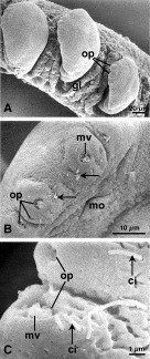
Scanning electron micrographs have confirmed that the openings of the anterior adhesive apparatus are located in the antero-lateral region of the worms and that there are 6 pits as observed with the phase-contrast microscope (Fig. 2A,B). Each anterior adhesive pit is seen to be cup-like containing short microvilli (Fig. 2C) that occasionally project from the pits. Uniciliated structures were observed in the body tegument in the anterior region of the worm and were more abundant in the vicinity of the openings of the anterior adhesive apparatus (Fig. 2B,C). Approximately 8 pairs of uniciliated structures were found in between the medial pair of anterior adhesive pits (Fig. 2B).

Fig. 2. Scanning electron micrographs of adult Bravohollisia gussevi, (A) located between the secondary gill lamellae (gl). Openings of anterior adhesive apparatus (op). (B) The anterior region of B. gussevi showing 3 pairs of openings of the anterior adhesive apparatus (op), uniciliated structures (arrows), microvilli (mv) and mouth (mo). (C) The microvilli (mv) projecting from the pits with the uniciliated structures (ci) located outside the openings of the anterior adhesive apparatus (op) of B. gussevi.
Transmission electron microscopy (TEM)
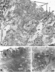
Transmission electron micrographs of the uniciliated structures associated with the openings of the anterior adhesive apparatus show them displaying a typical ‘9+2’ microtubule arrangement and anchored to the body tegument by an annular septate desmosome (Fig. 3A). Granular vesicles occupy the subjacent cytoplasm of these structures that extends into the parenchyma layer (Fig. 3A). The uniciliated structures are usually associated with an opening of a duct containing granular, electron-dense, membrane-bound bodies leading to gland cells located below the tegument (Fig. 3B,C). These membrane-bound bodies are roughly spherical and range in size from 150 to 270 nm in diameter. The gland cells producing these inclusion bodies have an ovoid euchromatic nucleus, granular cytoplasmic matrix and a membranous array of granular endoplasmic reticulum (GER) (Fig. 3C). No Golgi complexes were observed in these gland cells.

Fig. 3. Transmission electron micrographs of a longitudinal section showing (A) uniciliated structures (ci) associated with the anterior adhesive apparatus of Bravohollisia gussevi. The ‘9+2’ microtubule arrangement (ci) of a uniciliated structure is clearly visible together with a cell body (cb). Duct (d), nucleus (n), septate desmosome (sd), rod-like bodies (S1) and general body tegument (t). (B) Uniciliated structures (ci) and an associated opening (op) of a duct containing granular, electron-dense secretory bodies (sb). Granular endoplasmic reticulum (GER), muscle (mu), oval bodies (S2) and general body tegument (t). (C) Cell body associated with the uniciliated structures of B. gussevi, showing granular endoplasmic reticulum (GER), nucleus (n) and abundant granular, electron-dense secretory bodies (sb).
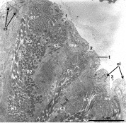
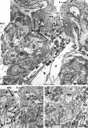
The floor of the adhesive pit is lined by tegument bearing microvilli interspersed with duct openings (Fig. 4). The tegumental cell bodies are located in the parenchyma layer below the pit (Fig. 5A). This microvillous tegument is demarcated from the general body tegument by a septate desmosome and can be readily distinguished from the general body tegument by having a greater preponderance of electron-dense vesicles and by the absence of electron-lucent vesicles commonly found throughout the general body tegument (Fig. 5B). A cross-section of ‘9+2’ microtubules of a presumed uniciliated structure was also observed amongst the microvilli in the pit (Fig. 5C). In the pits of the anterior adhesive apparatus, the ducts are connected to the microvillous tegument by an annular septate desmosome and their walls are lined with microtubules (Fig. 5B, C).

Fig. 4. Transmission electron micrograph of a longitudinal section through anterior adhesive pits (1–3) of Bravohollisia gussevi. Abundant rod-like bodies (S1) and oval bodies (S2) located in the ducts leading from the gland cells. Uniciliated structure (ci), muscle fibre (mu), microvilli (mv) and general body tegument (t).

Fig. 5. Transmission electron micrograph of a longitudinal section through (A) an anterior adhesive pit of Bravohollisia gussevi. Ducts (d) containing oval-bodies (S2) penetrate through the microvillous tegument (mgt) with a cell body (cb). Golgi (Go), muscle (mu), microvilli (mv), nucleus (n) and general body tegument (t). (B) An anterior adhesive pit of B. gussevi. Duct (d), electron-dense vesicles (edv), electron-lucent vesicles (elv), microvillous tegument (mgt), microtubules (mt), muscle (mu), microvilli (mv), opening of the anterior adhesive pit (op), septate desmosome (sd), oval bodies (S2) and general body tegument (t). (C) Transverse section through an anterior adhesive pit of B. gussevi. Within the microvilli (mv) the presumed uniciliated structures (ci) with the ‘9+2’ microtubule arrangement are present. Note microtubules (mt) are present in the ducts transporting the rod-like bodies (S1), which contain vesicles (v). Oval bodies (S2) and interlinking band-like structures (arrow).
A group of ducts was observed just posterior to each of the adhesive pits (Figs 4 and 6A). An average of 10–11 ducts per group was observed, containing rod-like (S1) bodies and oval (S2) bodies. In each group of ducts, 6–7 ducts usually contained S1 bodies, while 3–4 ducts contained S2 bodies (Fig. 6A). Occasionally, empty ducts were observed. Branching of S1 ducts occurs just below the adhesive pit (Fig. 6B). Such branching was not observed for S2 ducts. Bundles of muscle fibres and associated nerves were observed in the vicinity of the anterior adhesive apparatus (Figs 5B and 6A).

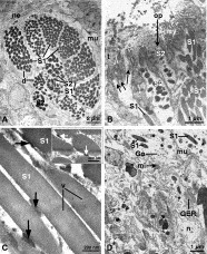
Fig. 6. Transmission electron micrograph of (A) a group of ducts situated just posterior to a microvillous pit of the anterior adhesive apparatus of Bravohollisia gussevi. Rod-like bodies (S1) and oval bodies (S2) are transported in their respective ducts (d). Muscle (mu) and nerve (ne). (B) An anterior adhesive pit of B. gussevi showing an opening of a duct (op) containing oval bodies (S2). Duct transporting the rod-like bodies (S1) branched (arrows) in the microvillous (mv) tegument. General body tegument (t). (C) Rod-like bodies (S1) in a duct of the anterior adhesive apparatus of B. gussevi. Interlinking band-like structures (arrows) between the rod-like bodies (S1), which contain less electron-dense vesicles (v). (Inset) Similar interlinking band-like structures (arrows) between the oval bodies (S2). (D) S1 gland cell of B. gussevi. Granular endoplasmic reticulum (GER), Golgi (Go), muscle (mu), nucleus (n), developing rod-like bodies (*).
The rod-like S1 bodies contain numerous vesicles (80±13 nm in diameter) in an electron-dense matrix (Figs 5C and 6C). The vesicles are less electron-dense than the matrix (Figs 5C and 6C). The oval S2 bodies display a comparatively more electron-dense matrix (Figs 4 and 6A). The number of S1 and S2 bodies in an average transverse section of ducts varies from 13 to 65 and 10 to 35, respectively (Fig. 6A). The S1 bodies are orientated parallel to the long axis of the ducts, measure 255±24 nm (n=10) in diameter, at least 6·5 μm in length and appear to be flexible such that they bend within the ducts. The S2 bodies, with a diameter of 308±48 nm (n=10), are more dispersed within the ducts. Interlinking membrane-like structures were observed between S1 bodies, especially in ducts near the pits (Figs 5C and 6C). Similar interlinking structures could also be observed between S2 bodies (Fig. 6C (inset)). However, interlinking membrane-like structures between the S2 bodies are less common. Under high magnification, these interlinking structures are seen as a series of 2–5 parallel bands occurring at intervals of approximately 9 nm.
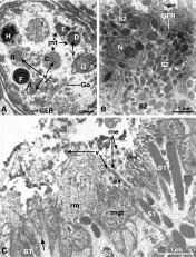
TEM micrographs of the glands located lateral to the pharynx reveal that the gland cells are uninucleated and flask-shaped with ducts extending distally to the adhesive pits (Figs 4 and 6D). These gland cells secrete the S1 and S2 bodies. The gland cell secreting S1 bodies has a moderately dense cytoplasmic matrix, a well-organized GER, numerous cristate mitochondria and a peripheral nucleus (Figs 6D and 7A). Several types of inclusions (A–I) occur in the cytoplasm of these S1-secreting gland cells (Fig. 7A). An irregularly shaped membrane-bound inclusion, A, contains small vesicles (diameter 29–43 nm) in a less electron-dense granular matrix occurring near the terminal cisterna of Golgi complexes (Fig. 7A). Inclusions B and C contain vesicles arranged in a honeycomb pattern in a less electron-dense granular matrix, while inclusions D and E consist of a core of vesicles in an electron-dense matrix (Fig. 7A). Vesicles within these membrane-bound inclusions (A, B, C, D and E) are similar in appearance and size to the vesicles found close to the terminal cisterna of the Golgi complex (Fig. 7A). A layer of microtubules (~23 in number) surrounds the outer surface of inclusions C, D and E (Fig. 7A). However, no microtubules were observed in the wall of the duct near the gland cell bodies (Figs 6D and 7A). In inclusions F to I, the contents could not be clearly identified (Fig. 7A).

Fig. 7. Transmission electron micrograph of (A) an S1 gland cell of Bravohollisia gussevi showing various membrane-bound inclusions (A–I) probably involved in the formation of the secretory bodies. Granular endoplasmic reticulum (GER), Golgi (Go), microtubules (mt) and vesicles (v). (B) An S2 gland cell of B. gussevi containing granular endoplasmic reticulum (GER), nucleus (n), and numerous oval-bodies (S2). (C) An anterior adhesive pit of B. gussevi showing electron-dense vesicles (v) present in the exuded rod-like, S1, bodies (ex) outside the microvillous tegument (mgt). Membrane of the exuded S1 bodies (me), microvilli (mv), released diffuse granular materials (rm), oval bodies (S2) and branching of S1 duct (arrow).
The gland cells producing the oval secretory bodies (S2) are found amongst the S1-secreting gland cells. Like the S1-secreting gland cells, the S2-secreting gland cells are also flask-shaped with a single peripheral nucleus (uninucleated) and with ducts extending to the adhesive pits. GER, numerous free ribosomes and few mitochondria are observed in the cytoplasm of the S2-secreting gland cells (Fig. 7B). The cytoplasm of these gland cells is more electron-dense than that of the S1-secreting gland cells. In the present study, no Golgi complexes or microtubules associated with the S2 bodies were observed in the S2-secreting gland cells in all 5 specimens of worm examined.
S1 bodies were observed to exit through the apertures of the branching ducts in the adhesive pit (Fig. 7C). Figure 7C shows the ultrastructure of an anterior adhesive pit with S1 bodies within the duct as well as outside the tegument. Outside the tegument, the membrane of the S1 bodies is clearly evident with small vesicles (diameter from 75 to 100 nm) within it (Fig. 7C). The S2 bodies were also observed to exude through the duct apertures in the microvillous tegument in the pit (Fig. 7C). A diffuse granular electron-dense matrix is seen just outside the aperture of the S2 ducts (Fig. 7C).
DISCUSSION
Anterior adhesive apparatus is present in all known monogenean species of the subclass Polyonchoinea Bychowsky, 1937 (see Bychowsky, 1957) but studies on them are limited to the 20 species listed in Table 1. Basically, the anterior adhesive apparatus of monogeneans consists of gland cells located lateral to the pharynx, with ducts (usually in bundles of 10–11 ducts per adhesive pit in B. gussevi) leading to openings in the anterior region where the secretions are released. The composite diagram (Fig. 1B) of the morphology of the adhesive pit is based on the information obtained from the SEM and TEM studies. Each of the 6 adhesive pits is lined with microvillous tegument interspersed with numerous duct apertures through which the membrane-bound, rod-like (S1) and oval (S2) bodies are extruded.
The main differences in the anterior adhesive apparatus of the different monogeneans are in the shape (circular to slit-like) and number of openings (2–40) as well as in the morphology of the anterior part of the anterior adhesive apparatus (microvillous pits to microvillous areas in exposed pads). In B. gussevi (present study) and Bychowskyella pseudobagri there are 6 distinctly separate circular openings and each opening is essentially a pit with microvillous tegument interspersed with apertures of ducts. Microvillous pits with slit-like openings are observed in Decacotyle lymmae (2 slits) and Monocotyle spiremae (16 slits). In the capsalids, such as Entobdella soleae and Benedenia lutjani, the anterior part of the anterior adhesive apparatus is in the form of 2 raised exposed pads, each with 3 microvillous areas.
Although 3 types of secretions have been identified in some monogeneans, only 2 types of secretions, S1 and S2 were observed in this study. The membrane-bound S1 bodies are the most abundant secretion in the anterior adhesive apparatus of B. gussevi and probably contain the major adhesive component in the droplets or ‘print’ as observed at the site of attachment during the locomotion of live specimens. The less electron-dense vesicles in the matrix of S1 bodies probably contain the adhesive materials. These vesicles are most likely crucial to the adhesion process by sequestering the adhesive material and thus facilitating its transport through the matrix of the rods without occluding the ducts. The presence of vesicles in the S1 bodies is also reported for Dactylogyrus amphibothrium and D. hemiamphibothrium by El-Naggar and Kearn (1983), but the vesicles in the S1 bodies of these species are more electron-dense than the matrix.
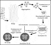
The inclusions (A to E) observed in the gland cells of B. gussevi are postulated to be different stages of development of the S1 bodies, which is the major secretion in B. gussevi. The formation of the S1 body, as summarized in Fig. 8, begins with the synthesis of the secretory products in the GER. The secretory product is transported to a Golgi complex to be packaged into vesicles. The vesicles begin to aggregate and coalesce resulting in a new developing S1 body with numerous vesicles (inclusions A and B). Coalescence of vesicles also occurs during the formation of S1 bodies of D. amphibothrium and D. hemiamphibothrium (see El-Naggar and Kearn, 1980). Next, microtubules begin to form around the developing S1 body (inclusion C). The ringlet of microtubules probably assists in transporting and strengthening the structure of the developing S1 body. Condensation of the matrix in the developing S1 body results in the matrix becoming more electron-dense (inclusions D and E). Smaller vesicles may coalesce forming larger vesicles in the developing S1 body. As the S1 body develops it loses its microtubules. Fully formed S1 bodies without microtubules are routed into ducts. In the ducts, the S1 bodies are observed linked together by membrane-like structures, which probably function to keep the various S1 bodies apart and prevent them from sticking together. The microtubules lining these ducts may assist in the movement of the rod-like bodies within the ducts.

Fig. 8. Schematic diagram illustrating the development of the S1 bodies of Bravohollisia gussevi based on the TEM observations. Granular endoplasmic reticulum (GER) and direction of process (arrows). (Not drawn to scale).
The membrane-bound S1 body on reaching the aperture of the duct in the microvillous pit discharges its dense matrix first, as indicated by the presence of S1 bodies without a matrix but with the intact vesicles. The microvilli in the pit probably assist in mixing the released substances, especially the vesicles and matrix of the S1 bodies, resulting in a sticky adhesive and spreading them over the host surface. The microvilli may also provide a large contact area for instant and strong attachment of the anterior adhesive apparatus to the surface of the gill. S2 bodies also discharge their contents into the microvillous pit resulting in a diffuse material outside the opening of ducts associated with S2 bodies. However, they are not usually observed in the microvillous pits. This may be due to the fact that fewer S2 bodies are produced compared to the S1 bodies. This observation was also noted by Kearn and Evans-Gowing (1998) who suggested that this was due to the S2 bodies being released first. The role of the S2 secretions in B. gussevi is not clear as no experimental work was done to examine their role. However, studies on the attachment and detachment mechanisms of the anterior adhesive pads of E. soleae by Kearn and Evans-Gowing (1998) showed that the S2 bodies may assist in adhesion while the secretions from the specialized tegument lining the adhesive pads probably help to sever the adhesion. Similarly the S2 bodies and the electron-dense vesicles in the specialized microvillous tegument in the anterior adhesive pits of B. gussevi may be involved in adhesion and severance of adhesion.
We have also observed that the uniciliated structures around the openings of the anterior adhesive apparatus are closely associated with gland cells producing spherical secretory bodies. These gland cells are probably similar to the marginal gland cells described by El-Naggar and Kearn (1983), who hypothesized that the secretions from these gland cells are for recognition of potential mates. However, more studies are needed to clarify the function of the secretion associated with the uniciliated structures of B. gussevi.
This study is part of a Ph.D. project (W.L.W.) carried out both in the University of Malaya and Queen's University Belfast and supported by Research & Development grant (Project no: 01-02-03-0387), Vote F (F 0182/2002B), a grant from the Committee for International Cooperation in Higher Education (CICHE) (Programme 353) and a post-graduate scholarship from the University of Malaya and a Commonwealth Split-site Doctoral Scholarship (MYCN2004-35) to W.L.W.