





Introduction
Podapolipid mites (Trombidiformes: Heterostigmatina: Podapolipidae) are permanent parasites of insects, particularly Coleoptera and Orthoptera and occasionally other taxa such as cockroaches, true bugs, and bees (e.g., Husband and Sinha Reference Husband and Sinha1970; Husband Reference Husband1984; Kurosa and Husband Reference Kurosa and Husband1994; Seeman and Katlav Reference Seeman and Katlav2024; Katlav et al. Reference Katlav, Seeman, Shaw and Riegler2026). Sexual transmission is likely the most common means of host transfer, especially for species that parasitise beetles (e.g., Hurst et al. Reference Hurst, Sharpe, Broomfield, Walker, Majerus, Zakharov and Majerus1995; Abbot and Dill Reference Abbot and Dill2001; Nahrung and Clarke Reference Nahrung and Clarke2007), although this mode remains unproven for many genera, including the largest genus of podapolipids, Eutarsopolipus Berlese, 1913. Eutarsopolipus includes 101 named species, all from carabid beetles (Seeman and Katlav Reference Seeman and Katlav2024). This large genus is arranged into ten species groups originally formed by Regenfuss (Reference Regenfuss1968, Reference Regenfuss1974), has been added to by works such as Husband and Husband (Reference Husband and Husband2009), and was honed most recently by Seeman (Reference Seeman2019a). Currently, the species groups are as follows: acanthomus (14 spp.), biunguis (11 spp.), lagenaeformis (1 sp.), leytei (7 spp.), megacheli (1 sp.), myzus (26 spp.), ochoai (28 spp.), pterostichi (11 spp.), secundus (1 sp.), and stammeri (1 sp.; Seeman Reference Seeman2019a). Four of the five larger species groups show a biogeographic pattern, with the acanthomus, biunguis, and myzus species dominating the Northern Hemisphere fauna and the ochoai group dominating the Australian fauna. Little is known of the South American, East Asian, and African fauna, although the four known Eutarsopolipus species from the Neotropical region are all members of the ochoai group (Seeman and Katlav Reference Seeman and Katlav2024).
The exception to these patterns is the pterostichi group, an enigmatic assemblage of 11 species found in all biogeographic zones other than South America and Antarctica (Seeman and Katlav Reference Seeman and Katlav2024). All of its members share the absence of both a respiratory system and setae on genua II–III but vary considerably in the expression of pretarsal claws (Katlav et al. Reference Katlav, Hajiqanbar, Riegler and Seeman2021). Within other species groups, the claws are, with rare intraspecific exceptions (Seeman Reference Seeman2019b), either present on all legs or absent from all legs, but in the pterostichi group, claws can be absent, present only on leg I, or present on all legs (either rudimentary or well developed).
This work describes two new species of Eutarsopolipus collected from carabid beetles from Elk Island National Park, Alberta, Canada – one from the pterostichi species group and one from the biunguis species group. Previously, the genus was known in Canada from five described species in the biunguis and myzus species groups (Seeman and Katlav Reference Seeman and Katlav2024): E. brevichelus Husband and Husband, 2003, E. elzingai Husband, 1998, E. lindquisti Husband, 1998, E. platyni Husband and Husband, 2002, and E. quebecensis Husband, 1998.
Materials and methods
The carabids we examined were bycatch in pitfall traps set to collect carrion-associated arthropods for the Master’s research study of B.G. (Grappone Reference Grappone2025). Traps were set throughout spring and summer 2023 at Elk Island National Park, Alberta, Canada (coordinates: 53.62, –112.85). The traps consisted of 4-L plastic buckets partly filled with 500 mL of propylene glycol-based antifreeze with a few drops of dish soap to break surface tension. A square of cardboard was hung over the top of the bucket to keep out rain, and the trap was surrounded by chicken wire to protect it from disturbance by vertebrates. After each bucket was collected, the contents were sieved through 500-µm mesh to remove the antifreeze. Material retained in the sieve was transferred into 70% EtOH for storage.
Carabid beetles were removed from a subset of samples by N.M.B., and their elytra were opened so that the wings and subelytral spaces could be examined with the aid of a dissecting microscope. All mites found under elytra were removed and stored in 70% EtOH. Before mounting them on slides, intact mites were either cleared in lactic acid for 24–48 hours or those that contained large masses of opaque white excretory product (guanine) were placed in a water and potassium hydroxide (KOH) solution for 10–15 minutes and then briefly into water and lactic acid to remove the KOH. After clearing, mites were mounted on glass slides in polyvinyl alcohol mounting medium and cured for four days at approximately 40 °C on a slide warmer. Co-author N.M.B. examined the cured slides using a Leica DMLB microscope (Wetzlar, Germany) with differential interference contrast lighting and identified mites to genus and species groups using Husband and Husband (Reference Husband and Husband2008, Reference Husband and Husband2009). The best of the slide-mounted specimens were sent to O.D.S. for description.
All illustrations of females of the new species are based on the holotype. Morphological terminology and abbreviations follow Lindquist (Reference Lindquist1986): ap 1–2, apodemes of coxisterna 1–2; appr, prosternal apodeme. Setal counts are given as femur–genu–tibia–tarsus; minute seta p′ is not included in counts or illustrations because it could not be seen in most specimens. Drawings were prepared by O.D.S. with the aid of a camera lucida on a Nikon Eclipse 80i microscope (Tokyo, Japan). All measurements are given in micrometres (μm) and were taken using calibrated ocular micrometers on the Eclipse 80i or a Leica DMLB (Wetzlar, Germany) microscope with phase-contrast lighting. For the female, the measurement of the holotype is provided first, followed by the range of paratypes. For the male and larva, measurements are given as ranges. Ventral photographs are provided instead of drawings because the venter of many Eutarsopolipus species, including those described herein, have little diagnostic value beyond the size and form of the coxal setae. Sejugal apodemes are absent in all Eutarsopolipus. In the descriptions, minute setae (m) refer to those shorter than 1 µm in length, sometimes being represented merely by a nubbin within the alveolus. Plate lengths were taken at their midpoints; widths are not given because plates are often split, flattened, or wrapped around the body; distance between setae represents the distance from setal insertion to setal insertion; body lengths of males do not include the genital capsule; body width of all stages is measured as maximum width. Higher classification in the Taxonomy section follows Lindquist et al. (Reference Lindquist, Krantz and Walter2009).
Holotypes and some paratypes of the new species are deposited in the Canadian National Collection of Insects, Arachnids and Nematodes (CNC; Ottawa, Ontario, Canada; accession numbers CNC1985249–CNC1985254), and the remainder of the paratypes are deposited in the Queensland Museum (QM; Brisbane, Australia). Carabid host specimens (symbiotypes) are deposited in the E.H. Strickland Entomological Museum at the University of Alberta (UASM; Edmonton, Alberta, Canada; accession numbers UASM440281–UASM440286). These beetles are also identified with ‘AB’ collection codes, as per Grappone (Reference Grappone2025).
Taxonomy
Cohort Heterostigmatina Berlese, 1899
Superfamily Tarsonemoidea Kramer, 1877
Family Podapolipidae Ewing, 1922
Genus Eutarsopolipus Berlese, 1913
Type species: Tarsopolipus lagenaeformis Berlese, 1911
Eutarsopolipus husbandi Seeman, Battle, Grappone, and Proctor sp. nov.
Eutarsopolipus husbandi sp. nov. adult female, dorsal idiosoma. Legs and gnathosoma shown in outline. Morphological terminology and abbreviations follow Lindquist (Reference Lindquist1986).

Figure 1. Long description
A diagram of the dorsal idiosoma of an adult female Eutarsopolipus husbandi sp. nov. mite. The diagram shows the legs and gnathosoma in outline. The labeled parts include v1, sc1, sc2, c1, c2, ia, d, im, f, and h. The diagram is annotated with morphological terminology and abbreviations following Lindquist (1986).
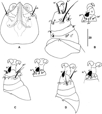
Eutarsopolipus husbandi sp. nov. adult female: A, gnathosoma, dorsal aspect; B, leg I; C, leg II; and D, leg III. Tarsi shown as separate dorsal and ventral aspects. Morphological terminology and abbreviations follow Lindquist (Reference Lindquist1986).

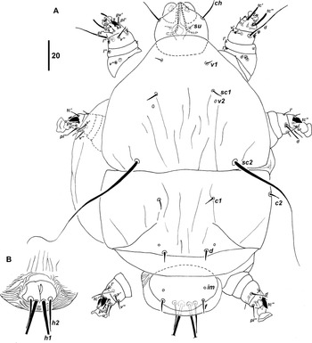
Eutarsopolipus husbandi sp. nov. adult male: A, dorsal aspect, left-hand side legs are shown as their ventral aspect; B, ventral aspect genital capsule. Morphological terminology and abbreviations follow Lindquist (Reference Lindquist1986).

Figure 3. Long description
Panel A: A diagram of the dorsal aspect of Eutarsopolipus husbandi sp. nov. adult male, with the left-hand side legs shown in their ventral aspect. The diagram includes various labeled parts such as ch, su, tc, pv, pl, d, v, sc1, sc2, c1, c2, ia, pd, tm, f, and h. Panel B: A diagram of the ventral aspect of the genital capsule of Eutarsopolipus husbandi sp. nov. adult male, with labeled parts including tc, pv, pl, d, v, and f.
Eutarsopolipus husbandi sp. nov. larva: A, dorsal aspect, left-hand side legs are shown as their ventral aspect; B, ventral caudal region, plate H. Morphological terminology and abbreviations follow Lindquist (Reference Lindquist1986).

Eutarsopolipus husbandi sp. nov. photographs (differential interference contrast lighting): A, adult female, coxisternal region I–II; B, adult female, ventral caudal lobes; C, larva, ventral aspect. Morphological terminology and abbreviations follow Lindquist (Reference Lindquist1986).

ZooBank Nomenclature Act: urn:lsid:zoobank.org:act:2C1ACD15-A740-4B00-8798-8E26FB582B16
Diagnosis. Species group: pterostichi. ALL LIFE STAGES: idiosomal setae unmodified, either slender or minute; alveolus v2 positioned posteriad seta sc1; femora I–III 3-0-0; genua I–III 2-0-0; tibia I lacking seta k. FEMALE: pretarsi I–III with strong paired claws (male pretarsus I with single claw; larva pretarsus I with bifurcate claw); tarsus II with solenidion; cheliceral stylets 36–38; seta sc1 7–8; distance between setae d–d (120–135) much greater than distance c1–c1 (49–60) and f–f (58–73); seta h 11–13; tarsus I solenidion (ω) about as long as wide; tibia III seta l′ shorter than ventral setae (v). MALE: idiosomal length 145–155, width 115–125; gnathosomal length 31–33; cheliceral stylets 22–23; distances between setae v1–v1 27–29, sc1–sc1 41–44; seta c1 4–5; seta f 4–5; tarsus I with seta pv′. LARVA: gnathosomal length 35–38; plate CD incised; plate H a pair of well-developed lobes; cheliceral stylets 26–28; seta v1 4–5; seta sc1 7–8; sc2 70–100; d 7–9; f 7–9; seta h2 20–22; coxal setae very short, m–2.
Material examined. HOLOTYPE. Female, ex Pterostichus adstrictus Eschscholtz, 1823 (AB127), Elk Island National Park, 25.vii.2023. Deposited in CNC (accession # CNC1985249). PARATYPES: 2 females, on same slide and same data as holotype (CNC); 5 females, 1 male, 6 larvae, ex P. adstrictus (AB89), 12.vi.2023, on two slides (CNC; accession #s CNC1985250-1985251; symbiotype accession # UASM440282); 1 female, 3 males, 1 larva, ex P. adstrictus (AB19), 15.v.2023, on two slides (QM; symbiotype accession # UASM440283).
Description. FEMALE (Figs. 1, 2). Gnathosoma: length 50 (50–56), width 45 (45–48). Palp lacking setae, with usual rudimentary distal sensory cluster. Cheliceral stylets 36 (36–38), ch 13 (11–15), su 7 (6–7), pharynx width 14 (14–15).
Idiosoma (Figs. 1, 5A, B): length 385 (320–460), width 255 (220–290), larger specimens moderately physogastric, swollen body elongate-ovate. Respiratory system absent. Prodorsal plate with setae v1 5 (4–5), v2 alveolar remnant, sc1 7 (7–8), sc2 40 (37–45); v2 posteriad sc1. Distance between setae v1–v1 37 (34–42), sc1–sc1 70 (66–69), v1–sc1 23 (24–27), v2–v2 72 (70–74), sc2–sc2 120 (120–130), sc1–sc2 58 (55–60). Plate C setae c1 8 (7–9), setae c2 5 (5–7), distance between setae c1–c1 60 (49–55). Plate D setae d 6 (6–7), d–d 120 (125–135), cupuli ia anteriad to anterolaterad setae d. Plate EF setae f 6 (6–7), f–f 58 (63–73), cupuli im ranging from anterolaterad to laterad of setae f. Plate H entire, bearing setae h 11 (11–13). Venter: coxisterna all weakly defined, more or less membranous; ap 1–4 and appr well defined, ap 2 reaching appr; coxal setae small, 1a 2 (2–3), 2a 3 (2–3), 3a 3 (3), 3b 3 (3). Alveoli of 1b usually indiscernible, visible asymmetrically in one paratype; 2b often indiscernible, visible symmetrically in one paratype, asymmetrically in another paratype.
Legs (Fig. 2): setal counts legs I–III: 3-2-5(+ϕ)-8(+ω), 0-0-4-7(+ω), 0-0-4-7. Pretarsi I–III with strong paired claws. Leg I: femur, d m (m), l′ 2 (2–3), thickened, v″ 2 (2–3), thickened; genu, l′ 2 (2–3), thickened, l″ 2 (2), thickened; tibia, d 33 (29–34), l′ 3 (3–4), thickened, l″ 2 (2), thickened, v′ 3 (3–4), v″ 10 (10–12), ϕ 3 (3–4), k absent; tarsus, tc′ 12 (12–13), tc″ 12 (13–14), pl′ 5 (6–7), pl″ 6 (7–8), pv′ 3 (3), pv″ 2 (2–3), ω 2 (2–3), squat, about as long as wide (sometimes slightly longer in paratypes), s 5 (5–6), u″ m (m). Minute seta p′ faintly visible in some specimens. Leg II: tibia, d 14 (15–19), l′ 5 (4–5), v′ 9 (9–11), v″ 7 (6–7); tarsus, tc′ 6 (6), tc″ 8 (7–8), pl″ 9 (10–13), pv′ 2 (2), pv″ 3 (3–4), ω 3 (3), u′ 7 (6–7), u″ m (m). Leg III: tibia, d 14 (14–17), l′ 5 (5–6), v′ 6 (7–10), v″ 10 (7–9); tarsus, tc′ 6 (6), tc″ 7 (6–7), pl″ 13 (13–17), u′ 6 (6–7), pv′ 2 (2–3), pv″ 3 (3), u″ m (m).
Large modified setae: tarsus I seta s and tarsi II–III setae tc″ and u′ thickened, large, spine-like, tips not obviously bifid (sometimes u″ tip slightly bifid in paratypes, perhaps depending on angle of viewing).
Variations: Paratype #4 lacks both idiosomal setae d in an unusual symmetrical anomaly.
MALE (Fig. 3). Gnathosoma: length 31–33, width 30–31. Cheliceral stylets 22–23, ch 9–12, su 3–4, pharynx width 10–11.
Idiosoma (Fig. 3): length 145–155, width 115–125. Prodorsal plate with setae v1 2–3, v2 alveolar remnant, sc1 5–6, sc2 62–68; v2 posteriad sc1. Distance between setae v1–v1 27–29, sc1–sc1 41–44, v1–sc1 14–16, v2–v2 44–47, sc2–sc2 63–67, sc1–sc2 35–38. Plate CD setae c1 4–5, c2 5–7, d 5, distance between setae c1–c1 41–44, c1–c2 30–32, d–d 32–36; cupuli ia ranging from anterolaterad to laterad of setae d. Plate EF with setae f 4–5, distance f–f 18–20; cupuli im anterolaterad setae f. Genital capsule length 25–26, width 24–26, setae h m. Venter: ap 1–4 and appr weakly defined, ap 2 reaching appr. Coxal setae 1a 2, 2a 2, 3a m–2, 3b 2.
Legs (Fig. 3): setal counts legs I–III: 3-1-5(+ϕ)-8(+ω), 0-0-4-7(+ω), 0-0-4-7. Pretarsus I with single claw, pretarsi II–III lacking claws. Leg I: femur, d m, l′ m–2, thickened, v″ m; genu, l′ m–2, l″ m–2; tibia, d 26–29, l′ m, l″ m, v′ m, v″ 6–8, ϕ 2–3, k absent; tarsus, tc′ 12–13, tc″ 13–14, pl′ 4, pl″ 5–6, pv′ 2–3, pv″ 2–3, ω 2, globular, s 4–5, u″ m, p′ absent or indiscernible. Leg II: tibia, d 7–9, l′ 3, v′ 5–6, v″ 3–4; tarsus, tc′ 5–6, tc″ 6–7, pl″ 6–8, pv′ 2, pv″ 3, ω 2, globular, u′ 5–6, u″ m. Leg III: tibia, d 9–10, l′ 2, v′ 4–5, v″ 2–3; tarsus, tc′ 5–6, tc″ 6–7, pl″ 7–8, u′ 5–6, pv′ 2, pv″ 3, u″ m–2.
Modified setae: same as female.
LARVA (Fig. 4). Gnathosoma: length 35–38, width 36–37. Cheliceral stylets 26–28, ch 15–16, ventral setae su 4–5.
Idiosoma (Figs. 4, 5C): length 170–195, width 115–140. Prodorsal plate with anterior part hood-like, covering about half of gnathosoma, with setae v1 4–5, v2 alveolar remnant, sc1 7–8, sc2 70–100; v2 posteriad sc1. Distance between setae v1–v1 28–31, sc1–sc1 36–39, v1–sc1 19–23, v2–v2 40–42, sc2–sc2 55–66, sc1–sc2 40–48. Plates C and D fused medially, incised posterolaterally. Plate CD setae c1 6–7, setae c2 8–9, distance between setae c1–c1 32–37, setae d 7–8, d–d 25–28, cupuli ia anterolaterad d. Plate EF rounded, setae f 8–9, f–f 24–27, cupuli im just anteriad f. Plate H bilobed, 23–24 wide, 16–17 long, bearing setae h1 > 150, h2 20–22. Venter: coxisterna 1–3 poorly defined and appr weakly defined, ap 2 reaching appr; coxal setae slender, 1a m, 2a m, 3a m–2, 3b m–2. Alveoli of 1b, 2b indiscernible.
Legs (Fig. 4): setal counts legs I–III: 3-2-5(+ϕ)-8(+ω), 0-0-4-6(+ω), 0-0-4-6. Pretarsus I with small paired claws; pretarsi II–III lacking claws. Leg I: femur, d m, l′ 2, thickened, v″ m; genu, l′ m, l″ m; tibia, d 25–34, l′ 2–3, l″ 3, v′ 2, v″ 9–10, ϕ m–2, k absent; tarsus, tc′ 9–11, tc″ 10–11, pl′ 4–5, pl″ 5, pv′ 2, pv″ m–2, ω m–2, s 4–5, u″ m. Minute seta p′ m visible in some specimens. Leg II: tibia, d 11–13, l′ 4, v′ 7–8, v″ 5–7; tarsus, tc′ 4–5, tc″ 6–7, pl″ 7–8, pv′ absent, pv″ 4, ω m, u′ 6, u″ m. Leg III: tibia, d 16, l′ 4–5, v′ 7–8, v″ 6; tarsus, tc′ 4–5, tc″ 6–7, pl″ 8–9, pv′ absent, pv″ 4–5, u′ 6–7, u″ m.
Modified setae: same as female.
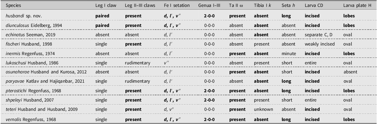
Differential diagnosis. The new species is close to Eutarsopolipus pterostichi Regenfuss, 1968 and Eutarsopolipus vernalis Regenfuss, 1968, which were collected from Pterostichus species in Germany and Ukraine (Regenfuss Reference Regenfuss1968; Eidelberg Reference Eidelberg1994; Seeman and Katlav Reference Seeman and Katlav2024). These species are the only members of the pterostichi species group with females having a long seta h and two setae on genu I (Table 1). Females are most easily separated, with the main feature distinguishing the species being the paired claws on leg I and the distance between the dorsal opisthosomal setae, with c1 and f being closer together and the distance between setae d being greater in the new species. The visual effect is that setae d are almost in a longitudinal line in E. pterostichi, but setae d are obviously far outside the line made between c1-e in E. husbandi sp. nov. As measurements, these are c1–c1 79, d–d 97, f–f 90 in E. pterostichi (as estimated from the drawings of Regenfuss (Reference Regenfuss1968)), compared with c1–c1 49–60, d–d 120–135, and f–f 58–73 in E. husbandi sp. nov. Other differences are that the tarsal solenidion (ω) is squat (versus elongate in E. pterostichi), genu I seta l″ is thorn-like (versus slender in E. pterostichi), and tibia III seta l′ is shorter than setae (v) (versus l′ as long as setae (v) in E. pterostichi). Tarsal seta pv′ is not depicted for E. pterostichi in Regenfuss (Reference Regenfuss1968), but this is probably overlooked because the tiny tarsal setae are difficult to see, especially without differential interference contrast microscopy. The original description of E. vernalis is less detailed than that of E. pterostichi in that it focuses on diagnostic differences from E. pterostichi, but the general characteristics should also apply to this species.
Taxonomically significant features in the pterostichi group. All are for females unless stated otherwise. A key to species other than Eutarsopolipus husbandi sp. nov. is available in Katlav et al. (Reference Katlav, Hajiqanbar, Riegler and Seeman2021). Features in bold font are shared with E. husbandi sp. nov. Morphological terminology and abbreviations follow Lindquist (Reference Lindquist1986).

Table 1 Long description
The table compares various species based on their morphological features. It has 12 rows and 9 columns. The columns are labeled as Species, Leg I claw, Leg II-III claws, Fe I station, Genua I-III, Ta II ω, Tibia I k, Seta h, Larva CD, and Larva plate H. The rows list different species and their corresponding features. For example, Row 1: Species, husbandi sp. nov.; Leg I claw, paired; Leg II-III claws, present; Fe I station, d, l′, v″; Genua I-III, 2-0-0; Ta II ω, present; Tibia I k, absent; Seta h, long; Larva CD, incised; Larva plate H, lobes. Row 2: Species, diunculosus Eidelberg, 1994; Leg I claw, paired; Leg II-III claws, present; Fe I station, d, l′, v″; Genua I-III, 0-0-0; Ta II ω, absent; Tibia I k, absent; Seta h, incised; Larva plate H, lobes. Row 3: Species, echinatus Seeman, 2019; Leg I claw, absent; Leg II-III claws, absent; Fe I station, d, l′; Genua I-III, 0-0-0; Ta II ω, absent; Tibia I k, absent; Seta h, separate C, D; Larva plate H, oval. Row 4: Species, fischeri Husband, 1998; Leg I claw, single; Leg II-III claws, present; Fe I station, d, l′; Genua I-III, 0-0-0; Ta II ω, absent; Tibia I k, present; Seta h, weakly incised; Larva plate H, oval. Row 5: Species, inermis Regenfuss, 1974; Leg I claw, absent; Leg II-III claws, absent; Fe I station, d, l′; Genua I-III, 0-0-0; Ta II ω, present; Tibia I k, absent; Seta h, minute; Larva CD, incised; Larva plate H, lobes. Row 6: Species, lukoschusi Husband, 1986; Leg I claw, single; Leg II-III claws, rudimentary; Fe I station, v′; Genua I-III, 0-0-0; Ta II ω, absent; Tibia I k, present; Seta h, short; Larva CD, entire; Larva plate H, oval. Row 7: Species, osunaharae Husband and Kurosawa, 2012; Leg I claw, absent; Leg II-III claws, absent; Fe I station, d, l′; Genua I-III, 0-0-0; Ta II ω, present; Tibia I k, absent; Seta h, short; Larva CD, incised; Larva plate H, absent. Row 8: Species, parayaoe Katlav and Hajiqanbar, 2021; Leg I claw, single; Leg II-III claws, rudimentary; Fe I station, d, l′; Genua I-III, 0-0-0; Ta II ω, absent; Tibia I k, absent; Seta h, long; Larva CD, incised; Larva plate H, oval. Row 9: Species, pterostichi Regenfuss, 1968; Leg I claw, single; Leg II-III claws, present; Fe I station, d, l′, v″; Genua I-III, 2-0-0; Ta II ω, present; Tibia I k, absent; Seta h, long; Larva CD, incised; Larva plate H, lobes. Row 10: Species, shpeyeli Husband, 2007; Leg I claw, single; Leg II-III claws, present; Fe I station, d, l′, v″; Genua I-III, 2-0-0; Ta II ω, present; Tibia I k, present; Seta h, short; Larva CD, entire; Larva plate H, oval. Row 11: Species, tetrer Husband and Husband, 2009; Leg I claw, single; Leg II-III claws, present; Fe I station, d, v′; Genua I-III, 0-0-0; Ta II ω, present; Tibia I k, unknown; Seta h, incised; Larva plate H, oval. Row 12: Species, vernalis Regenfuss, 1968; Leg I claw, single; Leg II-III claws, present; Fe I station, d, l′, v″; Genua I-III, 2-0-0; Ta II ω, present; Tibia I k, absent; Seta h, long; Larva CD, incised; Larva plate H, lobes.
Males and larvae of E. husbandi sp. nov. and E. pterostichi are more difficult to differentiate. Male E. husbandi sp. nov. have longer setae f (4–5 versus m–2), shorter distances between setae v1 (27–29 versus 45) and sc1 (41–44 versus 55), and a shorter gnathosoma (length 31–33 versus 40). Larvae of E. husbandi sp. nov. have longer setae d and f (length 7–9 versus 3–4) and a slightly shorter gnathosoma (35–38 versus 42).
The only other species of the pterostichi species group with females possessing paired claws on leg I is Eutarsopolipus diunculosus Eidelberg, 1994. Paired claws in females is a rare but homoplasious character state because it also occurs in Eutarsopolipus biuncatus Seeman, 2022 and in Eutarsopolipus janus Seeman, 2022, both members of the leytei species group (Seeman Reference Seeman2022). Paired claws in females represent the larval condition being expressed in the adult. The homoplasious nature of this feature is also apparent when E. husbandi is compared with E. diunculosus, because the latter species lacks setae on genu I, lacks tarsus II solenidion (ω) and, in females, lacks seta h (Table 1). The only other species of Eutarsopolipus that has been reported from Pterostichus in North America is Eutarsopolipus shpeleyi Husband, 2007. This species is obviously different from E. husbandi sp. nov. in having females with a single claw and larvae with seta h2 short (2–4) and plates C and D entire, that is, lacking an incision (Table 1).
Remarks. The host beetle Pterostichus adstrictus is a widespread Holarctic species found throughout Canada, the Rocky Mountains in the United States of America, the British Isles, Scandinavia, northern Japan, and Vladivostok and its surrounds in eastern Russia (Bousquet Reference Bousquet2012). Although deutonymphal astigmatan mites have been reported previously from P. adstrictus in Canada in a survey of 554 carabid beetles from 11 species in Ontario (Olynyk Reference Olynyk1978; Olynyk and Freitag Reference Olynyk and Freitag1979), those authors found no podapolipid mites on any beetle, including on the 45 P. adstrictus specimens they examined, and found surprisingly few mites of any taxon (n = 6) in the subelytral spaces of the 554 beetles.
Etymology. We name this species in honour of the late Dr. Robert Husband, who was the world expert on Podapolipidae for much of his life, described numerous species of Eutarsopolipus, and was extremely generous to anyone who became interested in the family.
Eutarsopolipus paraplatyni Seeman, Battle, Grappone, and Proctor sp. nov.
Eutarsopolipus paraplatyni sp. nov. adult female: A, dorsal idiosoma with separate detail of setae d and f; B, ventral gnathosoma; C, leg I; D, leg II; E, leg III. Tarsi shown as separate dorsal and ventral aspects. Morphological terminology and abbreviations follow Lindquist (Reference Lindquist1986).

Eutarsopolipus paraplatyni sp. nov. male, dorsal aspect, left-hand side legs are shown as their ventral aspect. Morphological terminology and abbreviations follow Lindquist (Reference Lindquist1986).

Eutarsopolipus paraplatyni sp. nov. larva: A, dorsal aspect; B, ventrocaudal region; C, gnathosoma; D, leg I, dorsal leg I and ventral tibia-tarsus; E, leg II, tibia and tarsus, dorsal and ventral aspect; F, leg III, tibia and tarsus, dorsal and ventral aspect. Morphological terminology and abbreviations follow Lindquist (Reference Lindquist1986).

Figure 8. Long description
Panel A: A diagram of the dorsal aspect of the Eutarsopolipus paraplatyni sp. nov. larva, showing various labeled anatomical features. Panel B: A diagram of the ventrocaudal region of the larva, highlighting specific structures. Panel C: A diagram of the gnathosoma, detailing its components. Panel D: Diagrams of leg I, showing dorsal and ventral views of the tibia-tarsus. Panel E: Diagrams of leg II, showing dorsal and ventral views of the tibia and tarsus. Panel F: Diagrams of leg III, showing dorsal and ventral views of the tibia and tarsus.
Eutarsopolipus paraplatyni sp. nov. photographs (differential interference contrast lighting): A, adult female, coxisternal region I–II; B, adult female, posterior venter; C, larva, coxisternal region I–II. Morphological terminology and abbreviations follow Lindquist (Reference Lindquist1986).

ZooBank Nomenclature Act: urn:lsid:zoobank.org:act:FF763A6C-ECE5-4A38-9D85-7B3BA081FD3A
Diagnosis. Species group: biunguis. ALL LIFE STAGES: idiosomal setae unmodified, either slender or minute; femora I–III 2-0-0; genua I–III 0-0-0; tibia I lacking seta k. FEMALE: plate C divided; pretarsi I–III lacking claws (male pretarsus I with single claw; larva pretarsus I with bifurcate claw); tarsus II lacking solenidion; alveolus v2 positioned anteromesad seta sc1; cheliceral stylets 44–46; setae v1 2, sc1 2–3, sc2 25–37, c1 2, d 2–3, f 2–3; h absent. MALE: cheliceral stylets 19–20; seta c1 2; setae f positioned close to posterior margin of plate EF; distances between setae v1–v1 27–30; sc1–sc2 33–38; v1–sc1 15–19. LARVA: plate CD incised, almost fully separating plates C and D; plate H a pair of weakly developed lobes; cheliceral stylets 30–31; setae v1 2, sc1 2–3, sc2 35–46, c1 3, c2 3–4, d 3, h2 31–38.
Material examined. HOLOTYPE. Adult female, from Elk Island National Park, ex Platynus decentis (Say, 1823), collection AB103, 26.vi.2023. Deposited in CNC (accession # CNC1985252; symbiotype accession # UASM440284). Paratypes. 6 physogastric adult females, 1 teneral female, 3 males, 5 larvae from Elk Island National Park, ex Platynus decentis, as follows: 4 females, 2 larvae, on same slide as holotype (CNC); 2 females, 1 teneral female, 3 males, 3 larvae, collection AB98, 26.vi.2023, on two slides (CNC; accession #s CNC1985253–1985254; symbiotype accession # UASM440285); 5 females, collection AB99, 26.vi.2023 (QM; symbiotype accession # UASM440286).
Description. FEMALE (Figs. 6, 9A, B). Gnathosoma: length 58 (57–60), width 56 (58–66), wider gnathosoma in more flattened specimens. Palp lacking setae, with usual rudimentary distal sensory cluster. Cheliceral stylets 44 (44–46), ch 12 (12–14), su 2 (2–3), pharynx width 16 (16–17).
Idiosoma (Figs. 6, 9A, B): length 295, width 225, larger specimens moderately physogastric, most larger specimens damaged in slide-mounting and not measurable. Respiratory system present, trachea without atrium. Prodorsal plate with setae v1 2 (m–2), v2 alveolar remnant, sc1 2 (2–3), sc2 37 (25–37); v2 anteromesad sc1. Distance between setae v1–v1 46 (46–48), sc1–sc1 88 (92–110), v1–sc1 32 (31–41), v2–v2 69 (70–74), sc2–sc2 94 (95–99), sc1–sc2 48 (48–51). Plate C divided, setae c1 2 (m–2), setae c2 4 (4–5), distance between setae c1–c1 100 (100–105). Plate D setae d 2 (2–3), d–d 66 (68–79), cupuli ia anteriad to anterolaterad setae d. Plate EF setae f 2 (2–3), f–f 58 (65–84), greater in larger flattened specimens, cupuli im anteriad setae f. Plate H rudimentary, lacking setae. Venter: coxisterna all weakly defined, membranous; ap 1–4 and appr well defined, ap 2 reaching appr; coxal setae small, 1a 2 (2–3), 2a 3 (3), 3a 5 (5–7), 3b 4 (4–5). Alveoli 1b, 2b indiscernible.
Legs (Fig. 6): setal counts legs I–III: 2-0-5(+ϕ)-8(+ω), 0-0-4-7, 0-0-4-7. Pretarsi I–III lacking claws. Leg I: femur, d m (m), l′ 3 (2–3), thorn-like; tibia, d 27 (19–27), l′ 5 (5–6), l″ 2 (2–3), v′ 3 (3), thorn-like, v″ 4 (4–5), thickened, ϕ 4 (4–6); tarsus, tc′ 13 (12–14), tc″ 10 (11–12), pl′ 5 (5–6), pl″ 6 (5–6), pv′ 2 (2–3), thickened, pv″ 2 (2), thorn-like, ω 3 (3–4), s 6 (6–7), u″ m (m), p′ absent. Leg II: tibia, d 7 (6–7), l′ 7 (7–9), v′ 8 (8–10), v″ 5 (5–6); tarsus, tc′ 5 (4–5), tc″ 10 (10–11), pl″ 9 (9–10), pv′ m (m), pv″ 3 (3–4), u′ 10 (10–12), tip undivided, u″ 2 (m–2). Leg III: tibia, d 6 (6–7), l′ 5 (6–7), v′ 6 (6–7), v″ 7 (7–9); tarsus, tc′ 6 (5–6), tc″ 9 (9–10), pl″ 13 (11–13), pv′ m (m), pv″ 4 (4–5), u′ 9 (9–10), u″ m (m).
Large modified setae: tarsus I seta s and tarsi II–III setae tc″ and u′ thickened, large, spine-like, tips not obviously bifid.
MALE (Fig. 7; n = 3). Gnathosoma: length 31–34, width 36–37. Cheliceral stylets 19–20, ch 7–9, su m, pharynx width 7–8.
Idiosoma (Fig. 7): length 160–165, width 125–130. Prodorsal plate with setae v1 m, v2 alveolar remnant, sc1 m–2, sc2 33–39; v2 posteromesad sc1. Distance between setae v1–v1 27–30, sc1–sc1 53–58, v1–sc1 15–19, v2–v2 37–42, sc2–sc2 61–67, sc1–sc2 33–39. Plate CD setae c1 2, c2 2–3, d 2, distance between setae c1–c1 75–76, c1–c2 17–20, d–d 36–43; cupuli ia indiscernible. Plate EF with setae f 2, distance f–f 29–32; cupuli im anteromesad setae f. Genital capsule length 26–28, width 30–31, setae h m. Venter: ap 1–4 and appr weakly defined, ap 2 reaching appr. Coxal setae 1a m–2, 2a 2, 3a 2–3, 3b 3.
Legs (Fig. 7): setal counts legs I–III: 2-0-5(+ϕ)-8(+ω), 0-0-4-6, 0-0-4-6. Pretarsus I with single claw, pretarsi II–III lacking claws. Leg I: femur, d m, l′ 2–3, thickened; tibia, d 11–12, l′ 2–3, thorn-like, l″ m–2, thickened, v′ 2, thorn-like, v″ 3–4, ϕ 3–4; tarsus, tc′ 10–11, tc″ 10–11, pl′ 3–4, pl″ 4–5, pv′ m–2, pv″ m–2, ω 3–4, s 5, u″ m, p′ absent. Leg II: tibia, d 4–5, l′ 4–5, v′ 5–6, v″ 2–3; tarsus, tc′ 3–4, tc″ 7–8, pl″ 8–9, pv′ absent, pv″ m–2, u′ 6–7, u″ m. Leg III: tibia, d 3–4, l′ 4, v′ 4–6, v″ 2–3; tarsus, tc′ 3–4, tc″ 7, pl″ 8–9, pv′ absent, pv″ m–2, u′ 6–7, u″ m.
Large modified setae: same as female.
LARVA (Figs. 8, 9C). Gnathosoma: length 35–39, width 41–44. Palp lacking setae, with usual rudimentary distal sensory cluster. Cheliceral stylets 30–31, ch 14–16, su m–2, pharynx width 9–10.
Idiosoma (Figs. 8, 9C): length 230–270, moderately to greatly engorged, width 205–220. Prodorsal plate with setae v1 2, v2 alveolar remnant, sc1 2–3, sc2 35–46; v2 mesad to posteromesad sc1. Distance between setae v1–v1 24–25, sc1–sc1 65–72, v1–sc1 21–24, v2–v2 47–49, sc2–sc2 60–62, sc1–sc2 38–41. Plates C and D fused medially, incised laterally, fused portion 28–30 wide. Plate CD setae c1 2–3, setae c2 3–4, distance between setae c1–c1 62–69, setae d 2–3, d–d 26–29, cupuli ia anteriad d. Plate EF rectangular, setae f 2–3, f–f 31–33, cupuli im anteriad to anteromesad f. Plate H comprising two small, poorly defined platelets, bearing setae h1 80–85, h2 31–38, distally appressed to setae h1. Venter: coxisterna 1–3 poorly defined, membranous, ap 1, ap 2 and appr weakly defined, ap 2 reaching appr; coxal setae slender, 1a 2, 2a 2–3, 3a 5–7, 4a 4–5. Alveoli of 1b, 2b indiscernible.
Legs (Fig. 8): setal counts legs I–III: 2-0-6(5+ϕ)-8(+ω), 0-0-4-6, 0-0-4-6. Pretarsus I with small paired claws. Pretarsi II–III lacking claws. Leg I: femur, d m, l′ m–2; tibia, d 27–28, l′ 3–4, l″ 2, thickened, v′ 2, v″ 3, ϕ 4–5; tarsus, tc′ 10, tc″ 9–10, pl′ 3–4, pl″ 4–5, pv′ m, pv″ m, ω 3, s 5, u″ m, p′ absent. Leg II: tibia, d 4–5, l′ 7, v′ 8–9, v″ 3–4; tarsus, tc′ 5–6, tc″ 8–9, pl″ 10–11, pv′ absent, pv″ m–2, u′ 8, u″ m. Leg III: tibia, d 6–7, l′ 6–7, v′ 7–9, v″ 3–4; tarsus, tc′ 5–6, tc″ 7–8, pl″ 9–10, pv′ absent, pv″ 2, u′ 7–8, u″ m.
Large modified setae: same as female.
Differential diagnosis. Eutarsopolipus paraplatyni sp. nov. is very close to E. platyni and E. agonobius, and females of the new species will be reached in their couplet in the key of Mortazavi et al. (Reference Mortazavi, Hajiqanbar and Kamali2014) due to sharing the combination of large but divided plates C, entire plate D, and long cheliceral stylets (>30 μm). Eutarsopolipus paraplatyni sp. nov. is distinguishable from E. platyni as follows: female E. paraplatyni sp. nov. have very small dorsal idiosomal setae compared to those of E. platyni, with most setae barely extending beyond their alveolus in E. paraplatyni sp. nov., whereas these setae clearly extend beyond the alveolus in E. platyni. As measurements, these setae are v1 (2 versus 3–5), sc1 (2–3 versus 5–6), c1 (2 versus 3–5), and d (2–3 versus 5). The species also differ in the position of the alveolar remnant of seta v2, which is anterior to setae sc1 in E. paraplatyni sp. nov. but is posterior to setae sc1 in E. platyni. Males are more difficult to distinguish, partly due to the surprising body size variation in E. platyni. However, seta f of males of E. paraplatyni sp. nov. are clearly positioned close to the posterior margin of plate EF, whereas in males of E. platyni, these setae are closer to the anterior margin of plate EF. Males may also differ in the distance between prodorsal setae, with a greater distance between setae v1–v1 (27–30 versus 20–24) and sc1–sc2 (33–38 versus ca. 28) and shorter distance between v1–sc1 (15–19 versus ca. 25). Larvae differ by the weakly developed plate H in E. paraplatyni sp. nov., with two poorly defined lobes bearing setae h1 and h2, which is instead a single well-developed plate in E. platyni. Larvae of E. paraplatyni sp. nov. have short but well-developed setae sc1 (2–3), c1 (3), c2 (3–4), and d (3) that obviously reach just beyond their alveolus, whereas these setae are minute in E. platyni. However, they are drawn as reaching beyond the alveolus in the illustration of Husband and Husband (Reference Husband and Husband2002), suggesting their lengths may be similar.
Eutarsopolipus agonobius Regenfuss, 1968 is known only from one female collected from the primarily European carabid species Agonum sexpunctatum (Linnaeus, 1758). Eutarsopolipus paraplatyni sp. nov. differs from E. agonobius by its larger gnathosoma (length 55–60 × width 52–60 versus 49 × 46 in E. agonobius), very short setae v1, sc1, c1, d, and f (barely extending beyond alveolus versus obviously longer in E. agonobius), and shorter setae d on tibia II (6–7 versus 15) (Husband and Husband Reference Husband and Husband2002; Mortazavi et al. Reference Mortazavi, Hajiqanbar and Kamali2014).
Remarks. The host beetle Platynus decentis is found throughout much of Canada and the United States of America (Bousquet Reference Bousquet2012). In comparison, the host beetle of the closely related species E. platyni, Platynus brunneomarginatus (Mannerheim, 1843), is found primarily in the western United States of America (Bousquet Reference Bousquet2012), particularly in California and Arizona, overlapping the distribution of P. decentis only in the southern Rocky Mountains. The holotype of E. platyni was collected from a beetle in Marin County, California, but some paratypes were collected from beetles collected in Nanaimo, Vancouver Island, British Columbia, Canada, which is the northernmost extent of this host species.
Etymology. The species name paraplatyni uses the Latin prefix para- (beside) to allude to the similarity between the new species and E. platyni.
Acknowledgements
The authors thank staff at Elk Island National Park, who granted the research permit and facilitated collecting. Danny Shpeley (retired collections manager at the E.H. Strickland Entomological Museum) kindly helped with identification of carabid species. Hannah Stormer (technician, Proctor Lab, University of Alberta) assisted in organising and labelling specimens and proofread the manuscript. The authors are also grateful to two anonymous reviewers for their helpful comments on a previous version of this article. This research was supported by a Natural Sciences and Engineering Research Council of Canada (NSERC) Discovery Grant to H.C.P.
Competing interests
The authors declare that they have no competing interests.





 Open access

