Introduction
Prosper Ménière first identified the disease that bears his name in 1861, and the term Ménière's disease was coined in 1872.Reference Ménière1, Reference Duplay2 The otopathology associated with Ménière's disease was first identified in 1938, and characterised as a prominently dilated endolymphatic space.Reference Hallpike and Cairns3 It was speculated by these authors that the observed dilation must be the effect of either hypersecretion or failure of the normal resorptive mechanism. The authors also noted that the findings were reminiscent of hydrops labyrinthi.Reference Witmaack, Henke and Lubarsch4 An alternative term ‘labyrinthine dropsy’ was introduced shortly thereafter in 1942,Reference Lindsay5 and the term ‘endolymphatic hydrops’ had gained parlance by 1946.Reference Williams6
From the beginning, the prominent hydropic appearance of the endolymphatic space led to the disease being viewed as based on endolymphatic fluid homeostasis. The dramatically distended and sometimes collapsed membranes have been assumed to be collateral damage from a disordered endolymph.Reference Schuknecht, Benitez and Beekhuis7 This view eclipsed any potential role that the membranes themselves might play in the overall process. Ironically, it is the membranes must be examined to detect the presence of endolymphatic hydrops.Reference Schuknecht and Gulya8
This raises the question of how the hydropic process interacts with the membranes to produce the pathological picture characteristic of Ménière's disease. Given Pascal's Law of uniform pressure distribution in a fluid, a disordered endolymph might be presumed to generate a random distribution of lesions throughout the membranous labyrinth. If, however, membrane properties such as structure and tensile strength determine vulnerability, then a skewed, non-random distribution of lesions should be encountered. To examine this issue and better understand the respective roles that disordered endolymph and membrane properties play in the generation of lesions, an analysis of existing temporal bone data on the otopathology associated with Ménière's disease was undertaken.
Materials and methods
Temporal bone reports on 93 specimens demonstrating endolymphatic hydrops from patients with a history of Ménière's disease and 91 specimens from those without such a history were identified in the scientific literature. Temporal bone data were compiled and collated to analyse the development, evolution and distribution of lesions.
Lesion data were drawn from reports based on four temporal bone collections. Three of these are independent temporal bone collections at centres of the Otopathology Research Collaboration Network. The network is a co-operative agreement between the National Institute for Deafness and Other Communication Disorders and the Otopathology Laboratories at the Massachusetts Eye and Ear Infirmary in Boston, Massachusetts, the Temporal Bone Laboratory at the House Research Institute in Los Angeles, California, and the Otopathology Laboratory at the University of Minnesota in Minneapolis, Minnesota. Studies on some of these collections had been reported several times.Reference Antoli-Candela9, Reference Yamashita and Schuknecht10 To avoid duplication of specimens, a single major report on the otopathology associated with Ménière's disease from each centre was used in the analysis. The fourth temporal bone collection consists of specimens originally reported on an individual basis in 14 early reports and summarised in a later publication.Reference Altmann and Kornfeld11 These specimens were considered a virtual collection of unique temporal bones and referred to as the Early Reports Collection.
This study focused primarily on otopathological findings. Limitations, omissions or special circumstances were noted for each report. Clinical considerations are beyond the scope of this study. Hearing loss, vertigo, tinnitus and aural fullness have long been assumed to be associated with the pathological findings, but the nature of the connection has been debated in recent reports.Reference Merchant, Adams and Nadol12
Results
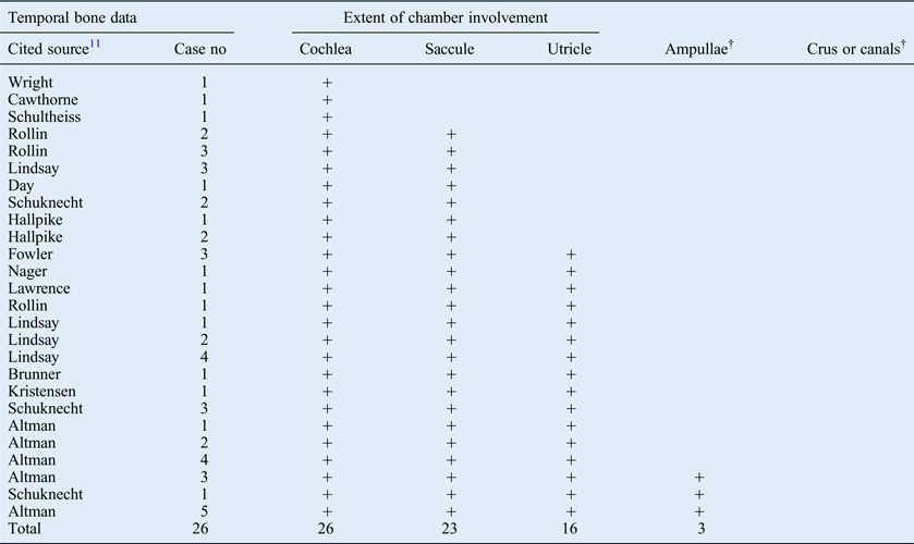
The Early Reports Collection consists of 26 temporal bone specimens displaying otopathology associated with Ménière's disease. Since these specimens were originally described in separate reports, data was available on a case-by-case basis and described the extent to which each chamber was affected in each specimen. All reports included the status of the cochlea and saccule, most reported the status of the utricle, but only a few reported the status of the ampullae and semicircular canals. Early Reports Collection data are summarised in Table I. The case sequence reflects the extent of lesions and shows that they predominately affected the cochlea. In fact, the cochlea was affected in every case, and in three cases cochlear lesions were the only lesions. Lesion progression was clear and orderly: lesions were not scattershot and haphazard. There were no isolated lesions in any chamber beyond the cochlea. Thus, all specimens conformed to a chain of lesions paradigm, indicating a process starting in the cochlea and progressing to involve the saccule, the utricle and the ampullae, in that order.
Ménière's disease data from the early reports collection*

* Findings in 26 specimens with a history of Ménière's disease, ordered by disease extent. †Lesions in ampullae and canals were generally not reported, with the exception of those noted. no = number
The California Collection findings were based on 19 temporal bones exhibiting otopathology associated with Ménière's disease.Reference Fraysse, Alonso and House13 These specimens were from 16 individuals, implying 3 bilateral cases. Sampling of individual case reports was used to exemplify disease details. Tabulation of findings for every specimen was not provided. Lesions were of the dilation and herniation types, with no ruptures reported, suggesting that California Collection cases were generally of a less severe variety. However, cases with saccular collapse might be presumed to involve rupture. Any rupture in the vestibular membrane, if present, would have had to be less than 200 µm in diameter (i.e. the distance between stained sections) to escape detection. All 19 specimens exhibited dilation of the cochlear duct, 17 specimens exhibited saccular dilation or collapse and 5 specimens showed utricular dilation. The lesion status of the ampullae and semicircular canals was not reported. These data are presented in Table II.
Ménière's disease data from the california collection*
* Findings in 19 specimens with a history of Ménière's disease, aggregated and ordered by disease extent. †Not reported.
Details of disease extent were presented for the cochlear duct and the saccule. In the cochlea, dilation of the vestibular membrane in the apical turn was present in all 19 specimens: 16 involved lower turns, 8 involved the hook region and 3 involved the vestibular cecum. In the saccule, 17 specimens exhibited pathology: 4 showed limited dilation without contacting nearby structures, 10 were sufficiently dilated to be in contact with the stapes footplate, and 3 showed saccular collapse. Utricular dilation was present in five specimens and noted to be mild.
An apparent link between lesion progression in the cochlear duct and the saccule was also reported. In three cases, in which dilation was limited to the apical turn, there was no alteration in the saccule. However, when the entire cochlear duct was dilated, saccular dilation extended to the footplate. In bilateral cases, the side with the greater cochlear duct dilation had the greater saccular dilation. No scattershot lesions were reported. Instead, an orderly and progressive chain of lesions was observed. This indicates a process that was most limited in the cochlear apex. Increasing dilation throughout the cochlear duct occurred in parallel with increasing saccular dilation.
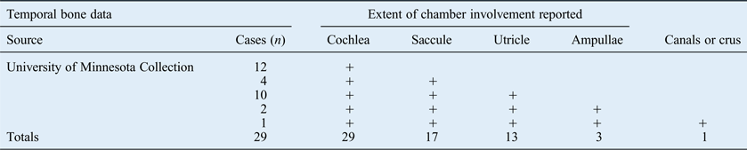
The Minnesota Collection findings are based on a total of 29 temporal bones exhibiting otopathology associated with Ménière's disease.Reference Sperling, Paparella, Yoon and Zelterman14 These were from 19 patients with a history of the disease, implying 10 bilateral cases. The report did not provide a bone-by-bone description of each specimen, but instead provided aggregate data (summarised in Table III). Lesions in the canal system were assumed to be ampullary, except in a single case specified as a crus rupture. However, the frequency with which the various chambers were affected, the frequency of ruptures exhibited, and a quantitative index for the degree of hydropic change in the cochlea were reported. It was noted that ruptures were more apt to occur in the superior labyrinth, while distension was more likely to occur in the inferior labyrinth.
Ménière's disease data from the minnesota collection*
* Temporal bone findings in 29 specimens with a history of Ménière's disease, aggregated and ordered by disease extent.
An index of hydropic severity in the cochlea was computed for each case based on the degree of bulging of Reissner's membrane averaged over the three turns: 0, normal configuration; 1, slight bulging without contact of the bony vestibular wall; 2, moderate bulging with bony contact but with an angle of less than 90° from the osseous spiral lamina; and 3, pronounced bulging of greater than 90°. Ruptures were associated with a cochlea severity index of 2.5, whereas distension without rupture was associated with a cochlea severity index of 1.8. This suggests that the degree of distension in the cochlear duct can predict disease severity in the other parts of the membranous labyrinth.
No cases of isolated lesions in chambers other than the cochlear apex were reported, all lower turn lesions were accompanied by lesions in the cochlear apex and all lesions in the vestibule were accompanied by lesions in the cochlear duct. Thus, no scattershot or haphazard lesions were encountered. Rather, an orderly chain of lesions was observed: the cochlear apex was the most frequently affected area, with decreasing involvement of saccule, utricle, ampullae and canals, in that order.
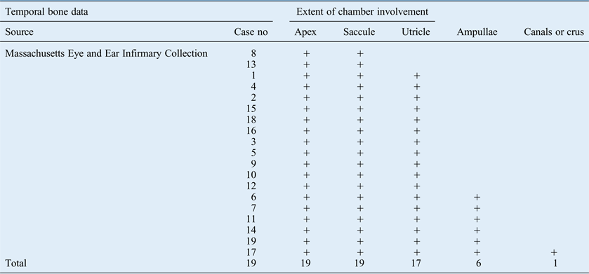
Massachusetts Collection findings were based on 19 temporal bones exhibiting otopathology associated with Ménière's disease.Reference Antoli-Candela9 Lesion data were reported in graphic detail and a case-by-case analysis of all 19 specimens was provided. The lesion type and disease extent for the entire labyrinth, including the ampullae, the crus commune and all canals, were described (summarised in Table IV). Ruptures were most apt to occur at points of maximal distension in Reissner's membrane and the saccule. Saccule rupture occurred in both cases with severe distension, in 8 out of 10 cases with moderate distension and in only 1 out of 7 cases of mild distension. The reinforced area of the saccule was generally neither displaced nor ruptured.Reference Perlman15 Healed blebs were generally found on the superior surface of the utricle and in the ampullae, where the capsular bone was in close proximity and may have facilitated healing. Open ruptures were generally found on the inferior surface of the utricle facing the open space of the perilymph cistern, as well as in the saccule and vestibular membranes. Chamber lesion involvement is summarised in Table IV (the order of the original cases was rearranged according to disease extent). In the Massachusetts Collection data, there were no specimens with disease limited to the cochlear apex, suggesting that the overall degree of pathology was greater in this group of specimens. All specimens had lesions of the cochlea and saccule, but only two specimens had lesions confined to these chambers. All other specimens showed increasing disease extent, with the next 11 specimens showing disease extending only to the utricle, the succeeding 5 had disease extending only to the ampullae and a final single case had disease extending to the canal system.
Ménière's disease data from the massachusetts collection*
* Findings in 19 specimens with a history of Ménière's disease, ordered according to disease extent. no = number
These cases illustrate that the cochlea and saccule are affected in every case: the least extensive disease is limited to these structures in the inferior labyrinth. Successive levels of more extensive disease first involve the utricle, then the ampullae and eventually the canal system. No lesions were found in any non-adjacent chambers, implying that scattershot haphazard lesions do not exist. Thus, all specimens conformed to an orderly chain of lesions paradigm, indicating a process that starts in the inferior labyrinth, and progressively involves the utricle, ampullae and canals.
The Minnesota Collection findings also included 13 temporal bone specimens demonstrating endolymphatic hydrops from asymptomatic patients with no otological history of Ménière's disease (data summarised in Table V).Reference Sperling, Paparella, Yoon and Zelterman14 Asymptomatic cases had an average cochlea severity index of 0.6, indicating very low overall distension in the cochlear duct. Two cases involved the saccule, and none involved the utricle or ampullae. (A single rupture in the canal system in a non-hydropic area may represent an artefact.) These data are consistent with mild disease that predominantly affects the cochlear apex, with very limited involvement of the saccule. Findings in asymptomatic cases are consistent with an early phase of endolymphatic hydrops, since there must necessarily be an initial symptom-free phase to a progressive disease process.Reference Machiavelli16
Asymptomatic hydrops data from the minnesota collection*
* Findings in 13 specimens with no history of Ménière's disease, aggregated and ordered according to disease extent.
The Massachusetts Collection included 78 specimens from patients without a history of Ménière's disease whose temporal bones displayed endolymphatic hydrops in the cochlear apex only.Reference Yamashita and Schuknecht10 These temporal bones were from patients with presbycusis, otosclerosis and otitis media, but not clinical Ménière's disease. Of the 78 cases, 24 specimens demonstrated mild dilation of Reissner's membrane, and 47 demonstrated moderate dilation. Only seven cases demonstrated pronounced distension of the apical turn with no distension in other turns (data summarised in Table VI). Pathology limited to the cochlear apex with no history of symptoms was construed to be a static condition.Reference Schuknecht and Gulya8
Asymptomatic hydrops data from the massachusetts collection*
* Findings in 78 specimens with no history of Ménière's disease, aggregated and ordered according to disease extent. †Cases selected on the basis disease limited to the cochlear apex.
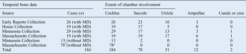
The combined collections provide an overview of the distribution of lesions encountered in endolymphatic hydrops. Of a total of 184 temporal bone specimens, 93 were from patients with a history of Ménière's disease and 91 were from patients without such history (combined data presented in Table VII). There were no scattershot lesions and the cochlear apex was affected in all cases.
Combined data from all temporal bone collections*
* Combined collections temporal bone findings in 93 specimens with a history of Ménière's disease and 91 specimens with no such history, aggregated and ordered according to disease extent. †Cases were selected on the basis of disease limited to the cochlear apex. MD = Ménière's disease
Discussion
Individual temporal bone specimens can document the accumulated effects of disease but cannot provide insight into the disease evolution, provide a timeline for lesion generation, help determine whether lesions appear simultaneously or in a sequential manner, or reveal whether lesions are distributed randomly or in an orderly pattern. However, a collection of temporal bones from Ménière's disease patients, acquired randomly and in sufficient numbers, ought to provide an overview of disease evolution and may thus indicate its overall trajectory.
In this regard, the various collections cited herein complement one another and present a more comprehensive view of this disease than might be gained from any single collection. Most early reports do not mention the status of the ampullae, crus commune or canals, leaving some question of just how extensive the disease might be. The Minnesota Collection specimens indicate that a lesser degree of cochlear damage is predictive of dilational lesions in the vestibule, while pronounced disease in the cochlear duct indicates a more severe disease characterised by ruptures. The preponderance of dilational lesions in the House Collection suggests that its specimens generally represent earlier phases of the disease process, whereas the prominent ruptures extending into the canal system in the Massachusetts Collection suggest that its cases represent later phases of disease. Data from the asymptomatic cases suggests that they represent very early disease that may not have yet reached a clinical threshold. Thus, each collection gives a somewhat different perspective on the otopathology associated with Ménière's disease, while the perspective offered by the combination of all collections is truly panoramic and gives a more coherent and robust understanding of how the disease process operates.
Endolymphatic hydrops associated with symptomatic Ménière's disease was found to affect all parts of the labyrinth, including the cochlear duct, saccule, utricle, crus commune, ampullae and semicircular canals. Extensive lesion distribution data from this meta-analysis of 93 symptomatic cases of Ménière's disease otopathology are presented in Figure 1. Lesions are not scattershot, but are instead distributed in an orderly pattern. Lesion data appear remarkably consistent across all temporal bone specimens reported.
Lesion distribution in 93 cases of endolymphatic hydrops in Ménière's disease
The 91 asymptomatic cases involved distension of Reissner's membrane, primarily in the apex of the cochlear duct and generally of mild-to-moderate degree. This suggests that substantial numbers of possibly pre-symptomatic endolymphatic hydrops cases never progress beyond a mild asymptomatic stage. There were only a few cases in which apical distension was pronounced (7/78, or 9 per cent, in one series and 1/6 or 17 per cent in the other) but not associated with rupture or known symptoms.Reference Yamashita and Schuknecht10, Reference Sperling, Paparella, Yoon and Zelterman14 These few may have been on the verge of showing symptoms since there obviously has to be a pre-symptomatic state in a progressive pathology. Certainly, such a spectrum was observed in clinical cases. In fact, a severity rating of cochlear disease based on the degree of cochlear duct distension correlated with the degree and character of disease in the rest of the labyrinth. The average severity rating was 0.6 for mild cases without symptoms, 1.8 for cases with dilation and 2.5 for cases with rupture.
• A meta-analysis of 184 endolymphatic hydrops cases revealed that lesions are not randomly distributed
• Lesions appear in an orderly sequence, beginning in the cochlear apex and then involving the saccule, utricle, ampullae and canals
• An otopathological staging system in endolymphatic hydrops is proposed based on disease extent
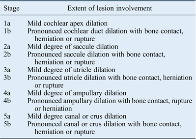
The orderly pattern of lesions further indicates that the disease proceeds in stages beginning with a mild degree of distension in the cochlear apex, progressing serially through adjacent chambers with more pronounced degrees of membrane distension, herniation and rupture, and eventually reaching the canal system (summarised in Table VIII). Such a staging approach offers a clearer appreciation of the chain of lesion paradigm operant in endolymphatic hydrops and can standardise pathological reporting, especially in gauging the success of therapeutic measures. It also provides foundational support for the proposed radiological grading system based on magnetic resonance imaging of the endolymphatic space.Reference Nakashima, Naganawa and Pyykko17
Proposed otopathology staging system for endolymphatic hydrops
The origin of this lesion is uncertain, but some speculation seems in order. The driving mechanism appears to be hydraulic. Potential aetiologies include barotraumas,Reference Antonelli, Parell, Becker and Paparella18 dark cell secretion,Reference Kimura19 aquaporin activity in the cochlear apex,Reference Eckhard, Gleiser, Arnold, Rask-Andersen, Kumagami and Muller20 resorptive defects in the endolymphatic sacReference Guild21 and obstructive phenomena in the endolymphatic duct.Reference Becvarovski and Zappia22 Membrane resistance to hydraulic pressure probably depends on structural characteristics such as configuration, composition and inherent strength.
The initial physiological interaction between endolymphatic pressure and membrane resistance is likely to be an elastic one that results in reversible dilation in all chambers. However, as intensity mounts, plastic deformation appears to start in the apical portion of Reissner's membrane, while the saccule, utricle, ampullae and canal are still operating within their elastic limits. As pressure rises further, these same chambers appear to undergo sequential permanent deformation. Early pathology appears to be limited to modest plastic dilation in a given chamber, which then can progress to bone contact, herniation and rupture if the hydraulic process continues. The overall process then appears to result in irreversible membrane deformity. This would be the simplest explanation for the various gradations encountered in each chamber, as well as the sequential spread to the other chambers of the membranous labyrinth.
Conclusion
The findings herein indicate that otopathology associated with Ménière's disease occurs in a predictable manner. The lesion pattern suggests a graded interaction between the hydropic process and membrane resistance. The disease stage, gauged by the extent to which the membranous chambers are affected in a serial manner, appears to reflect the intensity of the driving hydropic process.
These considerations suggest that the concept of this disease should be expanded from one based solely on hydropic considerations to one that encompasses a graded membrane resistance. Only differential membrane vulnerability can account for the orderly pattern of lesions in the face of an otherwise stochastic process such as hydrops. This perspective offers a new way to think about this disease and its remediation.