Introduction
Conduct disorder (CD) is diagnosed in children and adolescents who display a pattern of behaviour in which societal rules and the rights of others are violated (American Psychiatric Association, 2013). Although the lifetime prevalence of CD is higher amongst males than females (by a ratio of approximately 2.4:1), it is increasingly prevalent in adolescent females. Individuals with CD have poor prognoses with negative adult outcomes that include criminality, alcohol abuse, unemployment and poor mental and physical health. CD is one of the main reasons for referral to child and adolescent mental health services, and places a high burden on the affected individuals, families and society in general. Therefore, CD can be considered a major mental and public health priority and gaining a better understanding of its neurodevelopmental underpinnings is critical.
It has been proposed that limbic system dysfunction may underlie antisocial behaviour. Brain regions that make up the limbic system include the anterior cingulate cortex (ACC) and posterior cingulate cortex (PCC), orbitofrontal cortex, ventromedial prefrontal cortex (vmPFC), hippocampus, hypothalamus, amygdala and medial temporal lobe (Rolls, Reference Rolls2004, Reference Rolls2015). The limbic system is involved in emotion processing and regulation, reward-related decision-making and a range of other cognitive functions (Blair, Reference Blair2008). Evidence implicating limbic brain structures in antisocial behaviour comes from a number of sources. Two structural magnetic resonance imaging (MRI) meta-analyses concluded that the most robust abnormalities in grey matter volumes observed in this population are in limbic brain structures, such as the amygdala, ACC and vmPFC (Aoki et al., Reference Aoki, Inokuchi, Nakao and Yamasue2014; Raschle et al., Reference Raschle, Menks, Fehlbaum, Tshomba and Stadler2015; Rogers and De Brito, Reference Rogers and De Brito2016). In line with this, a recent meta-analysis of fMRI studies reported that CD individuals consistently displayed underactivation in the ACC and vmPFC during tasks involving emotion processing, and ‘hot’ (motivationally relevant) executive functions, and in dorsolateral prefrontal cortex (dlPFC), dorsal ACC and hippocampus during ‘cool’ (non-affective) executive function tasks (Alegria et al., Reference Alegria, Radua and Rubia2016).
Given this evidence for structural alterations and abnormal neural activity in limbic regions in individuals with CD, it is possible that the structural connections linking these regions are also compromised. The major limbic system white matter (WM) pathways include the fornix, the cingulum and the uncinate fasciculus (UF; Catani et al., Reference Catani, Dell’ Acqua and Thiebaut De Schotten2013). Structural connectivity and the micro-structural properties of brain tissue are frequently assessed using diffusion tensor imaging (DTI) techniques (Catani and Thiebaut de Schotten, Reference Catani and Thiebaut De Schotten2012). However, previous DTI studies in youths with CD and related disorders have had several limitations. First, the majority of DTI studies in CD have focused on males (although see Menks et al., Reference Menks, Furger, Lenz, Fehlbaum, Stadler and Raschle2017). Therefore, possible sex differences in the microstructural integrity of limbic system-related tracts have not been investigated. This is important as the neurobiological basis of CD has been shown to differ in several respects between males and females (Fairchild et al., Reference Fairchild, Hagan, Walsh, Passamonti, Calder and Goodyer2013; Decety et al., Reference Decety, Yoder and Lahey2015; Smaragdi et al., Reference Smaragdi, Cornwell, Toschi, Riccelli, Gonzalez-Madruga, Wells and Fairchild2017). Only one small study directly compared males and females with CD (n = 14 and 13, respectively) in terms of WM microstructure (Zhang et al., Reference Zhang, Gao, Shi, Huang, Wang, Situ and Yao2014). It investigated fractional anisotropy (FA) values of the UF using deterministic tractography (which investigates specific anatomical pathways). Interestingly, the authors found higher FA values in the UF in males, but not females, with CD. These preliminary findings suggest that WM microstructural alterations in temporo-frontal regions might be specific to males with CD.
Additionally, aside from one very recent study (Sethi et al., Reference Sethi, Sarkar, Dell'Acqua, Viding, Catani, Murphy and Craig2018), previous studies using DTI-based tractography methods in individuals with CD have largely focused on the UF tract (Passamonti et al., Reference Passamonti, Fairchild, Fornito, Goodyer, Nimmo-Smith, Hagan and Calder2012; Sarkar et al., Reference Sarkar, Craig, Catani, Dell ’Acqua, Fahy, Deeley and Murphy2013; Zhang et al., Reference Zhang, Gao, Shi, Huang, Wang, Situ and Yao2014). The fact that these studies focused on this tract may have been due to earlier studies in adults with antisocial personality disorder (ASPD; an adult condition of which CD is an antecedent) and psychopathy, finding lower FA in the UF in these individuals compared with healthy controls (Craig et al., Reference Craig, Catani, Deeley, Latham, Daly, Kanaan and Murphy2009). However, opposite findings have been reported in youths with CD, who show higher FA values in the UF relative to healthy controls (Passamonti et al., Reference Passamonti, Fairchild, Fornito, Goodyer, Nimmo-Smith, Hagan and Calder2012; Sarkar et al., Reference Sarkar, Craig, Catani, Dell ’Acqua, Fahy, Deeley and Murphy2013; Zhang et al., Reference Zhang, Gao, Shi, Huang, Wang, Situ and Yao2014). Similarly, a study investigating the dorsal and ventral components of the cingulum tract reported lower radial diffusivity (RD) in the dorsal cingulum bundle in individuals with CD compared with controls (Sethi et al., Reference Sethi, Sarkar, Dell'Acqua, Viding, Catani, Murphy and Craig2018). This was opposite to the pattern observed in adults with ASPD (Sethi et al., Reference Sethi, Gregory, Dell'Acqua, Periche Thomas, Simmons, Murphy and Craig2015). Several authors have suggested that the opposite patterns observed in WM microstructural measures in youths and adults might be due to abnormally accelerated maturation of WM tracts in individuals with CD (Passamonti et al., Reference Passamonti, Fairchild, Fornito, Goodyer, Nimmo-Smith, Hagan and Calder2012; Sarkar et al., Reference Sarkar, Craig, Catani, Dell ’Acqua, Fahy, Deeley and Murphy2013; Zhang et al., Reference Zhang, Gao, Shi, Huang, Wang, Situ and Yao2014). Although these previous studies were important first steps in understanding CD-related alterations in structural connectivity, the focus of research needs to be expanded to consider additional limbic system tracts. It is also important to test whether alterations in WM microstructure are common or distinct across males and females with CD.
An additional limitation of prior studies is that they used either tractography methods or a characterisation of WM diffusivity exclusively based on the diffusion tensor model. Although the diffusion tensor model is most frequently used to reconstruct WM tracts and characterise diffusivity in WM (Basser et al., Reference Basser, Pajevic, Pierpaoli, Duda and Aldroubi2000), this approach has several limitations. First, it is not well-suited for studying complex fibre configurations such as crossing fibres, branching regions or intra-voxel combinations of different tissue types (e.g. WM fibres and grey matter). Second, while FA is the most commonly used index to quantify water diffusivity in studies using tensor-based models, it is calculated at a voxel level and is determined by several microstructural and macrostructural features, such as myelination of WM fibres, size and packing density of cells and number of crossing fibres (Vanderauwera et al., Reference Vanderauwera, Vandermosten, Dell'Acqua, Wouters and Ghesquière2015). Thus, partial volume effects (i.e. not fibre- or tissue-specific) can affect DTI indices (e.g. FA, RD), and voxel-average diffusion MRI parameters such as FA, lack within-voxel single-fibre population specificity (Dell'Acqua et al., Reference Dell'Acqua, Simmons, Williams and Catani2013; Raffelt et al., Reference Raffelt, Smith, Ridgway, Tournier, Vaughan, Rose and Connelly2015). Novel non-tensor models such as constrained spherical deconvolution (SD) have the potential to overcome these limitations and more accurately characterise the underlying architecture of specific WM tracts (Dell'Acqua et al., Reference Dell'Acqua, Scifo, Rizzo, Catani, Simmons, Scotti and Fazio2010). In addition, the hindrance-modulated orientational anisotropy (HMOA) index that can be derived using SD algorithms provides greater sensitivity in terms of detecting microstructural changes in specific WM tracts than FA (Dell'Acqua et al., Reference Dell'Acqua, Simmons, Williams and Catani2013). Finally, most previous DTI studies included relatively small samples – typically groups of 15 participants or fewer (Finger et al., Reference Finger, Marsh, Blair, Majestic, Evangelou, Gupta and Blair2012; Passamonti et al., Reference Passamonti, Fairchild, Fornito, Goodyer, Nimmo-Smith, Hagan and Calder2012; Haney-Caron et al., Reference Haney-Caron, Caprihan and Stevens2014; Zhang et al., Reference Zhang, Gao, Shi, Huang, Wang, Situ and Yao2014; Sethi et al., Reference Sethi, Sarkar, Dell'Acqua, Viding, Catani, Murphy and Craig2018).
The present study addresses a number of these limitations, and extends previous findings by, first, examining sex differences in the relationship between CD and WM microstructure. Second, by examining two key limbic WM tracts overlooked in prior studies: the fornix and the cingulum bundles – the retrosplenial (RSC), parahippocampal (PHC) and subgenual cingulum (SGC; Jones et al., Reference Jones, Christiansen, Chapman and Aggleton2013) – as tracts plausibly involved in the pathophysiology of CD. Third, by enhancing statistical power and the robustness of our results by substantially increasing the sample size compared with previous studies. Finally, by employing a novel method – constrained SD. Recent studies have compared tensor v. non-tensor models in clinical samples and suggested that the latter approach provides more accurate and robust results (Auriat et al., Reference Auriat, Borich, Snow, Wadden and Boyd2015). However, to increase comparability with previous studies, we also estimated indices of FA – the most widely used parameter in previous structural connectivity research.
We hypothesised that differences between CD and control groups would be most evident in limbic tracts involved in socio-emotional processes (i.e. SGC, RSC and UF) in comparison with posterior and lateral limbic WM tracts (e.g. PHC). We also hypothesised that CD-related alterations in WM microstructure would be most evident in males (Zhang et al., Reference Zhang, Gao, Shi, Huang, Wang, Situ and Yao2014). In addition, recent DTI studies have shown that individuals with CD and elevated callous-unemotional (CU) traits may differ from those with low levels of CU traits in terms of WM microstructural abnormalities (Sethi et al., Reference Sethi, Sarkar, Dell'Acqua, Viding, Catani, Murphy and Craig2018; Puzzo et al., Reference Puzzo, Seunarine, Sully, Darekar, Clark, Sonuga-Barke and Fairchild2017). Thus, we also investigated whether CU traits contributed to the WM microstructural alterations observed in CD. We also tested for correlations between WM measures and the grandiose-manipulative and impulsive-irresponsible subdimensions of psychopathy and CD symptoms.
Methods
Participants
Participants for this study were recruited at four different sites involved in the Neurobiology and Treatment of Female Conduct Disorder (FemNAT-CD; http://www.femnat-cd.eu) study – University of Southampton, University of Birmingham, University Hospital Aachen and University of Basel. All participants and the majority of their parents underwent a diagnostic interview that was based on DSM-IV criteria (the Kiddie-Schedule for Affective Disorders and Schizophrenia-Present and Lifetime; Kaufman et al., Reference Kaufman, Birmaher, Perel, Dahl, Moreci, Nelson and Ryan1997). At the UK sites, IQ was assessed using the two subtest forms of the Wechsler Abbreviated Scale of Intelligence (Wechsler, Reference Wechsler1999), whereas the German version of the Wechsler Intelligence Scale for Children (Wechsler, Reference Wechsler2003) was used at the other sites. The t and standard scores from all sites were transformed into z-scores and then combined to yield estimates of full-scale IQ.
The Match program (Van Casteren and Davis, Reference Van Casteren and Davis2007) was used to select an IQ-, age- and gender-matched sample from the subset of participants for whom diffusion MRI data were available (n = 325). There are dramatic changes in WM development across childhood and adolescence (Casey et al., Reference Casey, Jones and Hare2008), thus we excluded children aged 9–12 years (n = 57; see online Supplementary Material for more information). A total sample of 200 adolescents [101 with CD (52 females) and 99 healthy controls (50 females)] was included in the present analyses – all aged 13–18 years. The Youth Psychopathic traits Inventory (YPI; Andershed et al., Reference Andershed, Kerr, Stattin and Levander2002); a self-report questionnaire assessing overall psychopathic traits and sub-dimensions of psychopathy, and the parent-report Inventory of Callous Unemotional traits (ICU; Essau et al., Reference Essau, Sasagawa and Frick2006); a standardised measure including callous, uncaring and unemotional subscales, were used to assess psychopathic and CU traits, respectively.
Diffusion-weighted MRI data acquisition
Diffusion-weighted MRI data were acquired with the following parameters: repetition time (TR) = 8000 ms (Achieva), 8800 ms (Tim-Trio), 7500 ms (Prisma); echo-time (TE) = 87 ms (Achieva), 92 ms (Tim-Trio), 71 ms (Prisma) and a bandwidth of 1633.3 Hz/Px (Achieva) or 1776 Hz/Px (Tim-Trio & Prisma); echo-spacing = 0.75 ms (Achieva), 0.73 ms (Tim-Trio), 0.65 ms (Prisma); slice thickness = 2.0 mm; field of view (FOV) = 256 × 256 × 124 mm; acquisition matrix = 128 × 128; voxel-size = 2 × 2 × 2 mm; 62 contiguous axial slices (no slice gap). Images were acquired with diffusion gradients (b-value = 1500 s/mm2) applied in 64 non-coplanar and non-collinear directions and two b-value = 0 (s/mm2) volumes with reversed phase encoding (blip-up/blip-down), yielding pairs of images with distortions in opposite phase-encode directions to enable accurate estimation and correction for susceptibility-induced distortions.
Pre-processing
Datasets were corrected for head motion and eddy current distortions using FSL (Andersson and Sotiropoulos, Reference Andersson and Sotiropoulos2016). Distortions in the magnetic field were estimated. The estimated field was subsequently used, together with all the diffusion data, to estimate eddy current-induced distortions and subject movement (Andersson and Sotiropoulos, Reference Andersson and Sotiropoulos2016). Movement-induced signal dropout was identified and the lost signal was replaced by a non-parametric Q-space interpolation (Andersson et al., Reference Andersson, Graham, Zsoldos and Sotiropoulos2016).
Spherical deconvolution was calculated using the damped Richardson–Lucy algorithm (Dell'Acqua et al., Reference Dell'Acqua, Scifo, Rizzo, Catani, Simmons, Scotti and Fazio2010) with a fibre response parameter of α = 1.5, 400 algorithm iterations, threshold parameters of 0.06 and a harmonic order of 8 (lmax = 8). An absolute (0.1%) and relative (5%) threshold on the Fibre Orientation Distribution (FOD) amplitude were applied to exclude spurious local connections (Dell'Acqua et al., Reference Dell'Acqua, Simmons, Williams and Catani2013).
Whole brain deterministic tractography was performed using a step size of 0.5 mm with a limit set to display streamlines between 20 and 400 mm. The Euler algorithm was used to follow the orientation vector of least curvature (angle threshold of 45°). Spherical deconvolution and tractography analysis was performed using StarTrack software (https://www.mr-startrack.com/). Explore DTI was used for the tensor fit. Tensor-derived FA and HMOA values of WM microstructural organisation were exported to TrackVis. We report FA values in the present study for the purpose of increasing comparability with previous studies.
Delineation of regions of interest
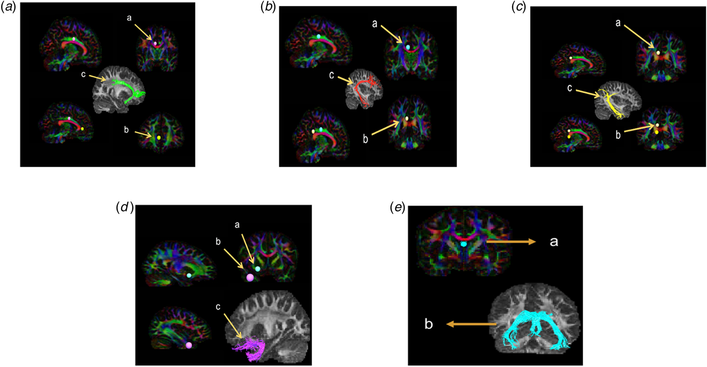
TrackVis was used to reconstruct the fornix, cingulum bundle subdivisions and UF. Reconstruction of these tracts has previously been described [fornix and UF: (Stieltjes et al., Reference Stieltjes, Brunner, Fritzsche and Laun2013), and CB subdivisions: (Jones et al., Reference Jones, Christiansen, Chapman and Aggleton2013)]. The Boolean logic (AND, and NOT gates) was employed to delineate the CB's subdivisions, the fornix and the UF (Fig. 1 shows the reconstruction for all of the limbic WM tracts).
Tractography reconstruction for all limbic white matter tracts. (A) Coronal tractography reconstruction of the subgenual cingulum (SGC): (a) First sphere of interest placed five slices anterior to the mid-sagittal view. (b) Target sphere placed three slices anterior to the genu of the corpus callosum. (c) Final reconstruction of the SGC. (B) Coronal tractography reconstruction of the retrosplenial cingulum (RSC): (a) First sphere of interest placed five slices posterior to the mid-sagittal view. (b) Target sphere placed 3–4 slices above the splenium. (c) Final reconstruction of the RSC. (C) Coronal tractography reconstruction of the parahippocampal cingulum (PHC). (a) First sphere of interest placed behind the splenium. (b) Target sphere placed 3–4 slices below sphere A. (c) Final reconstruction of the PHC. (D) Coronal tractography reconstruction of the uncinate fasciculus (UF): (a) First sphere of interest placed in the fronto-temporal junction. (b) Target sphere placed at the temporal pole. (c) Final reconstruction of the UF. (E) Coronal tractography reconstruction of the fornix. (a) One coronal sphere placed in the body of the fornix. (b) Final reconstruction of the fornix.

Statistical analysis
Matlab_R2016 was used to carry out statistical analysis. Shapiro–Wilk tests were used to verify normality of HMOA and FA values across subjects. Tract measures of HMOA and FA were analysed using a general linear model (GLM) to test for effects of diagnosis and sex-by-diagnosis interactions. The GLM included the following covariates which have been shown to be associated with WM microstructural integrity in adolescent studies: age (Asato et al., Reference Asato, Terwilliger, Woo and Luna2010), IQ (Dunst et al., Reference Dunst, Benedek, Koschutnig, Jauk and Neubauer2014) and site (coded as binary fixed effect). Where significant sex-by-diagnosis interactions were found, we followed these up by comparing FA and HMOA values between CD and healthy control males, and between CD and healthy control females.
Attention-deficit/hyperactivity disorder (ADHD) is a neurodevelopmental disorder that frequently co-occurs with CD, and previous DTI studies have shown that comorbid ADHD strongly modulates WM effects (Wang et al., Reference Wang, Horst, Kronenberger, Hummer, Mosier, Kalnin and Mathews2012). Thus, we repeated the GLM analysis while adding current ADHD symptoms (i.e. those displayed in the last year) as an additional covariate.
In addition, there is some evidence for structural differences between the childhood-onset (CO) and adolescent-onset (AO) variants of CD (Fairchild et al., Reference Fairchild, Toschi, Hagan, Goodyer, Calder and Passamonti2015). Accordingly, we used the same model to compare these subgroups, to assess the validity of combining these subgroups in our main analysis.
The significance threshold was adjusted using the Benjamin–Hochberg false discovery rate (FDR: q < 0.05) correction for multiple comparisons across each parameter independently. Effect sizes for diagnosis effects were calculated using Cohen's d and effect sizes for sex-by-diagnosis interactions were expressed as partial η 2 (ηp2).
In cases where significant main effects of diagnosis were observed, we followed these up by running a GLM analysis (only in the CD group), to test for associations between CD symptoms, psychopathy (YPI total), dimensions of psychopathic traits (grandiose-manipulative, and impulsive-irresponsible traits), CU traits (YPI subscale and ICU total), ICU subscales (callousness, uncaring, unemotional) and measures of HMOA and FA. Lastly, given that the CD sample included many individuals with comorbid ADHD, we also explored the relationship between ADHD symptoms and measures of HMOA and FA. These correlational analyses were run in a mixed-sex CD group (males and females with CD) as well as in each sex separately.
Results
Demographic variables
Individuals in the CD group had significantly more CD, ODD and ADHD symptoms than healthy controls. They also scored higher in overall psychopathic traits, as well as CU, grandiose and manipulative, and impulsive and irresponsible subdimensions of psychopathy (Table 1). There were no significant differences between males and females in the age-of-onset of CD (i.e. childhood-onset v. adolescent-onset).
Demographic and clinical characteristics of the sample

Note: CD, Conduct disorder; HC, healthy controls; ADHD, attention-deficit/hyperactivity disorder; ODD, Oppositional defiant disorder; PTSD, post-traumatic stress disorder; MDD, major depressive disorder; YPI, Youth Psychopathic traits Inventory; ICU, Inventory of Callous Unemotional traits. #Number of traumatic events were estimated from the K-SADS-PL interview data.
In terms of psychiatric comorbidity in individuals with CD, males and females differed only in rates of substance abuse (M > F); there were no other significant differences between males and females. Finally, there was an unequal sample distribution across the sites (see online Supplementary Table S1). To address this issue, we included site as a covariate of no interest.
Tractography results
There were no significant differences between the CO-CD and AO-CD subtypes in HMOA or FA in any WM tract.
Main effects of diagnosis
Relative to controls, individuals with CD had lower HMOA in bilateral RSC [right: t (190) = −2.22, p = 0.03, d = 0.10; left: t (190) = −2.27, p = 0.02, d = 0.16], and lower FA in the right RSC [t (190) = −2.91, p = 0.004, d = 0.28]. However, after correcting for multiple comparisons, only the effect on right RSC FA remained significant (pFDR = 0.03; Figure 2). There were no significant group differences in HMOA or FA in any of the other limbic WM tracts.
Main effects of diagnosis on fractional anisotropy of the right retrosplenial cingulum. Group differences are significant at p < 0.05, false discovery rate correction. Error bars show 95% confidence intervals of the mean. HC, healthy control; CD, conduct disorder; FA, fractional anisotropy.

Sex-by-diagnosis interactions
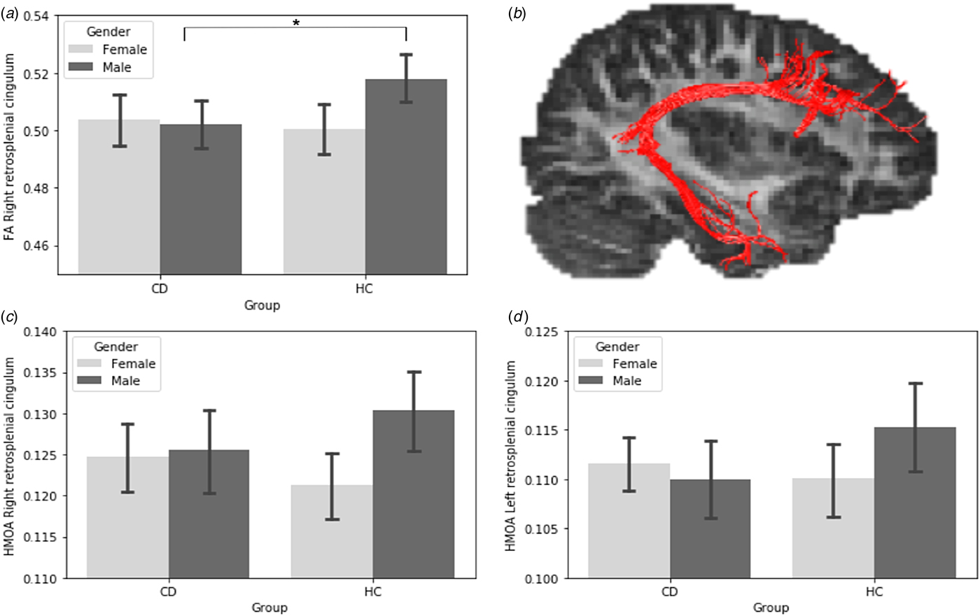
We observed sex-by-diagnosis interactions for HMOA in bilateral RSC [right: t (190) = 2.08, p = 0.04, ηp2 = 0.02: left: t (190) = 1.99, p = 0.05, ηp2 = 0.02], and FA in right RSC [t (190) = 2.75, p = 0.006, ηp2 = 0.04]. All interactions followed the same pattern: males with CD showed lower values than male controls, whereas females with CD showed higher values than female controls (Fig. 3). However, only the sex-by-diagnosis interaction for right RSC FA survived correction for multiple comparisons (pFDR = 0.05). No other significant interaction effects were found in the other limbic tracts (see online Supplementary Table S2). Post-hoc analysis showed that relative to male controls, CD males had lower HMOA in bilateral RSC [right: t (190) = −2.52, p = 0.04, d = 0.39; left: t (190) = −1.99, p = 0.04, d = 0.37] and lower FA in the right RSC [t (190) = −2.91, p = 0.01, d = 0.47]. However, after correcting for multiple comparisons, only the effect in right RSC FA remained significant (pFDR = 0.03). There were no significant differences between female CD and control groups.
Sex by diagnosis interactions in the retrosplenial cingulum tract. (a) Sex by diagnosis interaction on fractional anisotropy values in the right retrosplenial cingulum, *shows that the group difference is pFDR corrected < 0.05 whereas the other other group differences do not survive pFDR correction, which was significant at pFDR = 0.05. (b) Three-dimensional reconstruction of the retrosplenial cingulum tract. (c) Sex by diagnosis interactions in the Hindrance Modulated Orientational Anisotropy of the right retrosplenial cingulum. (d) Sex by diagnosis interactions in the Hindrance Modulated Orientational Anisotropy of the left retrosplenial cingulum. Error bars show 95% confidence intervals of the mean. HC, healthy control; CD, conduct disorder.

ADHD comorbidity as a potential confound
The main effects of diagnosis observed for FA in the right RSC (p = 0.03) and for HMOA in bilateral RSC (left: p = 0.05: right: p = 0.05) in CD v. healthy control males remained significant after factoring out current ADHD symptoms. However, only the group difference in the right RSC FA remained significant (pFDR = 0.03) after correcting for multiple comparisons. Moreover, significant main effects of diagnosis emerged in the right UF when factoring out ADHD symptoms: participants with CD showed lower FA [t (189) = 2.00, p = 0.05, pFDR = 0.05], and HMOA [t (189) = 2.07, p = 0.04, pFDR = 0.05; online Supplementary Fig. S1] relative to healthy controls. Unlike the findings for the RSC, this main effect of diagnosis in the UF was not qualified by a significant sex-by-diagnosis interaction.
Correlations between structural connectivity measures and CD symptoms, ADHD symptoms, and psychopathic or CU traits
Within the CD sample, there was a positive correlation between current CD symptoms and right RSC HMOA (r = 0.36, pFDR = 0.02; Fig. 4). There were no other significant correlations between CD, ADHD symptoms, overall psychopathic traits, the subdimensions of psychopathy, CU traits or the ICU subscales (Callousness, Uncaring, Unemotional) and measures of WM in other tracts.
Positive association between Conduct Disorder (CD) symptoms and hindrance modulated orientational anisotropy (HMOA) in the right retrosplenial cingulum in the CD group.

As effects of diagnosis were found in males, but not in females with CD, we conducted correlational analyses in male and female groups separately. A strong positive correlation between current CD symptoms (r = 0.45, pFDR = 0.002; online Supplementary Fig. S2), and a negative correlation between current ADHD symptoms (r = −0.31, pFDR = 0.03; online Supplementary Fig. S3) and right RSC HMOA was found in the male CD group. No effects of CU or psychopathic traits were observed in CD males. No significant correlations were found between clinical symptoms or CU/psychopathic traits or subscales and measures of structural connectivity in females with CD. Moreover, there were no significant sex-by-CD symptoms or sex-by-CU/psychopathic traits interactions for either HMOA or FA.
Discussion
Abnormalities in the limbic system have been consistently implicated in the pathophysiology of CD (Raschle et al., Reference Raschle, Menks, Fehlbaum, Tshomba and Stadler2015; Alegria et al., Reference Alegria, Radua and Rubia2016; Rogers and De Brito, Reference Rogers and De Brito2016). We extended the DTI literature by including female participants and a much larger sample than has been studied to date (N = 200). This allowed us to test whether females and males with CD show common or distinct alterations in limbic WM microstructure. We also investigated limbic WM tracts beyond the UF and capitalised on recent methodological advances in diffusion-weighted image processing by employing SD models. This approach provides a more reliable estimation of multiple fibres passing through a voxel with distinct orientations.
Our findings extend knowledge regarding alterations in limbic WM tracts in CD and support the hypothesis that abnormalities in fronto-limbic tracts are involved in the pathophysiology of this disorder. However, such abnormalities appear to be limited to males with CD – no such effects were found in females. More specifically, only males with CD showed lower FA in the right RSC relative to male controls. In fact, there was a suggestion that the opposite pattern was observed in females (females with CD appeared to show higher FA and HMOA values relative to control females) – although this was not statistically significant.
Previous DTI studies have found structural abnormalities in regions that overlap with the RSC. A recent study using a similar approach to the present study (i.e. region of interest-based tractography) investigated dorsal and ventral cingulum WM microstructure in male youths with CD (Sethi et al., Reference Sethi, Sarkar, Dell'Acqua, Viding, Catani, Murphy and Craig2018). Lower RD values were observed in bilateral dorsal cingulum in the CD group relative to controls (Sethi et al., Reference Sethi, Sarkar, Dell'Acqua, Viding, Catani, Murphy and Craig2018). FA values normally increase when RD decreases, and the opposite pattern seems associated with myelin loss and axonal abnormalities (Harsan et al., Reference Harsan, Poulet, Guignard, Steibel, Parizel, de Sousa and Ghandour2006). Although the anatomical delineation of the cingulum bundle differed between the two studies (i.e. dorsal and ventral in Sethi et al. v. RSC, PHC and SGC in the present study), the dorsal part of the cingulum overlaps most closely with the RSC tract compared with the other cingulum bundles – thus the findings are congruent in terms of location, but not in the direction of the effects. In addition, the Sethi et al. (Reference Sethi, Sarkar, Dell'Acqua, Viding, Catani, Murphy and Craig2018) study differs from the present study in the use of tensor-based models v. non-tensor models.
The RSC is composed of fibres that connect the medial prefrontal cortex, dlPFC, ACC, PCC, medial temporal lobe and angular gyrus together (Jones et al., Reference Jones, Christiansen, Chapman and Aggleton2013). These regions have been associated with social-emotional processing, self-reflection, executive functions and moral decision-making. They are key nodes of the default mode network (DMN) that is responsible for self-referential processing (Leech et al., Reference Leech, Braga and Sharp2012). Previous studies investigating DMN connectivity in youths with CD have reported reduced connectivity between core DMN regions including the medial PFC, PCC, precuneus and superior temporal gyrus, relative to controls (Zhou et al., Reference Zhou, Yao, Fairchild, Cao, Zhang, Xiang and Wang2016; Broulidakis et al., Reference Broulidakis, Fairchild, Sully, Blumensath, Darekar and Sonuga-Barke2016). It has been proposed that DMN dysfunction in CD may reflect delays in the development of brain circuits linked to self-awareness, regulating emotions, moral judgements and future planning (Zhou et al., Reference Zhou, Yao, Fairchild, Cao, Zhang, Xiang and Wang2016). Impairments in these processes have been reported in CD (e.g. White et al., Reference White, Clanton, Brislin, Meffert, Hwang, Sinclair and Blair2014). The RSC connects core regions that make up the DMN. Thus, the abnormal functional connectivity of the DMN observed in previous studies may have a structural basis in altered RSC connectivity.
Although group differences in FA in the UF only became significant after controlling for comorbid ADHD symptoms, our results are in contrast to findings reported by Zhang et al. (Reference Zhang, Gao, Shi, Huang, Wang, Situ and Yao2014). We did not observe any sex-by-diagnosis interactions in this WM tract. Both males and females with CD appeared to be equally affected in terms of showing lower UF FA. However, in line with Zhang et al. (Reference Zhang, Gao, Shi, Huang, Wang, Situ and Yao2014), we also observed sex differences in the RSC tract in youths with CD. Males with CD showed lower FA (and HMOA at an uncorrected level) relative to sex-matched healthy controls, whereas there were no significant differences between CD and control females.
Previous DTI studies of CD have observed higher FA values in male-only samples, suggesting accelerated maturation in individuals with CD. Here, we observed lower FA in males with CD compared with male controls. Although the results observed here were in a previously unstudied tract, it suggests that WM maturation is delayed in males with CD. Delayed maturation of WM is associated with poor inhibitory control (Simmonds et al., Reference Simmonds, Hallquist, Asato and Luna2014) – a key feature of CD.
Furthermore, our correlational analyses showed that CD symptoms were significantly (positively) correlated with HMOA of the right RSC tract in males, but not females. Therefore, the present study provides new evidence for sex differences in the neurobiological basis of CD – RSC WM abnormalities were observed in males but not females. We also observed a significant negative correlation between ADHD symptoms and HMOA in the right RSC tract in males but not females, indicating that ADHD comorbidity may have influenced the differences between CD and control males in the RSC. This is of significance due to the substantial overlap of ADHD and CD, and symptom dimensions related to ADHD such as impulsivity and hyperactivity have been associated with the development of antisocial behaviour in childhood (Barkley et al., Reference Barkley, Fischer, Smallish and Fletcher2004).
Several neuropsychological studies investigating aspects of executive functioning (i.e. assessing inhibition/attention and decision-making), are consistent with our findings by showing divergent results in males and females with CD. Males with CD exhibit deficits in reversal learning (Herpertz et al., Reference Herpertz, Huebner, Marx, Vloet, Fink, Stoecker and Herpertz-Dahlmann2008) and differ in terms of decision-making (e.g. making more risky choices) relative to control males, whereas CD females do not differ from control females (Sidlauskaite et al., Reference Sidlauskaite, González-Madruga, Smaragdi, Riccelli, Puzzo, Batchelor and Fairchild2018). In addition, our finding of a sex-by-diagnosis interaction in the RSC highlights the importance of taking sex into account when studying the neurobiology of CD, and the problems that might arise when combining males and females with CD in the same group (Smaragdi et al., Reference Smaragdi, Cornwell, Toschi, Riccelli, Gonzalez-Madruga, Wells and Fairchild2017). Future studies should investigate the functional consequences of altered RSC structural connectivity in males and females with CD by employing resting state functional connectivity methods, and by using neuropsychological tasks tapping decision-making and empathic processes in the same sample.
Strengths and limitations
The strengths of this study include the investigation of additional limbic WM tracts by using a more comprehensive approach – SD tractography. The main benefit of this approach is to resolve fibre-crossing issues. In addition, SD techniques improve the accuracy of fibre tracking compared with models based on the diffusion tensor alone (Dell'Acqua et al., Reference Dell'Acqua, Scifo, Rizzo, Catani, Simmons, Scotti and Fazio2010). Secondly, the comparatively large sample size in the present study (N = 200), which included males and females with and without CD, allowed us, for the first time, to comprehensively investigate sex differences in the relationship between CD and structural connectivity. Another strength is the fact that the CD group was assessed using standardised, semi-structured interviews based on DSM-IV criteria as well as obtaining detailed information about comorbid disorders and accounting for ADHD comorbidity in our statistical analyses.
However, our study also had several limitations. First, the sample ranged in age from 13 to 18 years. The CD and control groups did not differ in age; however, age is known to have an important effect on WM development. Thus, we included age as a covariate of no interest in all analyses. Second, the sex distribution across the sites was uneven (more girls were tested at some sites than others), and although quality control procedures were performed prior to starting data acquisition (e.g. matching acquisition parameters and going through a site qualification process), combining data from different sites and scanner manufacturers (Phillips and Siemens) may introduce unintended variability. However, to reduce the impact of this variability, all analyses included site as a covariate of no interest. Finally, although we used SD methods to reconstruct the WM tracts, indices of FA were derived from tensor-based models fitted to b = 1500 diffusion-weighted data and projected onto the SD-derived tracts. Hence there is a potential source of variability in terms of comparing the present FA measures with those reported in previous studies, although several earlier studies adopted a similar approach (e.g. Christiansen et al., Reference Christiansen, Aggleton, Parker, O'Sullivan, Vann and Metzler-Baddeley2016; Rojkova et al., Reference Rojkova, Volle, Urbanski, Humbert, Dell'Acqua and Thiebaut de Schotten2016).
In conclusion, we found that male adolescents with CD differed from healthy controls in RSC WM microstructure – showing lower FA and HMOA values in this tract. This effect was not seen in females with CD. These differences in structural connectivity may help explain sex differences in CD and its clinical presentation. Given the overlap of the RSC tract with brain regions that constitute the DMN, and its role in connecting these regions together, future studies should investigate whether there are sex differences in DMN functional connectivity in CD. This would improve our understanding of the pathophysiology of CD and could lead to improved diagnosis and treatments for both sexes.
Supplementary material
The supplementary material for this article can be found at https://doi.org/10.1017/S0033291718003951.
Author ORCIDs
Karen González-Madruga 0000-0002-7586-7536
Acknowledgments
We sincerely thank our participants and their families for taking part in this study. We also thank Dr Flavio Dell'Acqua for his guidance and advice in this study, and Dr Etta Howells for providing training in white matter tract dissection. Karen Gonzalez-Madruga was partly funded by a Ph.D. studentship from the National Council of Science and Technology (CONACYT), Mexico.
Financial support
This study was funded by the European Commission's Seventh Framework Programme for research, technological development and demonstration (FP7/ 2007–2013) under Grant Agreement no. 602407 (FemNAT-CD; coordinator: Professor Christine Freitag, Goethe University).
Conflict of interest
Professor Freitag receives royalties for books on attention-deficit/hyperactivity disorder and autism spectrum disorder. She has served as consultant to Desitin and Roche. Professor Sonuga-Barke has received speaker fees, consultancy, research funding and conference support from Shire Pharma. Speaker fees from Janssen Cilag, consultancy from Neurotech solutions, Aarhus University, Copenhagen University and Berhanderling, Skolerne, Copenhagen, KU Leuven. Book royalties from OUP and Jessica Kingsle. Edmund Sonuga-Barke has been awarded grants from the MRC, ESRC, Wellcome Trust, Solent NHS Trust, European Commission, Child Health Research Foundation New Zealand, NIHR, Nuffield Foundation, Fonds Wetenschappelijk Onderzoek-Vlan-deren (FWO) and MQ – Transforming Mental Health. Dr Fairchild has received funding from the European Commission, the UK Medical Research Council, the National Council for Science and Technology (CONACYT), the UK Economic and Social Research Council and Kids’ Company. Professor Konrad has received speaker fees from Shire Pharmaceuticals and Medice. Professor Stadler receives royalties for a book on aggression. Dr De Brito has received speaker fees from the Child Mental Health Centre and the Centre for Integrated Molecular Brain Imaging. All other co-authors declare no potential conflicts of interest.
Ethical standards
The authors assert that all procedures contributing to this work comply with the ethical standards of the relevant national and institutional committees on human experimentation and with the Helsinki Declaration of 1975, as revised in 2008.