Introduction
Seminal plasma free amino acids play an important role in semen quality and quantity. Free amino acids may serve as oxidizable substrates for aerobic metabolism by spermatozoa (Mann, Reference Mann1964); create a favorable condition for nucleic acids synthesis (Setchell et al., Reference Setchell, Hinks, Voglmayr and Scott1967); or enhance sperm survival (Tyler and Rothschild, Reference Tyler and Rothschild1951). Additionally, amino acids have been implied to play a role in progressive spermatozoal motility (Gassner and Hopwood, Reference Gassner and Hopwood1952), where the addition of any one of a number of amino acids and peptides to spermatozoa extends their duration of motility (Tyler and Tanabe, Reference Tyler and Tanabe1952). Furthermore, quantities of amino acids present in seminal plasma appear to be related to sperm concentration (Johnson et al., Reference Johnson, Pursel, Gerrits and Thomas1972).
Additionally, fatty acids play a major role in the metabolism of spermatozoa. The endogenous respiration of these cells depends largely on the oxidation of hydrolyzed acyl groups of plasmalogens (Hartree and Mann, Reference Hartree and Mann1961); endogenous fatty acids may also be utilized and are readily incorporated into the lipids of spermatozoa (Payne and Masters, Reference Payne and Masters1968; Mills and Scott, Reference Mills and Scott1969). Thus, lipids are basic components of semen, contributing to the membrane structure of spermatozoa, the metabolism of the sperm cells and to their ability to capacitate and fertilize the female gamete (Mann and Lutwak-Mann, Reference Mann and Lutwak-Mann1981). Functional roles of the lipids in the overall fertility and motility (Nissen et al., Reference Nissen, Kreysel and Schirren1981; Nissen and Kreysel, Reference Nissen and Kreysel1983) may be related to their general effects on the biophysical properties of the membranes, such as fluidity and permeability (Hammerstedt, Reference Hammerstedt1993).
In the present study, rabbits were chosen as an experimental animal owing to the fact that they are excellent model for reproductive toxicological studies. This is because the male rabbit is the smallest, least expensive animal that can be ejaculated with an artificial vagina, where serial semen samples could be obtained for biochemical and fertility evaluations (Foote and Carney, Reference Foote and Carney2000). The function of free amino and fatty acids and the factors affecting their content in rabbit seminal plasma have not been fully determined. Furthermore, no information is available comparing the amino and fatty acid content of rabbit seminal plasma with other species. Such a comparison might provide insight to the biological role of these acids in male reproduction.
Moreover, daily ingestion of gossypol provokes infertility in various animal species, including humans. The contraceptive effect of gossypol in man was discovered first in China, and still continues to be tested as a favorable candidate as a male contraceptive (Cui et al., Reference Cui, Xu, Yang, Xu and Xue2004). Studies on the effect of gossypol administration on the amino and fatty acid content of rabbit seminal plasma have not been reported. Accordingly, this discussion will focus on some free amino and fatty acids of interest, and offer some insight to their possible biological effect and their possible imbalance resulting from gossypol administration. Results in the rabbit can be used as a model to carry out further studies in larger animals or in humans.
Material and methods
The present experiment was carried out to study the effect of gossypol on free amino acids and total fatty acids of rabbit seminal plasma. Gossypol was extracted from cottonseeds and purified according to Boatner (Reference Boatner1948) as described by Taha et al. (Reference Taha, Shaaban, EL-Mahdy, EL-Nouty and Salem2006). Gossypol was firstly dissolved in acetone then in corn oil to give a concentration of 20 mg/ml; this mixture was freshly prepared on the day of dosing. Fifteen mature male New Zealand White rabbits aged 6 to 8 months and weighing 2.76 (s.e. 0.39) kg at the beginning of the experiment were used during the reproduction season (starting in September to avoid summer heat stress, as rabbit semen characteristics vary among seasons where increasing ambient temperature adversely affects semen quality (Hafez and Hafez, Reference Hafez and Hafez2000)). The rabbits were individually housed in cages. Feed and water were provided ad libitum. The animals were fed pellets consisting of (per kg) 330 g berseem (Trifolium alexandrinum) hay, 170 g soybean meal, 165 g grounded corn, 160 g barley, 120 g wheat bran, 38 g molasses, 10 g salt, 4 g dicalcium phosphate and 3 g vitamins. The chemical analysis of the diet according to Association of Official Analytical Chemists (AOAC, 1995) indicated that it contained (per kg) 175 g CP, 140 g crude fiber and 27 g fat. All animals were allowed to adjust to their new environment and tested for semen quality through 3 weeks before the experiment began.
The rabbits were randomly divided into three groups of five animals each, and were assigned at random to one of the following treatments: the first group served as control (the animals were given an equivalent dose of the vehicle consisting of corn oil + acetone); the second and third groups were used to study the effect of the low dose (1/100 LD50, 4 mg/kg BW) and high dose (1/20 LD50, 20 mg/kg BW) of gossypol. The proper dose for each rabbit was given orally with the help of a syringe directly into the esopharyngeal region. All animals were dosaged every other day throughout the treatment period, which extended for 8 weeks (i.e. 56 days, which is almost equal to the duration of spermatogenesis in the rabbit (52 days) (Swierstra and Foote, Reference Swierstra and Foote1965)). This period was followed by an 8-week recovery period where all drugs were withdrawn.
Semen collection was carried out weekly from all animals throughout the 16-week experimental period. Ejaculates were obtained using an artificial vagina and a teaser doe. Seminal plasma was separated from ejaculates by centrifugation at 5000 r.p.m. for 10 min. The recovered seminal plasma fraction was further centrifuged at 10 000 r.p.m. for 15 min at 4°C and the supernatant was stored at −20°C until analysis.
Free amino acid analysis in seminal plasma
Free amino acids were extracted according to the method described by Hamilton (Reference Hamilton1962). Seminal plasma, 1 ml (pooled samples of each group at the end of each period), was mixed with 50 mg of sulfosalicylic acid and centrifuged for 5 min at 3500 r.p.m. The supernatant was obtained and diluted at a rate of 1 : 1 with diluting citrate buffer of 2.2 pH. Individual free amino acids were then estimated by the method described by Spackman et al. (Reference Spackman, Stein and Morre1958) using a Beckman 119 CL amino acids analyzer (Spinco Division of Beckman Instruments, Inc., Palo Alto, CA, USA). The response of the amino acids analyzer was checked by analyzing a standard mixture of 17 commonly occurring amino acids in protein and ammonia, and the obtained recoveries were used to calculate the amounts of amino acids in various samples.
Total fatty acid analysis in seminal plasma
Total fatty acids of seminal plasma were extracted according to the procedure described by Folch et al. (Reference Folch, Lees and Sloane-Stanley1957). Seminal plasma, 5 ml (pooled samples of each group at the end of each period), was homogenized with 25 ml chloroform : methanol mixture (2 : 1, v/v) for 30 min in a 50 ml separatory funnel. The lower layer containing chloroform and fats was decanted in a beaker. The extraction of the residual upper layer containing methanol and the rest of the sample was repeated with another 25 ml of the same solvent mixture. The lower layer was also decanted to the same beaker. The beaker was left at room temperature to evaporate the chloroform.
Fatty acids methyl esters from seminal plasma total lipids were prepared according to the procedure of Radwan (Reference Radwan1978). They were then separated by gas liquid chromatography (GLC) using Shimadzu gas chromatograph GC-4CM (PFE) (Shimadzu Seisakusho Ltd., Analytical Instrument Plant, Kyoto, Japan) equipped with a flame ionization detector (FID) under the following conditions: an analytical glass column (3 m × 3 mm) packed with 5% diethylene glycol succinate (DEGS) on 80/100 chrom Q (SUPELCO, Inc., SUPELCO, S.A., Chemin du Lavasson 2, 1196 Gland, Switzerland) was used. Operating temperature, for column 180°C isothermal, for injector and detector 270°C. Gas flow rates (ml/min): nitrogen 30, hydrogen 1, air 0.5, Chart speed 0.5 mm/min.
A standard mixture of fatty acids methyl esters was analyzed under identical conditions prior to running the samples. The retention time of the unknown sample of methyl esters was compared with those of the standard. The proportions of methyl esters were calculated by the triangulation method. The quantitative evaluation of chromatograms was based on the area under each peak, calculated from the height multiplied by the width at half height. The area method has been recommended for calculating the percentage of each fatty acid (Kates, Reference Kates1972; Pomeranz and Meloan, Reference Pomeranz and Meloan1978).
Statistical analysis
Because there was no replication for the treatment, it was not possible to run analysis of variance for the data. Regression analysis of the response of the amount of amino acid or fatty acid to the concentration of gossypol was used. The model used was Y = a + bx, where Y is the concentration of amino or fatty acids, a is the intercept, b is the regression coefficient and x is the concentration of gossypol. The variation among the treatments was splitted to sum of squares (SS) due to regression and SS deviation from regression. The latter was used to calculate the variance of the deviation from regression with 1 d.f. and it was treated as M.S. of error. Because of the scarcity of d.f. of the error, a significant level of <0.34 was used as an indicator that the b value is greater than zero by 1 s.d. (Statistical Analysis Systems Institute (SAS), 1999).
Results
Amino acids
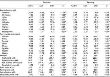
Changes in free amino acid concentrations in seminal plasma at the end of treatment and recovery periods of male rabbits with gossypol and their regression coefficients are shown in Table 1. At the end of treatment, results showed that all seminal plasma concentrations of free amino acids revealed negative regression coefficients as the gossypol dose increased. Regression coefficients of seminal plasma concentrations of total amino acids (TAA), total essential amino acids (EAA), total non-essential amino acids (non-EAA), (EAA/non-EAA) ratios, basic amino acids, acidic amino acids, neutral amino acids and basic/acidic amino acid ratios on dose of gossypol were significantly negative. Results showed that these concentrations decreased in a dose-dependent manner at the end of gossypol treatment. However, these concentrations regained their normal values after gossypol cessation except for TAA and total non-EAA where their regression coefficients were significantly positive at the end of the recovery period.
Changes in free amino acid concentrations (mg/100 ml) in seminal plasma at the end of treatment and recovery periods of male rabbits with gossypol and their regression coefficients

GLD = gossypol low dose; GHD = gossypol high dose.
*P < 0.34.
The deleterious effect on total AA concentrations was mainly due to the reduction in total EAA where its regression coefficient value (−5.56) was more pronounced than that of the non-EAA (−2.46). This relationship was reflected in a significantly negative regression coefficient of the (EAA/non-EAA) ratio. Gossypol treatment significantly reduced the concentrations of basic, acidic and neutral amino acids. The highest reduction was recorded for neutral amino acids (b = −5.94), while the concentrations of basic amino acids were markedly reduced (b = −1.79) than acidic amino acids (b = −0.29). This was reflected in significantly negative regression coefficient of basic/acidic amino acid ratios. During the recovery period, basic, acidic and neutral amino acid concentrations were nearly recovered.
Regarding the individual EAA concentrations, regression coefficients of all EAA (except for those of valine and arginine) on dose of gossypol were significantly negative. At the end of the recovery period, EAA concentrations showed almost complete recovery after gossypol cessation except for isoleucine and leucine (where they exhibited significantly positive regression coefficients) and for phenyalanine (where its negative regression coefficient on dose of gossypol was extended after gossypol cessation).
Regression coefficients of individual non-EAA on dose of gossypol were significantly negative only for both of glycine and alanine, while others were not significant. At the end of the recovery period, amino acid concentrations of glutamic, proline and tyrosine exhibited significantly positive regression coefficients, while serine and glycine showed significantly negative regression coefficients. Concentrations of other non-EAA showed almost complete recovery after gossypol cessation.
It is interesting to note that the levels of free amino acids in the control group were lower during the recovery period than during the treatment period. The decline was more pronounced in the total EAA (74% decline) than in the total non-EAA (48.2%) and in the basic amino acids (75.9% decline) than in the acidic (55.3%) or neutral amino acids (56.7%). These large differences were also noted in the treated groups between the two periods analyzed (i.e. treatment v. recovery) (Table 1). On the other hand, these large differences were not observed in the case of free fatty acids (Table 2). The decline in free amino acids (but not fatty acids) during the recovery period in the three experimental groups coincides with the withdrawal of all drugs (gossypol, corn oil and 5% acetone) during this period. However, due to the lack of information, these changes are difficult to explain at the present time and require further investigation.
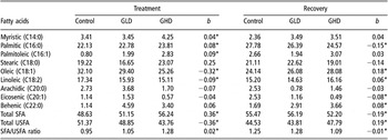
Individual fatty acid concentrations (% of the total peak area) in seminal plasma at the end of treatment and recovery of male rabbits with gossypol and their regression coefficients

GLD = gossypol low dose; GHD = gossypol high dose; SFA = saturated fatty acids; USFA = unsaturated fatty acids.
*P < 0.34.
Fatty acids
The individual fatty acid concentrations (% of the total peak area) in seminal plasma at the end of treatment and recovery periods of male rabbits with gossypol and their regression coefficients are presented in Table 2. Seminal plasma concentrations of total saturated fatty acids (SFA) showed significantly positive regression coefficient on the dose of gossypol, which was mainly due to the increase in myristic and palmitic acids, which represent 52% of the total SFA. On the other hand, regression coefficient of total unsaturated fatty acids (USFA) on the dose of gossypol was significantly negative due to the reduction in oleic and linoleic acids, which represent 98% of the total USFA. This relationship was reflected in a significantly positive regression coefficient of the (SFA/USFA) ratio as gossypol dose increased.
In general, gossypol administration showed increasing effect on SFA levels and decreasing effect on USFA levels. However, the (SFA) behenic and arachidic, and the USFA eicosenic were not affected by gossypol in a dose-dependent manner; thus their linear regressions could not be detected.
At the end of the recovery period, significant regression coefficients of total SFA, total USFA and SFA/USFA ratio, palmitic, oleic and linoleic on dose of gossypol were reversed to those observed at the end of the treatment period. Furthermore, in spite of undetectable linear regressions of eicosenic and behenic acids on the dose of gossypol at the end of the treatment period, significantly negative regression coefficient of eicosenic acid and significantly positive regression coefficient of behenic acid on the dose of gossypol were observed at the end of gossypol withdrawal.
Discussion
Amino acids
The testes and epididymis are the major sources of seminal amino acids (Hopwood and Gassner, Reference Hopwood and Gassner1962). Gossypol administration was reported to cause testicular destruction (Arshami and Ruttle, Reference Arshami and Ruttle1988; Chase et al., Reference Chase, Bastidas, Ruttle, Long and Randal1994), which could impair seminal amino acid synthesis. This would explain the marked reduction in total free amino acids observed in the present study on rabbits due to gossypol administration (Table 1). Our studies on the same rabbits showed that this gossypol-induced reduction of seminal amino acids was associated with poor-quality semen, i.e. low concentration, low motility, and increase in dead and abnormal sperm (Taha et al., Reference Taha, Shaaban, EL-Mahdy, EL-Nouty and Salem2006). This finding coincided with those of Hopwood and Gassner (Reference Hopwood and Gassner1962) who reported that increased concentration of seminal total free amino acids was related to the increased quality and fertility of semen, and that poor semen quality was related to amino acids imbalance. Furthermore, a quantitative reduction in the concentration of all seminal plasma free amino acids has been observed in cases of azoospermia (Chaudhury et al., Reference Chaudhury, Sharma, Jagannathan and Guha2002; Papp et al., Reference Papp, Grof and Menyhart1983) and oligospermia (Silvestroni et al., Reference Silvestroni, Morisi, Malandrino and Frajese1979).
In the current study, the reduction in total free amino acids was mainly due to the gossypol-induced depression in the concentration of EAA (Table 1). Since all animal groups were fed the same ration and their feed intake was not affected by treatment, the reduction in essential free amino acids in the gossypol-treated animals may reflect impaired metabolic efficiency. This suggestion is supported at least in part by the finding of Chadha et al. (Reference Chadha, Sanyal and Kanwar1988) who indicated a reduction in the intestine absorptive function of some amino acids in the rat after gossypol administration.
As far as the type of amino acids is concerned, the gossypol-induced reduction in basic amino acid concentrations was mainly due to the reduction in both lysine and histidine concentrations (Table 1). This decrease could be explained by the reported involvement of lysine (Morris et al., Reference Morris, Higgins and Matlin1986) and histidine (Javed and Waqar, Reference Javed and Waqar1995) in the metabolism of gossypol. On the other hand, acidic amino acid concentrations were less affected by gossypol administration due to the unchanged values of both glutamic and aspartic acid concentrations compared with control.
Several neutral amino acids (i.e. threonine, methionine, isoleucine, leucine, phenyalanine, glycine and alanine, which represent 76% of total neutral amino acids) decreased by gossypol treatment in a dose-dependent manner. This in turn was reflected by a net reduction in total neutral amino acids when gossypol dose increased. Gossypol treatment seems to exert its reducing effect on some neutral amino acids by direct and indirect manners. Directly, by reducing the intestinal absorptive function of leucine and alanine (Chadha et al., Reference Chadha, Sanyal and Kanwar1988). Vera et al. (Reference Vera, Brito and Burzio1987) reported that maximal incorporation of leucine into sperm outer dense fiber polypeptides occurred during spermatogenesis. This finding could support and explain the present results in which gossypol treatment resulted in a reduction in seminal plasma leucine concentration, which was associated with the disappearance of some of the outer dense fibers as depicted by electron microscopy (Shaaban, 2003). In addition, bovine spermatozoa were found to have functional mechanism for the active transport of glycine, which is enhanced at low hexose concentration (Dietz and Flipse, Reference Dietz and Flipse1966). Thus, a gossypol-induced reduction in fructose concentration, which we observed previously (Taha et al., Reference Taha, Shaaban, EL-Mahdy, EL-Nouty and Salem2006), may indirectly enhance glycine uptake by spermatozoa. Furthermore, using the male contraceptive RISUG™ was noted to induce sperm membrane rupture, which was associated with significant decreases in seminal plasma concentrations of isoleucine, leucine and alanine (Chaudhury et al., Reference Chaudhury, Sharma, Jagannathan and Guha2002).
The concentration of aromatic amino acids such as histidine and phenylalanine has been reported to decrease significantly in obstructive azoospermia (Chaudhury et al., Reference Chaudhury, Sharma, Jagannathan and Guha2002). This agrees with the present results in which the concentrations of these amino acids were reduced by treatment with gossypol in a dose-dependent manner (Table 1), which was accompanied by severe reduction in sperm concentration (Taha et al., Reference Taha, Shaaban, EL-Mahdy, EL-Nouty and Salem2006).
Fatty acids
Present results indicated that gossypol increased the percentages of seminal plasma total SFA and the SFA/USFA ratio, and induced a decrease in the percentage of linoleic acid (Table 2). These changes were reported by Fernandez-Real et al. (Reference Fernandez-Real, Broch, Vendrell and Ricart2003) to be associated with inflammatory activity. Furthermore, the activation of inflammatory cells were found to result in a higher ratio of palmitoleic acid to linoleic acid, an established index of essential fatty acid deficiency (Levy et al., Reference Levy, Rizwan, Thibault, Lepage, Brunet, Bouthillier and Seidman2000). This observation agrees with the present findings where palmitoleic acid increased while linoleic acid decreased, which in turn reflected an association of increasing ratio of palmitoleic acid to linoleic acid with increasing dose of gossypol administered (Table 2).
Additionally, the SFA/USFA ratio was reported to increase significantly, and the content of mono-USFA (predominantly oleic) was decreased after aldosterone treatment (Mrnka et al., Reference Mrnka, Novakova, Novak, Tvrzicka and Pacha2000). Thus, the present findings showing a gossypol-induced increase in the SFA/USFA ratio and a decrease in the percentage of oleic acid (Table 2) suggest a state of mineralocorticoid excess, which is in accordance with our previous observations on rabbits (Shaaban et al., Reference Shaaban, Taha, EL-Nouty, EL-Mahdy and Salem2008). However, the percentage of mono-USFA palmitoleic was increased (Table 2) probably due to the effect of mineralocorticoid excess on the inhibition of delta-6 desaturase activity (Marra and de Alaniz, Reference Marra and de Alaniz1990). Additionally, palmitoleic acid has been reported to increase by conditions of oxidative stress (Hara et al., Reference Hara, Yamashita, Fujisawa, Ishiwa and Ogawa1999), possibly as a consequence of gossypol action in generating reactive oxygen species.
Also, gossypol administration resulted in decreasing percentages of oleic and linoleic acids and in increasing percentage of palmitic (Table 2), which offered a suitable condition to induce cell apoptosis (Eitel et al., Reference Eitel, Staiger, Brendel, Brandhorst, Bretzel, Haring and Kellerer2002). On the other hand, oleic and linoleic acids were reported to be vulnerable to free radicals and reactive oxygen species and are easily peroxidized (Ouchi et al., Reference Ouchi, Ikeda, Nakamura, Harino and Kinoshita2002). Additionally, gossypol-induced hypothyroidism (Taha et al., Reference Taha, Shaaban, EL-Mahdy, EL-Nouty and Salem2006) acts synergistically to enhance cell apoptosis, where hypothyroidism leads to a decrease in oleic acid (Saha et al., Reference Saha, Ohinata, Ohno and Kuroshima1998). Collectively, these findings could support and explain our previous observations, showing that gossypol administration was followed by a reduction in sperm quality and quantity (Taha et al., Reference Taha, Shaaban, EL-Mahdy, EL-Nouty and Salem2006), which could be considered as a sign of cell apoptosis.
In conclusion, gossypol administration affected rabbit seminal plasma concentrations of amino and fatty acids in a dose-dependent manner. Gossypol reduced total AA, total EAA and total non-EAA. Additionally, gossypol caused decreases in total USFA and increases in total SFA. These deleterious effects were associated with poor-quality semen observed in our previous studies.