Common to all forms of chronic kidney disease is the progressive scarring of the tubulo-interstitial space. Although many different cell types are involved, accumulated fibroblasts are considered to be key mediators of interstitial fibrosis in the kidney, regardless of the underlying renal disease (Refs Reference Yang and Liu1, Reference Liu2). Some fibroblasts are recruited from circulating bone-marrow-derived stems cells, while others are acquired via the proliferation of resident renal fibroblasts. In addition, recent data demonstrate that in response to renal damage, a significant number of activated myofibroblasts can also arise from epithelial cells via epithelial to mesenchymal transition (EMT) (Ref. Reference Burns3). For example, in the unilateral ureteric obstruction (UUO) model of kidney fibrosis, over a third of interstitial myofibroblasts originate from the mesenchymal transition of tubular epithelial cells (Ref. Reference Iwano4). A similar accumulation of epithelial-derived myofibroblasts is observed in other chronic nephropathies, including chronic allograft nephropathy, glomerulonephritis, spontaneous lupus nephritis and diabetic kidney disease (Refs Reference Oldfield5, Reference Kalluri and Neilson6, Reference Rastaldi7). In each of these conditions, the accumulation of myofibroblasts is correlated with the degree of interstitial damage and the risks for the progression of renal disease (Refs Reference Oldfield5, Reference Kalluri and Neilson6, Reference Rastaldi7). This review examines in detail the molecular mechanisms of tubular EMT associated with renal scarring, looking specifically at the triggers of EMT in vitro and in vivo, their signalling mediators, and the potential for these pathways to be inhibited as a therapeutic target to prevent progressive kidney disease.
The process of EMT
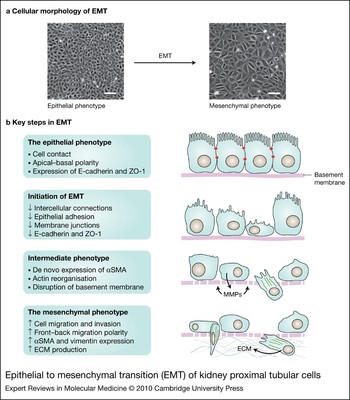
At its most basic, EMT is the phenotypic transition of epithelial cells, in response to environmental perturbations, that results in the progressive loss of their epithelial characteristics and the acquisition of features characteristic of a mesenchymal lineage (Ref. Reference Kalluri and Neilson6) (Fig. 1). EMT can be broadly categorised into three subtypes, based on the context in which it occurs, and the kind of cells that result from this transition and their respective biomarkers (Ref. Reference Zeisberg and Neilson8). Type 1 EMT is observed in embryogenesis when primitive epithelial cells acquire mesenchymal characteristics required for gastrulation, organogenesis and subsequent development of secondary epithelia (Ref. Reference Hay and Zuk9). Type 3 EMT represents an important stage of the evolution of epithelial cancers that leads to a more aggressive phenotype associated with uncontrolled growth, migration and invasion (Ref. Reference Acloque10). By contrast, type 2 EMT, which forms the specific focus of this review, sees the transition of specialised secondary epithelial cells, like those of the proximal tubule, into myofibroblasts with substantial profibrotic and pro-inflammatory activity.
Epithelial to mesenchymal transition (EMT) of kidney proximal tubular cells. (See previous page for figure.) The figure illustrates the change in cellular morphology and the key steps required in the process of tubular epithelial to mesenchymal transition (EMT). (a) Light microscopy of renal proximal tubular cells (NRK52-E) demonstrates the enlarged, spindle-shaped cells characteristic of the mesenchymal phenotype (top panel, right image), resulting from the addition of TGF-β1 at 10 ng/ml for 3 days. By contrast, untreated control cells (top panel, left image; control media for 3 days) have the cobblestone appearance of epithelial cells in culture. All images were photographed at magnification ×100; bar = 100 µm. (b) The schematics show successive stages in EMT, along the lines previously summarised in Ref. Reference Yang and Liu1. Reduced expression of membrane junction proteins such as E-cadherin and ZO-1 is accompanied by loss of cell–contact and apical–basal polarity. Subsequently, cells begin to express mesenchymal markers such as αSMA, which is reorganised into elongated ‘stress fibres’ (green lines denote actin filaments), and upregulated MMPs contribute to basement-membrane disruption. The mesenchymal phenotype is completed with the formation of the enlarged spindle-shaped myofibroblast, which expresses αSMA and vimentin, and has an enhanced capacity for cell migration via filopodia and lamellipodia across the disrupted basement membrane. Abbreviations: ECM, extracellular matrix; MMPs, matrix metalloproteinases; NRK52-E, normal rat kidney epithelial cells; αSMA, α-smooth muscle actin; TGF-β1, transforming growth factor β1; ZO-1, zonula occludens.

Several steps appear necessary to complete the process of type 2 EMT (Refs Reference Yang and Liu1, Reference Strutz, Muller and Neilson11). In tubular cells, the first phase of EMT is characterised by loss of cell–cell contact and apical–basal polarity, leading to separation of cells and cellular destabilisation (Fig. 1). This is associated with reduced expression of epithelial membrane junction and tight-junction proteins [such as E-cadherin (CDH1) and zonula occludens (ZO-1/TJP1)], as well as reduced function of ion and fluid transporters. There is then an intermediate phase, in which cells express a combination of epithelial and mesenchymal markers, including de novo expression of fibroblast-specific protein 1 (FSP1/ATL1) (Ref. Reference Strutz, Muller and Neilson11) and α-smooth muscle actin (αSMA/ACTA2). This stage is also associated with reorganisation of the cytoskeletal proteins, from a cortical bundle formation that supports the microvilli of epithelial cells into elongated ‘stress fibres’ containing αSMA that ultimately lead to ‘mesenchymal’ changes in cellular morphology. In addition, there is upregulation of matrix metalloproteinases (especially MMP2 and MMP9) (Ref. Reference Kalluri and Neilson6) and downregulation of MMP inhibitors (Ref. Reference Qi12), which contribute to the disruption of underlying basement membranes. The transition to mesenchymal phenotype is completed with the formation of the enlarged spindle-shaped myofibroblast, expressing mesenchymal markers [e.g. αSMA and vimentin (VIM)] and without membrane-associated expression of epithelial markers (e.g. E-cadherin and ZO-1). Myofibroblasts also have enhanced capacity for cell migration, including the expression of filopodia and lamellipodia required for invasion across the now disrupted basement membrane. In addition, these cells have a high synthetic capacity, with a prominent endoplasmic reticulum producing large amounts of extracellular matrix (ECM) protein (Ref. Reference Margetts13) important for both healing and scarring.
Mediators of type 2 EMT in the kidney
This complex phenotypic transition requires both the transcriptional activation (and derepression) of genes required to sustain mesenchymal-type structures and functions (e.g. vimentin, αSMA) alongside repression (or deactivation) of genes that act to maintain the epithelial phenotype (e.g. E-cadherin). Several factors have been suggested as potential initiators of EMT in different in vitro and in vivo models or settings (Refs Reference Yang and Liu1, Reference Kalluri and Neilson6, Reference Okada14, Reference Fan15, Reference Strutz16, Reference Ha and Lee17, Reference Lan18, Reference Lee19, Reference Zavadil and Bottinger20). With a few key exceptions [e.g. connective tissue growth factor (CTGF) – see below], each of these mediators requires the induction of transforming growth factor β (TGF-β) to complete the process of EMT (Refs Reference Yang and Liu1, Reference Liu2, Reference Lan18). This includes EMT induced by interleukin 1 (IL-1) (Ref. Reference Fan15), advanced glycation endproduct (AGE)-modified albumin (Ref. Reference Oldfield5), oxidative stress and angiotensin II (Ref. Reference Yang and Liu21). Similarly, disruption of the underlying basement membrane, achieved experimentally by inhibiting the assembly of type IV collagen (COL4) NC1 hexamers, induces EMT in tubular epithelial cells via a TGF-β-dependent pathway (Ref. Reference Zeisberg22). As each of these mediators results in the induction of TGF-β and the activation of common downstream pathways, these different initiators of EMT are able to act in a synergistic manner to promote the development and progression of phenotypic transition. For example, angiotensin II increases TGF-β's ability to induce EMT (Ref. Reference Yang and Liu21). Similarly, the induction of EMT by both fibroblast growth factor 2 and epidermal growth factor (EGF) is magnified when used in conjunction with TGF-β1 (Refs Reference Okada14, Reference Strutz16). This synergism has important implications in disease states, in which different pathogenic stimuli may result in the same phenomenon, or combine to accelerate pathological lesions.
The molecular pathways involved in the initiation of type 2 EMT in tubular cells by TGF-β1 (TGFB1) are complex and involve interactions on many levels (Refs Reference Massague and Wotton23, Reference Bottinger and Bitzer24). These may be different in different cells, potentially reflecting their overall sensitivity to EMT. For example, in a normal rat kidney epithelial (NRK52-E) cell model of proximal renal tubular epithelia, treatment with TGF-β1 was sufficient to induce EMT in a dose-dependent manner (Ref. Reference Fan25). However, early studies in primary renal proximal tubular epithelial cells (Ref. Reference Okada14) showed that while TGF-β1 could upregulate the expression of some markers of type 2 EMT (e.g. FSP1), it was not sufficient in itself to induce a complete EMT. By contrast, combining both TGF-β1 and EGF together led to the induction of FSP1 expression and EMT via an FSP1- dependent pathway (Ref. Reference Okada14).
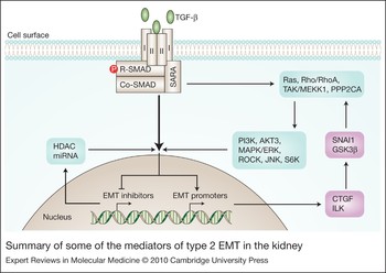
The first step in TGF-β1-dependent EMT in tubular cells is initiated by binding of TGF-β1 to a type II receptor (TGFBR2) dimer, which then recruits the type I receptor (TGFBR1) dimer, forming a large heterotetrameric complex with the ligand (Fig. 2). The type II receptor then phosphorylates serine residues of the type I receptor, which activates the protein and its kinase activity (Ref. Reference Wrana26). Activation of the type I receptor can be blocked by a specific inhibitor of the TGF-β receptor kinase, SB-431542, which by preventing TGF-β-dependent intracellular signalling is able to prevent the induction of EMT in tubular cells (Ref. Reference Wang27). Similarly, GW788388, a TGF-β type I receptor inhibitor with a better pharmacokinetic profile than SB-431542, effectively blocks TGF-β-induced EMT and fibrogenesis in vitro and in vivo (Ref. Reference Petersen28). However, the pleiotropic effects of TGF-β on immune, haematopoietic and vascular function mean that the upstream targeting of TGF-β and its receptor has significant limitations for the prevention of progressive kidney disease.
Summary of some of the mediators of type 2 EMT in the kidney. This simplified figure shows an amalgamation of various signalling pathways, some of which may be more important or active in some settings and in different cell types. Binding of TGF-β1 to a type II receptor dimer recruits the type I receptor dimer, forming a large heterotetrameric complex with the ligand. The type II receptor then phosphorylates serine residues of the type I receptor (not shown), which activates the protein and its kinase activity and recruits SMAD proteins to the complex. The TGF-β receptor complex then phosphorylates receptor-associated SMADs and promotes the recruitment of additional SMADs and other proteins. This activation also results in the translocation of a SMAD complex to the nucleus, where it activates the expression of some TGF-β target genes and represses the expression of others. Activation of TGF-β receptors also stimulates a range of pathways independent of and/or synergistic to classical SMAD-dependent gene activation including RhoA, MAPK and PI3K signalling cascades (see text for details). Abbreviations: AKT3/PKB, protein kinase B; CTGF, connective tissue growth factor; ERK, extracellular-signal-regulated kinase; GSK3β, glycogen synthase kinase 3β; HDAC, histone deacetylase; ILK, intracellular serine/threonine kinase – integrin-linked kinase; JNK, c-Jun N-terminal kinase; MAPK, mitogen-activated protein kinase; miRNAs, microRNAs; PI3K, phosphoinositide 3-kinase; PPP2CA, protein phosphatase 2; R1, type 1 TGF-β1 receptor dimer; R2, type 2 TGF-β1 receptor; RhoA, Ras homologue gene family, member A; ROCK, RhoA kinase; SARA, SMAD anchor for receptor activation; S6K, ribosomal S6 kinase; SNAI1, Snail; TAK, TGF-β-activated kinase 1; TGF-β1, transforming growth factor β1.

SMAD signalling and TGF-β-dependent EMT
SMADs are a class of transcription factors that interact with and modulate the activity of the TGF-β receptor complex (Refs Reference Massague and Wotton23, Reference Bottinger and Bitzer24, Reference Schnaper29). Ligand-induced activation of the TGF-β receptor complex phosphorylates receptor-associated SMADs and promotes the recruitment of additional SMADs and other proteins. This activation also results in the translocation of a SMAD complex to the nucleus, where it activates the expression of some TGF-β target genes and represses the expression of others (Fig. 2). The specificity of different transcriptional responses is partly determined by the combination of different elements that make up the TGF-β-receptor–SMAD complex.
SMADs can be loosely divided into three groups. The first group are the receptor-regulated SMADs (or R-SMADs), which include SMAD2 and SMAD3 (which complex the type I TGF-β receptor and activin receptors, respectively) and SMAD1, 5 and 8 [which complex bone morphogenic protein (BMP) receptors]. Expression and activation of R-SMADs are necessary for the induction of EMT by TGF-β in renal tubular cell, as inhibitory RNA against SMAD2/3 is able to block TGF-β1-induced EMT in vitro (Ref. Reference Phanish30). In addition, SMAD3-knockout mice are protected from induction of EMT by TGF-β (Ref. Reference Zavadil31) and from tubulo-interstitial fibrosis induced following UUO, a model associated with profound renal scarring and EMT (Ref. Reference Sato32). Notably, in this model system, decreased expression of E-cadherin appears to be SMAD3-dependent, while increased expression of MMP2 appears to be SMAD2-dependent (Ref. Reference Phanish30). Importantly, such findings suggest that, at least at a signalling level, EMT can be differentially regulated (Ref. Reference Phanish30).
The second group of SMADs are the inhibitory SMADs (I-SMADs: SMAD6 and 7). SMAD7 inhibits the TGF-β/activin signalling pathway by binding to the TGF-β receptor, thereby preventing its phosphorylation and the subsequent receptor-dependent activation of the R-SMADs (Refs 33, 34). SMAD7 is also able to promote the inactivation or degradation of the TGF-β receptor, by recruiting protein phosphatase 1 (PP1) and ubiquitin ligases, respectively. In addition, I-SMADs can disrupt the formation of the functional SMAD–DNA complex at the SMAD-recognition elements, and thus prevent TGF-β-regulated transcription. SMAD6 is also a regulator of BMP, itself an antagonist of TGF-β-dependent signalling (Ref. Reference Itoh35) (see below).
Disruption of SMAD7 function increases TGF-β/SMAD2/3-dependent renal fibrosis and inflammation, indicating that SMAD7 is a pivotal negative regulator of TGF-β/SMAD- dependent signalling (Ref. Reference Chung36). Overexpression of SMAD7 has been shown to antagonise TGF-β-mediated changes in various models of EMT. For example, overexpression of SMAD7 prevents SMAD3-dependent EMT following UUO (Ref. Reference Lan37) and may also prevent TGF-β1-induced EMT (Refs 38, Reference Li39) and angiotensin-II-induced EMT in vitro (Ref. Reference Carvajal40). Although reduced expression of SMAD7 has been observed in models of renal fibrosis (Ref. Reference Fukasawa41), overexpression of SMAD7 may also induce apoptosis in some systems, making its therapeutic potential problematic (Ref. Reference Schiffer42).
The final category of SMADs contains a single member, SMAD4, which is a common-partner SMAD (or Co-SMAD). SMAD4 oligomerises with R-SMADs (SMAD2 and/or SMAD3) after their phosphorylation by the TGF-β receptor, and therein facilitates the translocation of the SMAD complex to the nucleus (Ref. Reference Massague43). The role of SMAD4 in tubular EMT and renal fibrosis is less clear than for the other SMADs. However, increased cytoplasmic expression of SMAD4 may be involved in augmenting TGF-β1 signalling in the renal tubulo-interstitial cells associated with experimental renal fibrosis (Ref. Reference Goto44).
Connective tissue growth factor and EMT
CTGF is a matrix-associated, heparin-binding protein that plays an important role in the regulation of cell proliferation, apoptosis, embryogenesis, differentiation, and wound healing (Refs Reference Oemar and Luscher45, Reference Grotendorst46). In addition, CTGF has been shown to directly induce EMT. For example, infection of a tubular epithelial cell line (NRK52-E) with an adenovirus encoding CTGF causes a dose-dependent transition in cellular morphology (Ref. Reference Burns, Kantharidis and Thomas47). Similar changes were also seen in NRK52-E cells with the addition of exogenous CTGF (Ref. Reference Burns48). However, when compared with the complete transition to myofibroblasts induced by TGF-β1, EMT induced by CTGF, delivered as a recombinant protein or in an adenovirus, was only partial (Ref. Reference Burns48). Rather than observing a complete loss of E-cadherin expression, there was a rapid reorganisation of the pattern of expression, from a membrane-associated distribution characteristic of epithelial cells to a perinuclear pattern. This change may represent trafficking of E-cadherin to endosomes and/or lysosomes where it may be internalised and degraded. The functional significance of modified localisation remains to be established. However, given the key role of E-cadherin in maintaining the structural integrity and polarity of epithelial cells, this redistribution of E-cadherin may modify adherens junction formation and cell polarity, and contribute to the loss of cytoskeletal integrity and the acquisition of a mesenchymal phenotype (Refs Reference Liu2, Reference Kalluri and Neilson6, Reference Strutz, Muller and Neilson11). A similar change in E-cadherin distribution has been previously reported in response to TGF-β1, at an earlier time point (Ref. Reference Peinado, Quintanilla and Cano49), suggesting that CTGF is predominantly an early, rather than late, player in the EMT pathway.
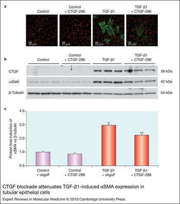
The expression of CTGF is induced by TGF-β1 (Ref. Reference Okada50), and other stimuli that trigger the expression of TGF-β1 (e.g. angiotensin II, AGEs) (Ref. Reference Burns48) – contexts in which it is broadly regarded as a key downstream mediator. For example, blockade of CTGF is able to attenuate TGF-β-dependent EMT in vitro and in vivo. However, although blockade of CTGF reduced the changes associated with EMT, it did not completely ameliorate the phenotypic changes caused by TGF-β1 treatment (Refs Reference Burns, Kantharidis and Thomas47, Reference Burns48) (Fig. 3). Nonetheless, this partial blockade may be sufficient to reduce renal fibrosis in vivo. In a separate study, CTGF blockade attenuated angiotensin-II-induced EMT (Ref. Reference Chen51), indicating that angiotensin II induces EMT partly through a CTGF-dependent pathway. In rodent models of chronic kidney fibrosis, CTGF appears to be required in addition to TGF-β1 to induce persistent injury (Ref. Reference Mori52). In addition, blockade of CTGF using CTGF antisense oligonucleotides attenuated increases in CTGF, fibronectin (FN1), collagen I (COL1) and αSMA gene expression and reduced the interstitial fibrotic areas normally seen in the UUO model or renal fibrosis, despite the sustained overexpression of TGF-β being unaffected (Ref. Reference Yokoi53). This supports the role of CTGF as a (partial) downstream mediator of the actions of TGF-β. Finally, diabetes-induced glomerular basement membrane thickening is attenuated in heterozygous Ctgf +/− mice (Ref. Reference van Nieuwenhoven54).
CTGF blockade attenuates TGF-β1-induced αSMA expression in tubular epithelial cells. NRK52-E cells were preincubated in transfection reagent (oligoF) in the presence or absence of small interfering RNA (siRNA) targeting CTGF (CTGF-286) (1nm) for 24 h prior to the addition of TGF-β1 (10 ng/ml) for 3 days. The altered protein expression of αSMA was demonstrated by confocal microscopy and western blotting. (a) Cells were immunostained for αSMA (green staining). The cell nuclei were stained with propidium idodide (red). All images are taken at magnification ×640. (b) Western blot demonstrating the expression of CTGF, αSMA and β-tubulin. (c) Quantitation of western blot data for αSMA protein expression (versus β-tubulin expression). The oligoF + control group was arbitrarily assigned a value of 1. Each bar represents the mean ± SEM for three (protein) samples per group (*P < 0.01 versus control + oligoF; #P < 0.01 versus TGF-β1 + oligoF). The figure is compiled from experiments and data reported in Ref. Reference Burns48. Abbreviations: CTGF, connective tissue growth factor; αSMA, α smooth muscle actin; TGF-β1, transforming growth factor β1.

The mechanism by which CTGF mediates its effects remains to be fully established. Certainly CTGF has a number of actions that may be considered independent to TGF-β (Refs Reference Yang and Liu1, Reference Lan18, Reference Okada50), as TGF-β1-neutralising antibodies do not prevent the induction of EMT by CTGF in tubular cells (Ref. Reference Burns48). As well as mediating some of the actions of TGF-β, CTGF may also enhance the effects of TGF-β by inhibiting the negative feedback loop of the TGF-β–SMAD signalling pathway (Ref. Reference Abdel Wahab and Mason55). For example, treatment of mesangial cells with CTGF rapidly inhibits the expression of the I-SMAD SMAD7 (Ref. Reference Wahab, Weston and Mason56), possibly via a tyrosine receptor kinase-A (TrkA/TRK1)- and TGF-β immediate early gene (TIEG/KLF10)-mediated pathway (Ref. Reference Wahab, Weston and Mason57). In addition, studies in a human kidney cell line (HK-2 cells) have shown that CTGF-induced EMT is also able to activate the mitogen-activated protein kinase (MAPK) and phosphoinositide 3-kinase (PI3K) signalling pathways implicated in the development and progression of EMT (Ref. Reference Liu58) (see below).
Several studies have investigated possible receptors for CTGF. However, a definitive CTGF-specific receptor remains to be identified. A crosslinking study in a human chondrocytic cell line (HCS-2/8) identified a 125I-labelled-CTGF–receptor complex with an apparent molecular mass of 280 kDa as a potential CTGF receptor (Ref. Reference Nishida59). CTGF has been shown to bind to the multiligand receptor LRP2 (low-density lipoprotein receptor-related protein 2) in fibroblasts (Ref. Reference Segarini60). However, a separate study has shown LRP2 plays a role in the endocytosis of CTGF, which leads to its degradation by endosomes (Ref. Reference Chen61). Therefore, it is possible that binding of CTGF to this putative receptor may facilitate clearance rather than lead to signal transduction. More recently, the receptor TrkA and the pan-neurotrophin receptor (NGFR/p75NTR) were shown to interact with CTGF in primary human mesangial cells (HMCs) (Ref. Reference Wahab, Weston and Mason57). These receptors are activated by nerve growth factor (NGF) and mediate the biological effects of neurotrophins. In further support of the finding that CTGF activates the receptor TrkA in HMCs, the TrkA-selective inhibitor K252a blocked the internalisation and sorting of the receptor–ligand complex and the CTGF-dependent induction of TIEG (Ref. Reference Wahab, Weston and Mason57); however, unlike blockade of CTGF, the blockade of TrkA does not prevent TGF-β1-mediated EMT in proximal tubular cells in vitro [Burns, W.C. (2009) PhD thesis. University of Melbourne, Australia], suggesting the actions of CTGF in this model are probably through alternative and as yet unknown signalling pathways.
ILK and Snail and TGF-β-dependent EMT
The induction of an intracellular serine/threonine kinase – integrin-linked kinase (ILK) – also plays a role in mediating TGF-β1- and CTGF-dependent EMT (Ref. Reference Liu62). TGF-β1 is a potent inducer of ILK in renal tubular epithelial cells, via activation of SMAD2/3 (Ref. Reference Li38) and CTGF (Ref. Reference Liu62). In experimental studies, activation of ILK is able to independently induce EMT (Ref. Reference Li38), while forced expression of a ‘kinase-dead’ ILK is able to block TGF-β1-induced tubular EMT (Ref. Reference Li38), suggesting that activation of ILK may be an important committed step for the induction of EMT. Consistent with this hypothesis, hepatocyte growth factor (HGF), an endogenous inhibitor of EMT (see section below), decreases the expression of ILK, paralleling its antifibrotic actions (Ref. Reference Li38).
ILK is thought to act by coupling integrins and growth factor receptors at the cell surface with intracellular signalling mediators, including protein kinase B (AKT3) and glycogen synthase kinase-3 (GSK3) through phosphorylation (Refs Reference Liu2, 38). Phosphorylation of GSK3 inhibits its functions, leading to the stabilisation of β-catenin and the zinc finger transcriptional repressor SNAI1 (Snail). Induction of SNAI1 significantly impacts on the development and progression of EMT (Refs Reference Acloque10, Reference Thiery63), including the transcriptional repression of E-cadherin and the tight-junction proteins claudins and ZO-1 (Ref. Reference Ikenouchi64). The expression of SNAI1 is partly regulated by other proteins with which it complexes, including SMAD3 and SMAD4 and the nuclear cofactor HMGA2 (high-mobility-group A2 protein) (Refs Reference Thuault65, Reference Thuault66, Reference Vincent67). SNAI1 expression also can be induced by TGF-β1 via MAPK- and PI3K-dependent pathways (see below) (Ref. Reference Peinado, Quintanilla and Cano49). Consistent with the important role of SNAI1 in TGF-β-dependent signalling, in experimental models overexpression of SNAI1 results in renal fibrosis and increased expression of MMP2 (Ref. Reference Yokoyama68) and MMP9 (Ref. Reference Jorda69).
Additional and downstream signalling pathways for TGF-β-dependent EMT
Activation of TGF-β receptors is also able to stimulate a range of pathways independent of and/or synergistic to classical SMAD-dependent gene activation (Refs Reference Zavadil and Bottinger20, Reference Derynck and Zhang70, Reference Kokudo71). Although most data indicate that SMADs are necessary to complete TGF-β-dependent EMT in tubular cells, activation of so-called non-SMAD pathways (Ref. Reference Zhang72) – including RhoA (RHOA; Ras homologue gene family, member A), MAPK and PI3K signalling cascades (Ref. Reference Derynck and Zhang70) – also contributes to the development and progression of the phenotypic transition (Refs Reference Zavadil and Bottinger20, Reference Kokudo71). Calling them ‘SMAD-independent’ is somewhat of a misnomer, as each can regulate TGF-β and SMAD expression and activity through feedback pathways and vice versa. Moroever, their actions with respect to EMT appear to be more cooperative than independent or alternative.
RhoA and TGF-β-dependent EMT
RhoA is a GTPase that is required for the formation of stress fibres and other cytoskeletal reorganisation needed for phenotypic transition. RhoA activates the serine/threonine-specific protein kinase ROCK I/II (RhoA kinase, p160), which triggers a downstream cascade that results in the wholesale reorganisation of the actin cytoskeleton of the cell and the instability of the adherens junctions (Refs Reference Bolos73, Reference Bhowmick74). In addition, RhoA phosphorylates other target proteins to regulate several different cellular functions, including kinase activity, contractile function, proliferation, cell division and remodelling. Activation of RhoA plays an important role in TGF-β-induced EMT. TGF-β1, CTGF and ILK are all able to induce RhoA and ROCK phosphorylation via a range of different pathways (depending on the cell line, context and time-point). For example, rapid activation of RhoA kinase is seen following exposure to TGF-β via non-SMAD pathways, with later and persistent changes sustained by SMAD-dependent pathways (Ref. Reference Kolosionek75). The potential importance of these signalling components is suggested by the finding that blockade of either RhoA or its downstream target ROCK inhibits EMT induced by TGF-β1 (Ref. Reference Bhowmick76), angiotensin II (Ref. Reference Rodrigues-Diez77) or media conditioned by activated mononuclear cells (Ref. Reference Patel78).
MAPK and PI3K pathways and TGF-β-dependent EMT
TGF-β1 is also able to activate the MAPK/ERK signalling pathways (Ref. Reference Rhyu79) rapidly via non-SMAD actions and more slowly via SMAD-dependent signalling. (Ref. Reference Rhyu79) This rapid activation may be through activation of upstream mediators, including Ras, TAK1/MAP3K7, CDC42 or MEKK1/MAP3K11 (Ref. Reference Zhang72). In addition, TGF-β1-induced oxidative stress may also play a role in TGF-β1 signalling upstream of p38 MAPK. For example, treatment with antioxidants prevented TGF-β1-dependent induction of p38 MAPK (MAPK11/12/13/14) and ERK (MAPK1/3) activity in cultured proximal tubular epithelial cells, and subsequently EMT (Ref. Reference Rhyu79). As for other downstream targets of TGF-β signalling, studies have also shown that inhibition of MAPK/ERK signalling is able to prevent EMT induced by TGF-β1 (Ref. Reference Rhyu79).
The PI3K signalling pathways also regulate EMT in several different cells types and systems. TGF-β1 can independently activate PI3K (Ref. Reference Bakin80), possibly via RhoA (Ref. Reference Zhu81) but also via EGF receptor activation (Ref. Reference Docherty82). Whether these actions are important in type 2 EMT in the kidney remains unclear. Certainly, PI3K inhibition has been shown to prevent TGF-β1-dependent EMT in HK-2 cells (Ref. Reference Docherty82). In addition, the expression and activity of the PI3K pathways is strongly associated with renal fibrosis in both experimental models and in the clincial setting (Ref. Reference Winbanks83).
Notch pathway and the induction of EMT
Another important downstream mediator of EMT is the so-called Notch signalling pathway (Ref. Reference Zavadil31). This pathway is a key regulator of cell–cell communication, controlling both intracellular signalling pathways as well as cytoskeleton organisation. Interactions between TGF-β1/SMADs and Notch signalling have been shown to occur in a range of tissues, including the kidney (Ref. Reference Zavadil31). For example, in cultured kidney tubular epithelial cells, TGF-β1 treatment results in increased expression of a subset of Notch target genes of the hairy/enhancer-of-split-related (H/Espl; HEY1/2) family and the Notch ligand JAG1 (jagged 1) (Ref. Reference Zavadil31). Notably, chemical inactivation of Notch or silencing of JAG1 in these cells attenuates TGF-β1-induced EMT (Ref. Reference Zavadil31). Some in vivo studies have also shown increased expression of JAG1 in tubular epithelium following UUO (Ref. Reference Morrissey84). In addition, JAG1 and HEY1 are upregulated and coexpressed in the same abnormal tubular cells in renal biopsies of patients with diabetic nephropathy (Ref. Reference Walsh85).
Other modulators of tubular EMT
The histone deacetylases (HDACs) also play an important role in TGF-β-mediated signalling and EMT (Ref. Reference Yoshikawa86). These enzymes remove acetyl groups from ɛ-N-acetyl lysine residues on histones, thereby modifying both their interaction with adjacent DNA and transcription by modulating chromatin structure. In so far as many of the actions of TGF-β require the interaction of SMADs with DNA, the activity of HDACs and availability of DNA sequences through remodelling of chromatin is important. Recent studies have shown that HDAC inhibitors can inhibit the morphological changes of TGF-β1-induced EMT (Ref. Reference Yoshikawa86). This may be partly due to the induction of endogenous inhibitors [ID2 (inhibitor of differentiation 2) and BMP7] following increased acetylation of histones (Ref. Reference Yoshikawa86). SKI and SnoN (SKIL) also regulate TGF-β signalling possibly by recruiting HDAC complexes to the promoter site (Refs Reference Deheuninck and Luo87, Reference Miyazono, Suzuki and Imamura88).
Recent evidence also demonstrates that expression of short noncoding microRNAs (miRNAs), which act as post-transcriptional repressors of gene function, are substantially altered by TGF-β1. Changes in the expression of specific miRNAs (e.g miRNA-200 and miRNA-205) have been suggested as a biomarker of EMT (Ref. Reference Zeisberg and Neilson8). Moreover, these changes potentially contribute to the regulation of some of the phenotypic changes observed with EMT. Although direct evidence for a causal roles for miRNAs exists only for type 3 EMT associated with tumour invasiveness/carcinogenesis (Ref. Reference Zavadil89), it is clear that miRNAs also participate in the molecular pathogenesis of kidney diseases. For example, miRNA-192 is enhanced in glomeruli from mice with diabetes as well as by TGF-β treatment of cultured mesangial cells, where it promotes fibrogenesis (Ref. Reference Kato90).
Finally, activation of the type V TGF-β receptor (also known as LRP1) also modulates the phenotypic response to TGF-β1 in concert with the activation of other signalling pathways. Although most evidence exists for the modulation of the growth response associated with carcinogenesis and type 3 EMT (Ref. Reference Huang and Huang91), activation of LRP1 promotes the generation and survival of renal myofibroblasts (Ref. Reference Hu92).
Endogenous inhibitors of EMT
Although most attention has focused on the positive signals that trigger EMT, this transition also requires repression of signals that prevent phenotypic transition and maintain homeostasis. These signals may also act to stimulate uncommitted cells to restore their epithelial phenotype (mesenchymal–epithelial transition, MET) (Ref. Reference Hay and Zuk9). The most well known of these homeostatic signals is BMP7 (Ref. Reference Wang, Lapage and Hirschberg93). BMP7 is a member of the TGF-β superfamily, which, like its other members, interacts with (and phosphorylates) R-SMADs and other associated proteins in heterogeneous complexes. In renal cell culture, BMP7 is able to prevent TGF-β1-induced EMT (Refs Reference Postlethwaite, Shigemitsu and Kanangat94, Reference Zeisberg95), possibly by inducing the expression of ID2 (Refs Reference Kondo96, Reference Kowanetz97), an antagonist of SMAD2/3 signalling (Ref. Reference Saika98) and a critical regulator of αSMA expression during EMT (Ref. Reference Valcourt99). In addition, BMP7 is able to directly induce MET in adult renal fibroblasts and thus potentially assist in the regeneration of an epithelial phenotype, particularly in uncommitted cells (Ref. Reference Zeisberg, Shah and Kalluri100). These beneficial actions are paralleled by experimental studies showing that BMP7 is also able to prevent renal fibrosis in a rat model of UUO or even partly reverse fibrosis when provided as a late intervention (Ref. Reference Zeisberg95). Similarly, treatment of diabetic mice with BMP7 is able to partially restore tubular function and normalise urinary albumin excretion (Ref. Reference Wang101). In support of these beneficial actions, the expression of gremlin, a secreted BMP antagonist that acts by binding BMP7 and neutralising its activity, is increased in cultured mouse proximal tubule cells treated with TGF-β1 (Ref. Reference Wang, Lapage and Hirschberg93). Similarly, in diabetic nephropathy, the renal expression of gremlin is upregulated as BMP7 levels are also reduced (Ref. Reference Wang, Lapage and Hirschberg93).
HGF has also been shown to inhibit TGF-β1-induced EMT in the diseased kidney (Refs Reference Yang and Liu21, Reference Yang, Dai and Liu102), possibly by preventing TGF-β1-induced ILK expression (Ref. Reference Li38) (see above), or via the induction of the SMAD transcriptional corepressor SnoN, which results in the formation of a transcriptionally inactive SnoN–SMAD complex (Ref. Reference Yang, Dai and Liu102). Overexpression of HGF is able to attenuate renal injury in experimental diabetes (Ref. Reference Dai103) or even induce regression of mesangial expansion and glomerulosclerosis in advanced disease (Ref. Reference Cruzado104). Notably, these beneficial effects have been associated with suppression of TGF-β1 and CTGF expression, paralleling a reduction in renal interstitial myofibroblasts (Ref. Reference Cruzado104). The effects of HGF in other systems and other types of EMT may not be the same as those observed in the kidney. In the original descriptions, HGF was known as scatter factor and was the first cytokine shown to cause cell scattering or EMT in different cancer types (Ref. Reference Brinkmann105), including renal cancer (Ref. Reference Weidner106).
A range of other transcriptional repressors involved in the regulation of TGF-β1-dependent signalling and EMT have been described, including SNAI2 (Slug) (Refs Reference Savagner, Yamada and Thiery107, Reference Zavadil108), FOS, TWIST 1, members of the Hairy/Enhancer-of-split family (Refs Reference Zavadil and Bottinger20, Reference Zavadil31), and the δEF family members δEF1 (DEFA1) and SIP1 (SFRS2IP) (Refs Reference Eger109, Reference Comijn110). These have largely been demonstrated in nonrenal models and are beyond the scope of the current review. However, it is likely that each will ultimately prove to play some role in EMT and renal fibrogenesis.
The role of EMT in renal pathophysiology
While EMT is not the only effector of chronic renal fibrosis, there is strong circumstantial evidence that it significantly contributes to the development and progression of many forms of chronic renal disease. As noted above, from a renal standpoint EMT is largely thought of as a pathogenic phenomenon, through its ability to increase the numbers and activity of interstitial myofibroblasts, the principal effector cells responsible for matrix production and renal fibrosis (Refs Reference Yang and Liu1, Reference Liu2). Again, there are several mechanisms that contribute to myofibroblast accumulation, including the activation/proliferation of local stromal cells and differentiation of bone-marrow-derived precursors, as well as the phenotypic transition of endothelial cells and vascular pericytes into fibroblasts. However, a significant proportion of interstitial burden of prosclerotic cells may be acquired through the tubular EMT (Refs Reference Oldfield5, Reference Strutz, Muller and Neilson11, Reference Burns48, Reference Guarino, Tosoni and Nebuloni111). For example, over a third of interstitial myofibroblasts associated with renal fibrosis in the UUO model originate from the mesenchymal transition of tubular epithelial cells (Ref. Reference Iwano4). Proof-of-concept studies have also shown that tubular-epithelial-selective changes can induce progressive kidney disease in experimental animals, pointing to a direct and specific role for EMT in renal fibrosis (Ref. Reference Cheng112). Finally, the presence of EMT has also been demonstrated in several studies of renal biopsies, including patients with different renal diseases (Refs Reference Rastaldi7, Reference Boutet113).
The impact of EMT in the kidney may also extend beyond the accumulation of myofibroblasts. The loss of epithelial structure and function associated with EMT may also contribute to renal function decline and progressive renal damage. In particular, functional nephron units can be lost as a result of disconnection of glomeruli from the proximal tubule. While it has been traditionally suggested that external compression by interstitial fibrosis is the primary cause of tubular–glomerular disconnection, other mechanisms may also be involved, including tubular EMT – both directly by stenosing the glomerulotubular junction and indirectly via promoting interstitial scarring. The resulting ‘atubular glomeruli’ are commonly observed in most forms of chronic kidney disease (Ref. Reference Marcussen114). For example, in patients with chronic renal allograft rejection, up to 18% of glomeruli are atubular (Ref. Reference Pagtalunan115), correlating with myofibroblast accumulation and interstitial fibrosis. Similarly, in diabetes over half of cortical glomeruli may be attached to dilated and atrophic tubules, and up to 17% of glomeruli may be completely disconnected from tubular structures (‘atubular’ glomeruli) (Ref. Reference Thomas, Burns and Cooper116).
In so far as phenotypic transition of epithelial cells changes the way cells behave and respond in a stable and persistent manner, EMT may also be a mediator of so called ‘metabolic memory’, whereby metabolic perturbations have long-lasting or persistent effects, long after the exposure has ended (Ref. Reference Chalmers and Cooper117). This is clinically illustrated by findings in patients with diabetes, in which transient periods of intensified glycaemic control appear to result in sustained protection against diabetic complications when compared with individuals who had received less intensive glycaemic control. This protection persists even though differences in the intensity of this control have been abolished for nearly a decade (Refs Reference Chalmers and Cooper117, Reference Holman118). This is more than just lead-time, as comparable persistence is not seen for blood pressure control, despite it being effective in preventing complications, but only while this control is being achieved (Ref. Reference Holman119). A number of hypotheses have been proposed to explain metabolic memory, including persistent epigenetic changes and post-translational modification of protein and DNA (Refs Reference Chalmers and Cooper117, Reference Ceriello, Ihnat and Thorpe120). However, in target organs like the kidney, the accumulation of interstitial myofibroblasts may represent an ongoing reminder of prior metabolic indiscretion as well as a contributor to progressive renal dysfunction.
Rather than simply a pathogenic phenomenon, EMT in the kidney should also be regarded as a form of metastable adaptation, albeit one that is ultimately maladaptive. Much in the same way that glomerular hyperfiltration acts to maintain renal function in the failing kidney, at the cost of glomerular hypertension and accelerated nephron dropout, it has been suggested that EMT could represent an expedient allowing cells threatened by a hostile environment to escape cell death (Ref. Reference Song121), albeit at cost. This neodarwinian (‘adapt or die’) hypothesis deserves further attention, particularly if the inhibition of EMT is to be considered a clinically relevant target for the prevention of renal fibrosis.
In conclusion, the mesenchymal transition of tubulo-epithelial cells in the kidney is associated with the development and progression of renal dysfunction in a range of renal disorders. While EMT is not the only outcome of fibrogenic stimuli like TGF-β1 and CTGF, there is now evidence that EMT plays a significant role in their clinical outcome. Although a range of inhibitors has been developed, to date each has been limited by pleiotropic actions across several physiologically important pathways. It is likely that, in the future, a greater understanding of EMT and its signalling mediators will lead to more-selective and clinically safe interventions for the prevention of renal failure. Moreover, as evidence grows for the role of type 2 EMT in other disorders, such as hepatic and cardiac fibrosis, a broader application of such interventions may also be seen.
Acknowledgements and funding
The authors are supported by competitive grants from the Australian NHMRC, Kidney Health Australia and the Bootle bequest. We thank the reviewers for their useful comments and criticism in the preparation of this manuscript.