Introduction
The Neotropical region has one of the largest populations of ichthyofauna in the world, due to high diversity of hydrographic systems that results in a great variety of fish. Approximately 4399 freshwater species have been described and organized into two main orders. There have been 1847 Characiforms species and 2147 Siluriformes species identified with 57 Characiforms and 91 Siluriformes being considered endangered (Froese & Pauly, Reference Froese and Pauly2015; Brasil, 2016) only in Brazil, which has one of the biggest ichthyofauna populations in the world. In Brazil, the Paraná River basin is an important basin showing a great variety of endemism (Galves et al., Reference Galves, Shibatta and Jerep2009) and endangered species, as is the case with Pseudopimelodus mangurus (Valenciennes, 1835). This species belongs to the Siluriformes order, and was recently included in the ‘red list’ of endangered species in the state of São Paulo (Bressan et al., Reference Bressan, Kierulff and Sugieda2009; São Paulo, 2014). This situation demands conservation strategies for this fish species.
Some reproductive biotechniques have been established for fish conservation, including surrogate propagation (Yasui et al., Reference Yasui, Fujimoto, Sakao, Yamaha and Arai2011) combined with sterile fish obtained by chromosome manipulation (Piferrer et al., Reference Piferrer, Beaumont, Falguiere, Flajshans, Haffray and Colombo2009; do Nascimento et al., Reference do Nascimento, Pereira-Santos, Piva, Manzini, Fujimoto, Senhorini, Yasui and Nakaghi2017). For the use of these techniques, prior knowledge about the stages of embryonic development is necessary (Fujimoto et al., Reference Fujimoto, Kataoka, Sakao, Saito, Yamaha and Arai2006). The developmental stage can be influenced by several factors, and temperature is the main limiting factor in early development (Dos Santos et al., Reference Dos Santos, Yasui, Xavier, de Macedo Adamov, do Nascimento, Fujimoto, Senhorini and Nakaghi2016). Temperature manipulation during early development is also a strategy for synchronizing embryonic development of different species. In germ cell transplantation techniques, the donor and host species must be synchronized at the same developmental stage, which can be achieved with controlled temperatures in both donor and host embryos.
Embryonic development in Neotropical fishes has not been fully investigated, especially in migratory fish species (Godinho & Godinho, Reference Godinho and Godinho2003). The aim of this study was to describe the developmental stages of two Siluriformes, to establish a protocol for germ cell transplantation. The donor species, the marbled catfish Pseudopimelodus mangurus, is a large bodied catfish (>8 kg) that was recently categorized as an endangered species (Bressan et al., Reference Bressan, Kierulff and Sugieda2009; São Paulo, 2014). For this species, domestication and reproduction are critical. As a host species, the spotted catfish Pimelodus maculatus (Lacépède, 1803) was selected as it is a small bodied catfish (~1 kg) for which domestication and breeding procedures are well established under artificial conditions. In addition, P. maculatus can then be used as a model host for germ cells for other endangered Siluriformes.
Materials and methods
Origin of broodstock and artificial fertilization
All procedures were conducted at the Laboratory of Fish Biotechnology (Cepta/ICMBio, Pirassununga City, São Paulo State, Brazil).
Adult males and females of Pimelodus maculatus were collected in the Mogi Guassu river using a cast net (5 cm mesh) and, then, transferred to 1000 m2 earthen tanks. The fish were fed to satiation with a 6-mm commercial fish pellet (45% crude protein, 3800 kcal kg−1) three times a week. These fish promptly accepted artificial feeding 5–10 days after collection. After a period of 3 to 6 months, the fish were selected for induced reproduction during the spawning season. Females are larger than males and present increased peritoneal areas, in addition to a reddish colour in the papillae area. Males were selected randomly based on the length reduced diameter of the body, in comparison with females. The females were anesthetized in clove oil 100 mg l−1 and induced with two doses of crude carp pituitary extract at 0.5 mg kg−1, followed by a second dose of 5 mg kg−1 after 165 hour-degrees. Males were induced to spermiation using a single dose of crude carp pituitary at 0.5 mg kg−1 at the same time as the second induction of the females. Reproduction induction was performed in 500 litre tanks with constant water flow. Gamete sampling was performed by hour-degrees methodology, which is performed by summing the water temperature (°C) for each hour until spawning, resulting in 165 hour-degrees for both species. The oocytes were extruded in a glass bowl. Males were euthanized with the use of anaesthetic clove oil (100 mg l−1) and sperm was collected by testicle maceration in a 15 ml tube containing 3 ml of calcium and magnesium-free Eagle's Minimum Essential Medium with the pH adjusted to 7.8 (E-MEM, Sigma, St. Louis, USA). The diluted sperm were added on the oocytes, and the gamete activation was achieved using 300 ml of hatchery water.
The marbled catfish Pseudopimelodus mangurus adults were collected from the Mogi Guassu river during the spawning season using hook-and-line fishing with a baitfish (hook > 5 cm). The fish were induced to spawn immediately after sampling using the same protocol described above for P. maculatus, but in doses of 0.6 mg kg−1 and 6 mg kg−1, and after induction, the broodstock was maintained in 8000 litre circular tanks with constant water flow. The females were selected by the same criteria described above, including increased redness of papilla and body volume. Males were selected based on the protuberant papillae and semen release after gently abdominal stripping.
Gametes were collected using the same procedure described above. As the marbled catfish sperm is commonly contaminated with urine, the sperm was directly collected in a 15 ml tube containing 5 ml of calcium and magnesium-free Eagle's Minimum Essential Medium with the pH adjusted to 7.8 (E-MEM, Sigma, St. Louis, USA), used as immobilizing medium.
Collection and embryonic development
After fertilization, each spawning was divided into three batches at 22°C, 26°C, and 30°C. For each temperature, a small aliquot (30–80 embryos) was collected randomly and fixed in 2.5% glutaraldehyde in Dulbecco's phosphate-buffered saline (D-PBS) (Sigma #D5773, St. Louis, USA). The samples were collected over the following time intervals: every 2 min post-fertilization until 16 min, every 5 min until 2 h 30 min post-fertilization; every 10 min until 4 h 30 min post-fertilization; every 15 min until 7 h post-fertilization; every 20 min until 11 h post-fertilization; and every 30 min until hatching.
Embryos aliquots from each batch were observed using a stereomicroscope (Nikon SMZ 1500, Nikon, Tokyo, Japan) with a charge coupled device (CCD) camera (Nikon DS-Fi, Nikon, Japan). Digital images were captured using NIS-AR Elements software (Nikon, Tokyo, Japan). The embryonic development of P. maculatus and P. mangurus was classified into zygote, cleavage, blastula, gastrula, segmentation, and hatching stages, and each period was subdivided into phases based on previous studies (Fujimoto et al., Reference Fujimoto, Kataoka, Sakao, Saito, Yamaha and Arai2006), (Dos Santos et al., Reference Dos Santos, Yasui, Xavier, de Macedo Adamov, do Nascimento, Fujimoto, Senhorini and Nakaghi2016) and (Kimmel et al., Reference Kimmel, Ballard, Kimmel, Ullmann and Schilling1995)
Statistics
Data are shown as the mean ± standard error. Data were obtained in triplicate for which different egg batches were considered as a replicate. Percentages of embryos in each of the developmental stages were analyzed with analysis of variance (ANOVA), followed by Tukey multiple range test. In all analyses, the software STATISTICA (7.0, StatSoft, USA) was used with the probability set at 0.05.
Results
Embryogenesis
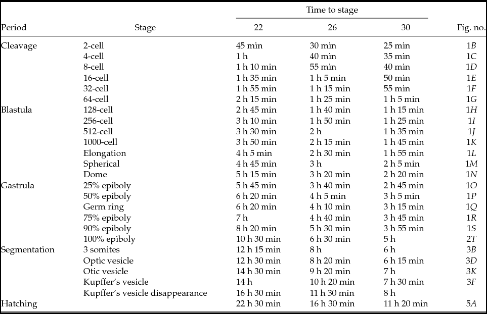
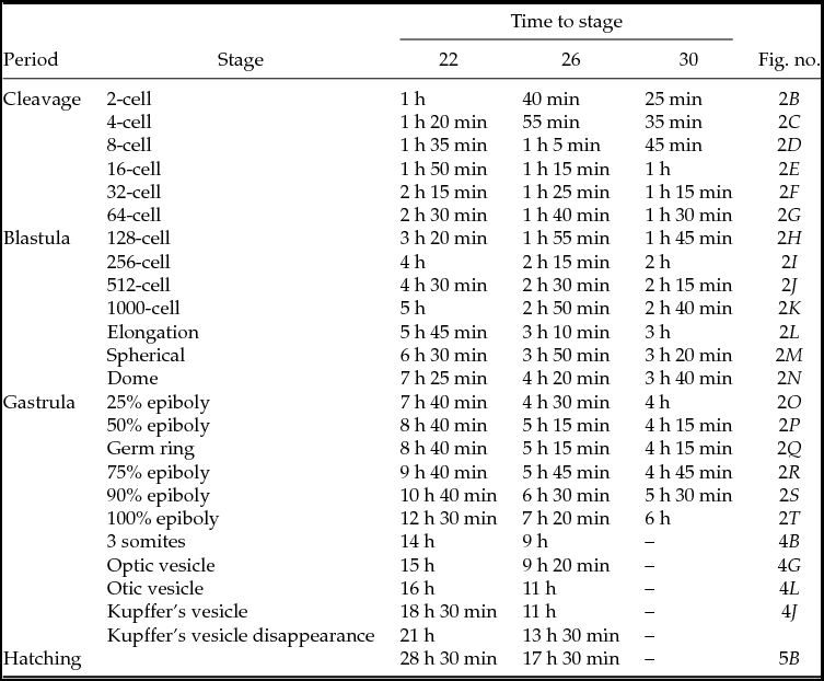
Developmental stages of P. maculatus and P. mangurus at different incubation temperatures are presented on Tables 1 and 2. In the following topics, a detailed description of the developmental stages for both species is shown according to each temperature.
Range of embryonic development for Pimelodus maculatus incubated at temperatures of 22°C, 26°C and 30°C

Range of embryonic development for Pseudopimelodus mangurus incubated at temperatures of 22°C, 26°C and 30°C

Cleavage period
After fertilization and hydration, the chorion begins to expand, inducing growth of the perivitelline space. The cytoplasm begins to migrate, forming the animal pole, initiating the formation of the blastocyst that covers the yolk in the animal pole, in which a meroblastic cleavage pattern occurs.
2-cell stage
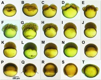
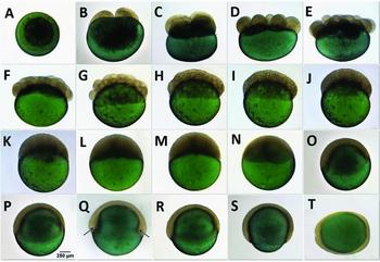
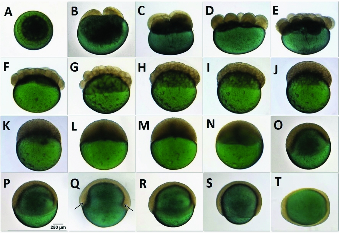
In P. maculatus, embryos reached the 2-cell stage at 45 min post-fertilization when incubated at 22°C, 30 min post-fertilization when incubated at 26°C, and 25 min post-fertilization when incubated at 30°C (Fig. 1B). For P. mangurus, cleavage occurred 1 h post-fertilization when incubated at 22°C, 40 min post-fertilization when incubated at 26°C, and 25 min post-fertilization when incubated at 30°C (Fig. 2B).
Embryonic development of P. maculatus, at period of cleavage, blastula, gastrula and initial segmentation. (A) Animal pole differentiation; (B) 2-cell stage; (C) 4-cell stage; (D) 8-cell stage; (E) 16-cell stage; (F) 32-cell stage; (G) 64-cell stage; (H) initial blastula stage with 128 blastomeres; (I) 256 blastomeres stage; (J) stage of 512 blastomeres; (K) stage with more than1000 blastomeres; (L) elongation stage; (M) spherical stage; (N) dome stage; (O) initial gastrula stage with 25% of epiboly; (P) 50% epiboly; (Q) germ ring stage (arrows indicate the germ ring); (R) 75% epiboly stage; (S) 90% epiboly stage; (T) initial segmentation stage (neurula stage). Scale bar indicates 250 µm.

Embryonic development of P. mangurus, at period of cleavage, blastula, gastrula and initial segmentation. (A) Animal pole differentiation; (B) 2-cell stage; (C) 4-cell stage; (D) 8-cell stage; (E) 16-cell stage; (F) 32-cell stage; (G) 64-cell stage; (H) initial blastula stage with 128 blastomeres; (I) 256 blastomeres stage; (J) stage of 512 blastomeres; (K) stage with more than1000 blastomeres; (L) elongation stage; (M) spherical stage; (N) dome stage; (O) initial gastrula stage with 25% of epiboly; (P) stage with 50% epiboly; (Q) germ ring stage (arrows indicate the germ ring); (R) 75% epiboly stage; (S) 90% epiboly stage; (T) initial segmentation stage (neurula stage). Scale bar indicates 250 µm.
4-cell stage
In this stage, the initial two cells (blastomeres) divide symmetrically giving rise to four cells 1 h post-fertilization when incubated at 22°C, 40 min post-fertilization at 26°C, and 35 min post-fertilization at 30°C in P. maculatus (Fig 1C). This occurred 1 h 20 min post-fertilization for P. mangurus when incubated at 22°C, 55 min post-fertilization at 26°C, and 35 min post-fertilization at 30°C (Fig. 2C).
8-cell stage
During the third stage of cleavage, the four cells of last stage divide symmetrically in a set of eight blastomeres, arranged in two 4-cell lines on the yolk. In P. maculatus, this stage occurs 1 h 10 min post-fertilization when incubated at 22°C, 55 min post-fertilization at 26°C, and 40 min post-fertilization at 30°C (Fig. 1D). For P. mangurus, eight cells were observed 1 h 35 min post-fertilization when incubated at 22°C, 1 h 5 min post-fertilization at 26°C, and 45 min post-fertilization at 30°C (Fig 2D).
16-cell stage
The fourth stage of the cleavage has 16 cells symmetrically arranged in an arrangement of four rows of four cells each. In P. maculatus, this stage begins 1 h 35 min post-fertilization when incubated at 22°C, 1 h 5 min post-fertilization at 26°C, and 50 min post-fertilization at 30°C (Fig. 1E). For P. mangurus, the fourth cleavage stage was observed 1 h 50 min post-fertilization when incubated at 22°C, 1 h 15 min post-fertilization at 26°C, and 1 h post-fertilization at 30° (Fig. 2E).
32-cell stage
The 16 cells divide again, and the blastocyst now consists of 32 blastomeres, arranged in an arrangement of four rows of eight cells each. The cluster of cells begins to overlap irregularly. For P. maculatus, this stage begins 1 h 55 min post-fertilization when incubated at 22°C, 1 h 15 min post-fertilization at 26°C, and 55 min post-fertilization at 30°C (Fig. 1F). For P. mangurus, this stage occurred after 2 h 15 min post-fertilization when incubated at 22°C, 1 h 25 min post-fertilization at 26°C, and 1 h 15 min post-fertilization at 30°C (Fig. 2F).
64-cell stage
In the last stage of cleavage, the 32 cells divide giving rise to a 64-cell blastocyst. For P. maculatus, it occurred 2 h 15 min post-fertilization when incubated at 22°C, 1 h 25 min post-fertilization at 26°C, and 1 h 5 min post-fertilization at 30°C (Fig. 1G). P. mangurus reached this stage 2 h 30 min post-fertilization when incubated at 22°C, 1 h 40 min post-fertilization at 26°C, and 1 h 30 min post-fertilization at 30°C (Fig. 2G).
Blastula period
The blastula period is divided into the following stages: 128 cells, 256 cells, 512 cells, over 1000 cells, elongation, spherical, and dome. The cells were now arranged irregularly and overlapping each other on the yolk.
128-cell stage
For P. maculatus embryos, this stage began 2 h 45 min post-fertilization, when incubated at 22°C, 1 h 40 min post-fertilization at 26°C, and 1 h 15 min post-fertilization at 30°C (Fig. 1H). For P. mangurus, the cells reached this stage 3 h 20 min post-fertilization when incubated at 22°C, 1 h 55 min post-fertilization at 26°C, and 1 h 45 min post-fertilization at 30°C (Fig. 2H).
256-cell stage
Pimelodus maculatus embryos achieve the second stage of the blastula after 3 h 10 min post-fertilization when incubated at 22°C, 1 h 50 min post-fertilization at 26°C, and 1 h 25 min post-fertilization at 30°C (Fig. 1I). For P. mangurus, the cells reached this stage 4 h post-fertilization when incubated at 22°C, 2 h 15 min post-fertilization at 26°C, and 2 h post-fertilization at 30°C (Fig. 2I).
512-cell stage
The embryos of P. maculatus started this stage 3 h 30 min post-fertilization at 22°C, 2 h post-fertilization at 26°C, and 1 h 35 min post-fertilization at 30°C (Fig. 1J). For P. mangurus, this stage was reached 4 h 30 min post-fertilization at 22°C, 2 h 30 min post-fertilization at 26°C, and 2 h 15 min post-fertilization at 30°C (Fig 2J).
1000-cell stage
Pimelodus maculatus embryos reached this stage 3 h 50 min post-fertilization when incubated at 22°C, 2 h 15 min post-fertilization at 26°C, and 1 h 45 min post-fertilization at 30°C (Fig 1K). Pseudopimelodus mangurus reached this stage 5 h post-fertilization when incubated at 22°C, 2 h 50 min post-fertilization at 26°C, and 2 h 40 min post-fertilization at 30°C (Fig 2K).
Elongation stage
Pimelodus maculatus embryos incubated at 22°C were observed 4 h 5 min post-fertilization, 2 h 30 min post-fertilization at 26°C, and 1 h 55 min post-fertilization at 30°C (Fig 1L). For P. mangurus, it took 5 h 45 min post-fertilization to reach this stage when incubated at 22°C, 3 h 10 min post-fertilization at 26°C, and 3 h post-fertilization at 30°C (Fig. 2L).
Spherical stage
The cell pellet was already organized on the yolk forming a ball shape. P. maculatus embryos reached this stage 4 h 45 min post-fertilization when incubated at 22°C, 3 h post-fertilization at 26°C, and 2 h 5 min post-fertilization at 30°C (Fig. 1M). Pseudopimelodus mangurus embryos took 6 h 30 min post-fertilization to reach this stage when incubated at 22°C, 3 h 50 min post-fertilization at 26°C, and 3 h 20 min post-fertilization at 30°C (Fig. 2M).
Dome stage
The group of cells began to cover the whole yolk in epiboly movement. For P. maculatus, this stage occurred 5 h 15 min post-fertilization when the embryos were incubated at 22°C, 3 h 20 min post-fertilization at 26°C, and 2 h 20 min post-fertilization at 30°C (Fig. 1N). For P. mangurus, embryos took 7 h 25 min post-fertilization to reach dome stage when incubated at 22°C, 4 h 20 min post-fertilization at 26°C, and 3 h 40 min post-fertilization at 30°C (Fig. 2N).
Gastrula period
During this period, epiboly were observed. The blastoderm converged and extended on the yolk. The stages in this period were divided according to the percentage of the yolk that was covered by the blastoderm.
25% epiboly stage
A quarter of the yolk was covered by the blastoderm. Pimelodus maculatus arrived at this stage 5 h 45 min post-fertilization at 22°C, 3 h 40 min post-fertilization at 26°C, and 2 h 45 min post-fertilization when incubated at 30°C (Fig. 1O). For P. mangurus, embryos were observed covering a quarter of the yolk 7 h 40 min post-fertilization when incubated at 22°C, 4 h 30 min post-fertilization at 26°C, or 4 h post-fertilization at 30°C (Fig. 2O).
50% epiboly stage
Half of the yolk was covered by the blastoderm in this stage. For P. maculatus, half of the yolk was covered 6 h 20 min post-fertilization at 22°C, 4 h 5 min post-fertilization 26°C, and 3 h 5 min post-fertilization when incubated at 30°C (Fig. 1P). Pseudopimelodus mangurus embryos were observed at this stage 8 h 40 min post-fertilization at 22°C, 5 h 15 min post-fertilization at 26°C, and 4 h 15 min post-fertilization at 30°C (Fig. 2P). During this stage, it was also observed that there was a germinative ring for P. maculatus and P. mangurus (Fig. 1Q, 2Q).
75% epiboly stage
Three-quarters of the yolk was covered by the blastoderm. Pimelodus maculatus embryos arrived at this stage at 7 h post-fertilization when incubated at 22°C, 4 h 40 min post-fertilization at 26°C, and 3 h 45 min post-fertilization when incubated at 30°C (Fig. 1R). Pseudopimelodus mangurus embryos reached this stage 9 h 40 min post-fertilization at 22°C, 5 h 45 min post-fertilization at 26°C, and 4 h 45 min post-fertilization at 30°C (Fig. 2R).
90% epiboly stage
At this stage, 90% of the yolk was covered by the blastoderm. The embryos of P. maculatus arrive at this stage at 8 h 20 min post-fertilization when incubated at 22°C, 5 h 30 min post-fertilization at 26°C, and 3 h 55 min post-fertilization at 30°C (Fig. 1S). P. mangurus embryos arrived at this stage 10 h 40 min post-fertilization at 22°C, 6 h 30 min post-fertilization at 26°C, and 5 h 30 min post-fertilization at 30°C (Fig. 2S).
100% epiboly stage
At this stage, 100% of the yolk was already covered by the blastoderm. P. maculatus embryos reached this stage 10 h 30 min post-fertilization when incubated at 22°C, 6 h 30 min post-fertilization at 26°C, and 5 h post-fertilization when incubated at 30°C (Fig. 1T). For P. mangurus, the embryos took 12 h 30 min post-fertilization to arrive at this stage at 22°C, 7 h 20 min post-fertilization at 26°C, and 6 h post-fertilization at 30°C (Fig. 2T).
Segmentation period
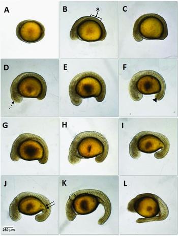
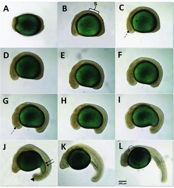
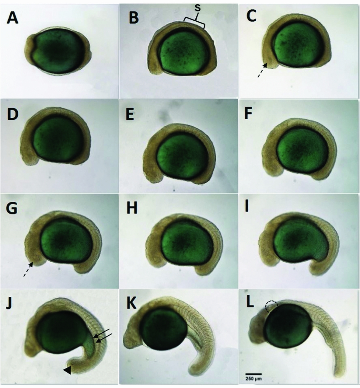
The segmentation period began in the neurula stage with the appearance of somites (Fig. 3A, 4A) and differentiation of head and tail (Fig. 3B, 4B), and it ended with hatching (Fig. 5A, 5B). During this stage, the embryo began to develop its morphological structures, such as the optic vesicle, otic vesicle, Kupffer vesicle, and the onset of somatogenesis. Somites development occurs from the trunk to the tail of the embryo. This period is defined by the structures observed and by the number of somites.
Embryos of P. maculatus during segmentation period, when incubated at 26°C. (A) Neurula stage; (B) 3-somite stage, brackets indicates the first somites (S); (C) 5-somite stage; (D) 8-somite stage, dashed arrow points the optic vesicle; (E) 10-somite stage; (F) 12-somite stage (arrowhead indicates the Kupffer's vesicle); (G) 14-somite stage; (H) 15-somite stage; (I) 20-somite stage; (J) 21-somite stage, double arrows indicate the elongation of the yolk; (K) 24-somite stage, dashed circle indicates otic vesicle; (L) 28-somite stage. Scale bar indicates 250 µm.
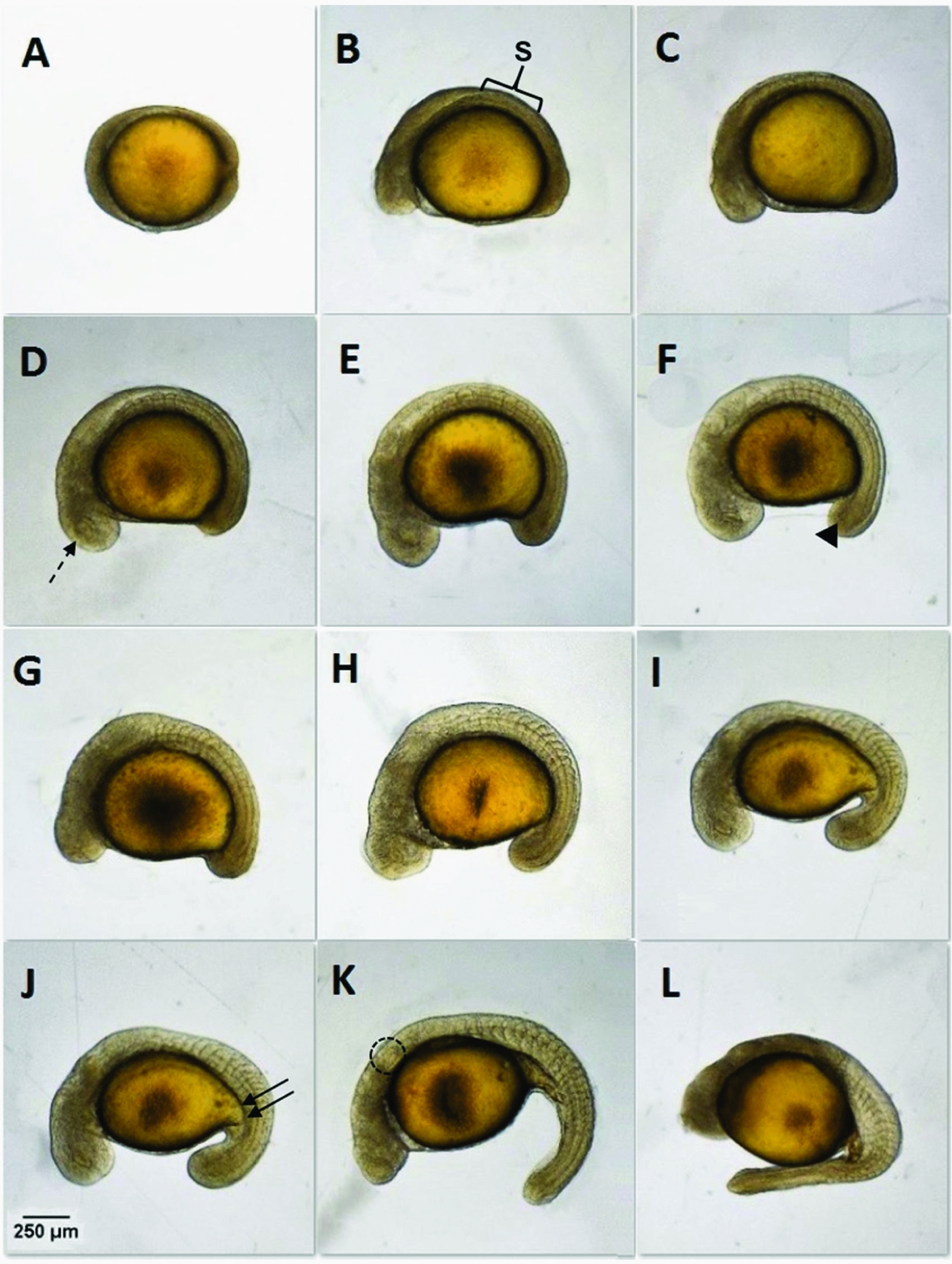
Embryos of P. mangurus during segmentation period when incubated at 26°C. (A) Neurula stage; (B) 3-somite stage, brackets indicates the first somites (S); (C) 5-somite stage; (D) 8-somite stage; (E) 10-somite stage; (F) 12-somite stage; (G) 14-somite stage, dashed arrow points the optic vesicle; (H) 15-somite stage; (I) 20-somite stage; (J) 21-somite stage, arrowhead indicates Kupffer's vesicle and double arrows indicate the elongation of the yolk; (K) 24-somite stage; (L) 28-somite stage, dashed circle indicates otic vesicle. Scale bar indicates 250 µm.
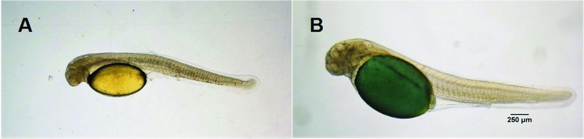
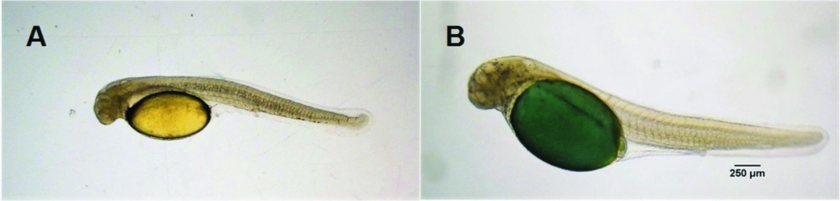
(A) Larvae of Pimelodus maculatus; (B) Pseudopimelodus mangurus after hatching. Scale bar indicates 250 µm.
During this period, a high mortality rate occurs during P. mangurus embryo incubation, resulting in 100% mortality when embryos are incubated at 30°C.
In the P. maculatus embryos, the first somites appeared 12 h 15 min post-fertilization when incubated at 22°C, 8 h post-fertilization at 26°C, and 6 h post-fertilization when incubated at 30°C. For the P. mangurus, the first somites appeared 14 h post-fertilization when incubated at 22°C and 9 h post-fertilization at 26°C.
The segmentation period ended at hatching. At this moment, the larvae broke out of the chorion and started free swimming
Hatching period
The embryonic development ended when the larvae broke out of the chorion. Larvae of P. mangurus incubated at 22°C hatched 28 h 30 min post-fertilization, presenting 37 somites; 17 h 30 min post-fertilization when incubated at 26°C with 35 somites; and no larvae hatched at 30°C (Fig. 5B). Pimelodus maculatus larvae hatched 22 h 30 min post-fertilization when incubated at 22°C, presenting 32 somites, 16 h 30 min post-fertilization when incubated at 26°C with 29 somites; and 11 h 20 min post-fertilization at 30°C, having 27 somites (Fig. 5A).
Oocyte size
For P. mangurus, the size of oocytes non-hydrated, hydrated and perivitelline space was 1217.16 ± 13.35, 1790.95 ± 18.38 and 356.87 ± 19.21 µm, respectively. While for P. maculatus it was 792.26 ± 18.69, 1134.76 ± 19.46, and 259.67 ± 12.89 µm, respectively.
Effect of temperature on embryonic development
There was a large difference in embryo development time between the two species when incubated at 22°C, 26°C, or 30°C. During P. maculatus incubation, the embryos incubated at 30°C hatched 11 h 10 min faster compared with the embryos incubated at 22°C and 5 h 10 min when compared with embryos incubated at 26°C (Table 1). In P. mangurus, the difference was 11 h between the temperatures of 22°C and 26°C (Table 2). Warmer temperatures (30°C) accelerated embryo development time in incubation, however, this led to a higher rate of abnormality among the P. maculatus larvae and caused the death of all P. mangurus embryos up to the segmentation period.
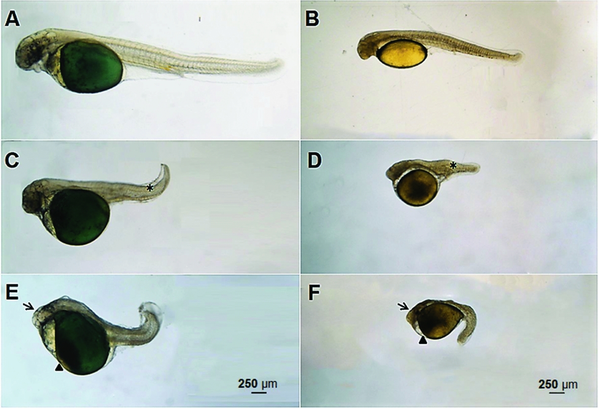
Temperature influenced embryonic development in P. maculatus and P. mangurus that were submitted to treatment, and higher and lower temperatures accelerate and decreased, respectively, embryonic development. Additionally, for both species, the larvae from the lower (22°C) and higher temperatures (30°C) also presented increased abnormality rates, as observed in the head (Fig. 6E, 6F), tail (Fig. 6C, 6D) and yolk regions (Fig. 6E, 6F). The lowest abnormality rate was observed at 26°C, which was considered the optimal incubation temperature for both species. P. mangurus embryos showed tolerance to temperatures with a 45.4 ± 21.02% survival and 2.3 ± 1.73% larvae abnormality when incubated at 22°C, and presented 68.08 ± 12.67% survival and 3.3 ± 1.86% larvae abnormality when incubated at 26°C. When incubated at 30°C, all embryos died before hatching (Table 3). Conversely, P. maculatus embryos hatched at all temperatures with a 20.13 ± 19.8% survival rate and 21.84 ± 2.37% larvae abnormality when incubated at 22°C. When incubated at 26°C, they presented a 57.57 ± 17.49% survival rate and 10.79 ± 4.17% larvae abnormality. At 30°C, they presented a 53.63 ± 16.27% survival rate and 20.76 ± 7.45% larvae abnormality (Table 4).
(A) Normal larvae of Pseudopimelodus mangurus. (B) Normal larvae of Pimelodus maculatus. (C) Larvae of Pseudopimelodus mangurus containing abnormality in caudal region (asterisk indicates abnormal tail). (D) Larvae of Pimelodus maculatus containing abnormality in caudal region (asterisk indicates abnormal tail). (E) Larvae of Pseudopimelodus mangurus containing abnormality in yolk and head region (arrow indicates the abnormal head and arrowhead indicates abnormal yolk region). (F) Larvae of Pimelodus maculatus containing abnormality in yolk and head region (arrow indicates the abnormal head and arrowhead indicates abnormal yolk region). Scale bar indicates 250 µm.

Percentage of main qualitative stages of embryonic development of Pseudopimelodus mangurus incubated at 22°C, 26°C or 30°C

a,bValues with different superscripts differ significantly.
Percentage of main qualitative stages of embryonic development of Pimelodus maculatus incubated at 22°C, 26°C or 30°C

Discussion
Defining the optimal temperature range for embryo incubation is a preliminary step for artificial propagation in fish species. Improvement in egg hatchability with less malformations is among the main parameters for evaluation of incubation temperatures (Pepin, Reference Pepin1991; Jordaan, Reference Jordaan2002). As ectotherms, fish embryos may develop over a wide range of temperatures, in which warmer temperatures increase the velocity of development and embryo formation (Dos Santos et al., Reference Dos Santos, Yasui, Xavier, de Macedo Adamov, do Nascimento, Fujimoto, Senhorini and Nakaghi2016). However, such a range of incubation temperature is well known to be species specific, as observed in Atractosteus tristoechus (26°C) (Comabella et al., Reference Comabella, Hurtado, Canabal and García-Galano2014), Lota lota (2°C) (Lahnsteiner et al., Reference Lahnsteiner, Kletzl and Weismann2012), Rhamdia quelen (21–30°C) (Galdino, Reference Galdino2013; Rodrigues-Galdino et al., Reference Rodrigues-Galdino, Maiolino, Forgati, Donatti, Mikos, Carneiro and Rios2010), Oncorhynchus nerka (10°C) (Velsen et al., Reference Velsen, Bernard, McKinnell and Jamieson1980), Brevoortia tyrannus (15–25°C) (Ferraro, Reference Ferraro1980), Limanda ferruginea (8–14°C) (Laurence & Howell, Reference Laurence and Howell1981), Anguilla anguilla (20°C) (Davidsen, Reference Davidsen2012), Cynopoecilus melanotaenia (20°C) (Arenzon et al., Reference Arenzon, Lemos and Bohrer2002), Coregonus clupeaformis (0.5–6°C) (Price, Reference Price1940), Hexagrammos otakii (12–16°C) (Hu et al., Reference Hu, Pan, Gao, Jian, Wang, Li, Zhang and Guo2015) and Ctenopharyngodon idella (26–28°C) (Korwin-Kossakowski, Reference Korwin-Kossakowski2008).
The emerald green oocytes of the P. mangurus were larger than the yellowish oocytes of the P. maculatus and other Neotropical Characiformes species such as Prochilodus scroffa (1111 µm non-hydrated) (Fenerich-Verani et al., Reference Fenerich-Verani, Godinho and Narahara1984), Brycon insignis (1175 µm non-hydrated) (Andrade Talmelli et al., Reference Andrade Talmelli, Kavamoto and Narahara2002) and Brycon cephalus (1001.6 µm non-hydrated) (Romagosa et al., Reference Romagosa, Narahara, Borella and Fenerich-Verani2001). However, P. mangurus oocytes were smaller than those observed for others Neotropical Siluriformes, such as Zungaro Jahú (1600 µm non-hydrated and 2400 µm hydrated) (Nogueira et al., Reference Nogueira, Azevedo, Canelhas, Bedore, Lopes and Godinho2012), R. quelen (1470 µm non-hydrated and 2640 µm hydrated) and P. charus (1660 µm non-hydrated and 2670 µm hydrated) (Vieira Sampaio & Yoshimi, Reference Vieira Sampaio and Yoshimi2006).
The presented data suggest that the moderate temperature of 26°C is more suitable for embryo development in the five studied species, as observed similarly in previous work with the yellowtail tetra Astyanax altiparanae (Dos Santos et al., Reference Dos Santos, Yasui, Xavier, de Macedo Adamov, do Nascimento, Fujimoto, Senhorini and Nakaghi2016). At 30°C, decreased hatching rates and a high number of abnormal embryos were observed. Surprisingly for the marbled catfish, all embryos died when incubated at 30°C. Most Neotropical fish species spawn during the rainy season, during which time the water temperature commonly reached a lethal limit. Therefore, this may explain the spawning behaviour of migratory fish in which rain is a main trigger to induce spawning in the reproductive season. After upstream migration, most Neotropical species wait for rain, and such behaviour synchronizes spawning of several species. Then, it is expected that rain triggers reproduction and improves egg hatchability with better water quality within an optimum range for incubation. In addition, synchronized spawning may improve survival for all fish species, including for carnivore species that have better food availability.
Only 4°C separates the optimal incubation temperature (26°C) from the temperature at which all P. mangurus embryos die (30°C) and this situation shows that the successful reproduction of fish species can be hampered by global warming (Ficke et al., Reference Ficke, Myrick and Hansen2007), leading to a decline in the populations of several species. Such evidence was observed in our study due to the increased embryo mortality and larval abnormality in P. maculatus when incubated at 30°C. Similar results was also evidenced for others Neotropical fish such as A. altiparanae (Dos Santos et al., Reference Dos Santos, Yasui, Xavier, de Macedo Adamov, do Nascimento, Fujimoto, Senhorini and Nakaghi2016), Brycon amazonicus (da Silva et al., Reference da Silva, dos Santos, Senhorini, Paes, Valentin, Fujimoto, do Nascimento, Yasui and Nakaghi2017) and R. quelen (Rodrigues-Galdino et al., Reference Rodrigues-Galdino, Maiolino, Forgati, Donatti, Mikos, Carneiro and Rios2010).
The establishment of incubation temperatures is interesting for application in studies involving germ cell transplantation. For blastomere transplantation, which involves embryo-to-embryo transplantation, both donor and host species must be at the blastula stage (Yamaha et al., Reference Yamaha, Mizuno, Hasebe, Takeda and Yamazaki1998). However, development of donor and host embryos may identify a specific temperature for incubation and a specific velocity of development. Although synchronizing both embryos to the blastula stage is challenging, temperature may be easily employed to manipulate embryo development. The marbled catfish P. mangurus and the spotted catfish P. maculatus are an interesting model for blastomere transplantation. However, based in these results, the blastula period is very short in both species, limiting the transplantation period to a few minutes. For instance, hatching in both Neotropical catfishes in this study took place within 11 to 28 h post-fertilization, although other transplanted species reported in the literature have longer hatching periods, e.g. medaka Oryzias latipes (10 days post-fertilization) (Shinomiya et al., Reference Shinomiya, Shibata, Sakaizumi and Hamaguchi2003; Iwamatsu, Reference Iwamatsu2004), loach Misgurnus anguillicaudatus (48 h) (Fujimoto et al., Reference Fujimoto, Kataoka, Sakao, Saito, Yamaha and Arai2006), zebrafish (48 h) (Kimmel et al., Reference Kimmel, Ballard, Kimmel, Ullmann and Schilling1995; Lin et al., Reference Lin, Long, Chen and Hopkins1992) and also in salmonids in which the embryonic development takes place after several weeks (Velsen et al., Reference Velsen, Bernard, McKinnell and Jamieson1980; Takeuchi et al., Reference Takeuchi, Yoshizaki and Takeuchi2001; Takeuchi et al., Reference Takeuchi, Yoshizaki and Takeuchi2003; Winckler-Sosinski et al., Reference Winckler-Sosinski, Schwarzbold and Schulz2005; Okutsu et al., Reference Okutsu, Shikina, Kanno, Takeuchi and Yoshizaki2007). As seen above, most germ cell transplantations have been performed in cold water species, in which the transplantation period is longer. In Neotropical species, the presented data suggest that the transplantation strategy should be different than that for cold water species, due to a shorter transplantation period. Thus, a lower number of transplants per each egg batch will be produced in Neotropical species, suggesting that an increased number of egg batches is then required to produce an adequate number of transplanted embryos.
The success of transplantation and subsequent production of germline chimera for surrogate propagation depends on the phylogenetic relationship between donor and host species, in which related species may increase the success of germline transmission (Yamaha et al., Reference Yamaha, Saito, Goto-Kazeto and Arai2007). For the spotted catfish P. maculatus, this is interesting because several species of catfish are considered endangered in the Neotropical region (Machado et al., Reference Machado, Drummond and Paglia2008). Siluriformes are considered to be the largest group in the Neotropical region, suggesting that this species may become an interesting model fish for surrogate propagation with subsequent utilization in both academic and aquaculture purposes.
In conclusion, the present study verified the effect of temperature in the embryonic development of two Neotropical catfishes. This exercise is useful for embryo transplantation in Siluriformes because endangered catfish species can be used as donors of PGCs, and a common catfish species can be used as a host. This is strategic for this species and also applicable for other endangered or aquacultured catfish from the Neotropical region.
Acknowledgements
The authors would like to thank the Institudo de Biociências (IBB) of Universidade Estadual Paulista (UNESP), Botucatu, São Paulo, Brazil, in addition to the Laboratório de Biotecnologia de peixes and CEPTA/ICMBio, Pirassununga, Brazil. We would also like to thank Michael James Stablein of the University of Illinois Urbana-Champaign for his translation services and review of this work.
Financial support
The author express his gratitude to the AES Tietê for financial support.
Ethical standards
The experiments were conducted in accordance with the Ethics Committee for the Use of Laboratory Animals of the National Center for Research Conservation of Aquatic Biodiversity (CEUA/CEPTA; #010/2015). The fish were collected in natural environments using the sampling licence according to Brazilian law (Sisbio #55725-1).