Introduction
Sperm polymorphism is the phenomenon by which males produce two or more types of sperm (Jamieson, Reference Jamieson1987; Swallow & Wilkinson, Reference Swallow and Wilkinson2002; Till-Bottraud et al., Reference Till-Bottraud, Joly, Lachaise and Snook2005). In most cases, the polymorphic sperm include fertilizable eusperm and non-fertilizable parasperm. In the former, the sperm cell and developed nuclei have typical sperm morphology, while in the latter the shapes of the cell and nuclei often exhibit irregular shape. For instance, the gastropod Semisulcospina lebertina produces parasperm with an excess chromatin (Okura et al., Reference Okura, Kohata, Harutsugu and Yasuzumi1988), whereas the gastropod Cinctiscala eusculpta produces large filament-like sperm with no chromatin. The silkworm Bombyx mori produces parasperm with a small amount of chromatin (e.g. Jamieson, Reference Jamieson1987; Yamashiki & Kawamura, Reference Yamashiki and Kawamura1997). These parasperm are produced through a constant developmental process. Although the constituents of DNA vary among the parasperm and although the parasperm themselves have no fertilizing ability, the roles of the parasperm in the reproduction of these animals is partially known: the parasperm of silkworms are considered to stimulate metabolism in order to supply energy to the fertile eusperm (Osanai et al., Reference Osanai, Kasuga and Aigaki1987), or to facilitate fertilization (Sahara & Kawamura, Reference Sahara and Kawamura2002), while the parasperm of white butterfly Pieris napi are considered to confer an advantage to kin-fertile eusperm in sperm competition at fertilization (Cook & Wedell, Reference Cook and Wedell1999).
In vertebrates, sperm polymorphism has been found in quite a limited number of species. It is known that sperm polymorphism occurs in cottoid fish (Hayakawa et al., Reference Hayakawa, Komaru and Munehara2002a; Hayakawa & Munehara, Reference Hayakawa and Munehara2004; Hayakawa, Reference Hayakawa2007). The marine cottoid fish Hemilepidotus gilberti has parasperm (Hayakawa et al., Reference Hayakawa, Komaru and Munehara2002a) that play an adaptive role in sperm competition (Hayakawa et al., Reference Hayakawa, Munehara and Komaru2002b). The eusperm of the species have a small head with a diameter of 1.6–2.0 µm with a haploid nucleus, a midpiece with a pair of mitochondria and a flagellum of about 20 µm in length. The parasperm are large cells being 5–7 µm in diameter with two elongated nuclei that are fused together. In most cases they have no flagellum (Hayakawa et al., Reference Hayakawa, Komaru and Munehara2002a), although sometimes they have one or two flagella with low motility. Both types of sperm are released simultaneously at spawning and eusperm approach the eggs with vigorous motility for fertilization. The parasperm without motility gather around the jelly-like ovarian secretion that is released with the eggs and form a physical barrier to prevent the eusperm of other males from accessing the eggs. As a result, the first male to spawn has a great reproductive advantage (Hayakawa et al., Reference Hayakawa, Munehara and Komaru2002b). Despite our knowledge of these interesting roles of parasperm and eusperm in the fertilization of species, it is still unknown how the two types of sperm differentiate during the process of spermatogenesis.
Spermatogenesis in the teleostean testes generally occurs in cysts that are formed by Sertoli cells (Pudney, Reference Pudney1993). Sertoli cells surround a single type B spermatogonium and form a cyst and the sequestered germ cell begins to proliferate. In H. gilberti, the morphological differences between eusperm and parasperm are first observed at the second meiotic division. Normal meiotic division occurs in some cells to establish eusperm and incomplete division occurs in other cells to develop parasperm. Interestingly, eusperm and parasperm differentiate within the same cyst from spermatocytes that are connected by a cytoplasmic bridge (Hayakawa et al., Reference Hayakawa, Komaru and Munehara2002a). Spermatogenesis in teleosts is commonly controlled by the interaction of germ cells and Sertoli cells under the influence of hormones. Moreover, differentiating germ cells are connected to each other by a cytoplasmic bridge and synchronously proliferate and differentiate into sperm. These raised the question of how the different pathways that progress towards both types of cottoid fish sperm can progress in the primary spermatocytes, under the same conditions for spermatogenetic control in a single cyst. To address this problem, we performed a cell culture system using H. gilberti, in which a part of spermiogenesis occurred from primary spermatocytes after DNA duplication without keeping contact with Sertoli cells. Using this system, we found out that differentiation toward both eusperm and parasperm could progress from the initial stage of primary spermatocytes independently of the influences of Sertoli cells and circulating hormones.
Materials and Methods
Animals
Male fish of Hemilepidotus gilberti were captured by angling in the coastal waters (50–90 m in depth) off Usujiri (41°57′N, 140°58′E), southern Hokkaido, Japan in May 2004. Some fish were kept in an aquarium with running seawater (10°C: almost the same temperature as in the natural spawning site) at the Usujiri Fisheries Station, Hokkaido University until spawning season (October).
Histology
Testes were dissected and fixed in Bouin's fixing fluid. They were dehydrated in an alcohol series and then embedded in paraffin. Sections of 6 µm thickness were prepared and stained with haematoxylin–eosin solution. Histological observations of testis were performed in May and October.
Cell culture
Cell culture of germ cells in H. gilberti was prepared fundamentally according to the culture system for Oryzias latipes (Saiki et al., Reference Saiki, Tamura, Matsumoto, Katowgi, Watanabe and Onitake1997; Sasaki et al., Reference Sasaki, Watanabe, Takayama-Watanabe, Suzuki, Abe and Onitake2005). Testes were dissected from males and cut into small pieces. They were treated with 0.1% trypsin in salt solution consisting of 204 mM NaCl, 4.5 mM KCl, (pH 7.4) at 10°C for 30 min. The solution was prepared according to the estimated concentrations of Na+ and K+ included in the male body fluid of H. gilberti (Hayakawa & Munehara, Reference Hayakawa and Munehara1998). The tissues were disrupted by gentle pipetting and then they were centrifuged at 400 g for 7 min. The supernatant was centrifuged again at 700 g for 7 min. The cell pellet was resuspended in the salt solution and a density gradient centrifugation was performed using 1.25× L-15 medium (Invitrogen) containing 1, 3, or 5% bovine serum albumin (BSA; Sigma–Aldrich Co.). A fraction of 5% BSA was collected and centrifuged at 100 g for 7 min and the cell pellet was resuspended in 1.25× L-15 medium containing 10% fetal bovine serum (FBS; Equitech Bio. Inc.), 50 IU/ml penicillin and 25 µg/ml streptomycin. On a culture dish (1007; BD Falcon, Becton–Dickinson), 2 × 10⁶ cells were spread and cultured at 10°C for up to 12 days. The condition of the cultured cells was observed daily with a microscope (CK40; Olympus Co.). The diameters of the cultured cells were estimated on day 0 and day 8 under a microscope using a micrometer. Motility of developing germ cells was recorded by time-lapse video recorder (AG-6720, Panasonic Inc.).
Incorporation and detection of bromodeoxyuridine
Germ cells were cultured in the medium adding bromodeoxyuridine (BrdU) at a final concentration of 5 µM for up to 8 days. Those cells were fixed in 10% formaldehyde in artificial male body fluid (ABF) consisting of 204 mM NaCl, 4.5 mM KCl, 2.1 mM CaCl2 and 2.1 mM MgCl2 (pH 7.4), which was prepared in accordance with the estimated concentrations of major ions in the male body fluid (Hayakawa & Munehara, Reference Hayakawa and Munehara1998). The fixed cells were washed in 0.01 M phosphate-buffered saline (PBS; pH 7.4) and treated with 0.3% Triton X-100 in PBS and then 1 N HCl in PBS for 5 min. After washing with PBS and blocking with 0.5% skimmed milk in PBS, they were treated with the anti-BrdU antibody (1:500 dilution; Chemicon International Inc.) for 30 min. After being washed and blocked again, the cells were treated with biotin-conjugated anti-mouse IgG (1:100 dilution; BioSource International) in PBS for 30 min. After being washed and blocked, the cells were treated with HRP-conjugated avidin (1:100 dilution; BioSource International) for 30 min. Finally, they were washed twice in PBS and stained with a solution containing 0.02% diaminobenzidine and 5 mM Tris–HCl (pH 7.4).
Mitochondrion distribution in the cultured cells
Eusperm of H. gilberti have an elongated, condensed nucleus in the sperm head, a pair of mitochondria in the midpiece and an axoneme that penetrates through the sperm head to the tail end (Hayakawa et al., Reference Hayakawa, Komaru and Munehara2002a). On the other hand, parasperm have two highly condensed nuclei in a large head and sometimes two axonemes in the tail. However, the localization of mitochondria in the sperm is unclear. In order to examine the mitochondrion distribution in the spermatids developed in the culture, Mitotracker FM (Invitrogen), a fluorescent dye for mitochondrial membrane, was added to the culture medium at a final concentration of 10 µM at day 4 and incubated for 1 h at 10°C. The staining was observed by a fluorescence microscope (CK40-URFLT50; Olympus Co.).
Scanning electron microscopy
Cultured cells were collected by centrifugation at 1000 g for 5 min. The obtained cells were suspended in ABF and spread on a 35 mm culture dish coated with 0.01% poly-l-lysine. After ABF was removed, cells attached to the bottom of the culture dish were fixed with 2.5% glutaraldehyde in 0.1 M phosphate buffer (pH 7.0) at 4°C overnight. They were then rinsed with the phosphate buffer, dehydrated and dried in a critical point apparatus (HCP-1; Hitachi Co.). Finally, the cells were coated with platinum using a magnetron sputterer (JUC-5000; JEOL Ltd) and observed with a scanning electron microscope (JSM-5400; JEOL Ltd).
Results
Histological observation of the testes
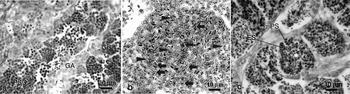
Hemilepidotus gilberti undergoes seasonal breeding and an apparent cycle of spermatogenesis is established in the testis (Hayakawa et al., Reference Hayakawa, Kobayashi, Munehara, Watanabe and Onitake2007). The testes were at the active spermatogenesis stage in May, when type B spermatogonia and primary spermatocytes were major components in the testes (Fig. 1a). Each of them was observed in a single cyst, suggesting that they were going to differentiate synchronously in every cyst, like those in other teleost species (reviewed in Pudney, Reference Pudney1993). In contrast, some cysts contained two types of cells that were small with a spherical shape and larger irregularly shaped cells (Fig. 1b). The cells of the former type have a nucleus with spherical shape whereas in the latter, two elongated nuclei were observed in parallel to one another. From the distribution and the morphological features, those cells would correspond to euspermatids that develop into eusperm and paraspermatids that develop into parasperm, respectively.
Sections of the testis of H. gilberti. (a) Active spermatogenesis stage. Type A and B spermatogonia (GA and GB) and primary spermatocytes were major germ cells in this period. (b) High magnification view of a single cyst. Arrowheads and arrows indicate small spherical cell with a single nucleus and larger cells with two nuclei, respectively. (c) A high magnification view of a cyst containing both eusperm (ES) and parasperm (PS) observed in the testis at the spermiation stage. Bar: 10 µm.

Germ cell differentiation in cell culture
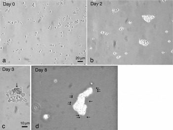
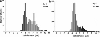
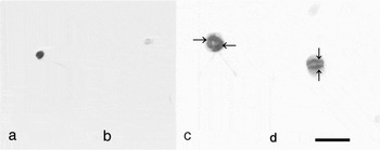
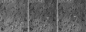
The majority of the cultured cells were spherical in shape, with no flagellum and about 5–7 µm in diameter in the preparation from the testes of H. gilberti in May (Figs. 2a, 3). Most of these cells did not adhere to the substratum of the culture dish. Small aggregates of the cells were observed 2 days after the beginning of cell culture (day 2) (Fig. 2b). The size of the aggregates gradually increased and in some aggregates, cells became retroflex shaped (Fig. 2c). In some cases, those types of cells occupied an entire aggregate. Many cells in the aggregates had a flagellum on day 8 (Fig. 2d). Some spherical cells were also observed when cells were dissociated from aggregates by gentle pipetting. Each spherical cell possessed a flagellum. The spherical cells were about 5 µm in diameter (Fig. 3). The BrdU-labelling of these cells with different morphologies demonstrated that most of the retroflux cells had two elongated nuclei that located in parallel to one another while most of the flagellated spherical cells had only one nucleus (Fig. 4). These corresponded to the features of paraspermatids and euspermatids confirmed by histological observation. Thus, the cells with the former morphology and the latter were paraspermatids and euspermatids, respectively, which were going to develop separately from the prepared germ cells during day 2 to day 8 in the cell culture. Some of the euspermatids showed forward motility on day 11 (Fig. 5).
Cell culture of germ cells of H. gilberti. Germ cells were prepared from the testes at active spermatogenesis stage and cultured in 1.25× L-15 medium containing 10% FBS. (a) Day 0: most cells had spherical shape. Small cells connected to the other cells by cytoplasmic bridge. (b) Day 2: small aggregates of germ cells were observed. (c) High magnification view of the aggregates observed on day 3. Arrow indicates cells with a parasperm-like morphology. (d) Day 8: large aggregates of germ cells were observed. Moving flagella (arrows) were observed in many of the cells with parasperm-like morphology. Bars: 20 µm (a, b, d), 10 µm (c).

Changes of distribution of germ cell diameter in cell culture. Cultured cells were observed under a microscope. In five randomly selected views, the diameters of all germ cells except for those that were aggregated were measured on day 0 (a) and day 8 (b). Three peaks were observed at day 0, while the two larger peaks disappeared at day 8. n = total number of measured germ cells.

BrdU-labelled spermatids of H. gilberti in cell culture. Germ cells obtained from the testes at the active spermatogenesis stage were cultured in 1.25× L15 medium containing 10% FBS, 5 µM BrdU. They were fixed in day 8 and BrdU incorporated into germ cells was detected by immunohistochemistry. (a) A BrdU-labelled euspermatid. (b) BrdU-not incorporated euspermatid. (c, d) BrdU-labelled paraspermatid. Arrows indicate the BrdU-positive nuclei. Bar: 10 µm.

Motile spermatids of H. gilberti developing in cell culture. Motility of cultured cells was recorded with time-lapse videocassette recorder at day 11. (a): View of cultured cells observed under the microscope. (b, c): Views taken 5 and 15 s after that in (a). Arrows indicate spermatids swimming. Bar: 20 µm.

The paraspermatids were similar to parasperm in their retroflux shape and elongated nuclei, although, in the culture, those with fused nucleus were not observed. Moreover, flagella were typically observed in euspermatids in culture while many parasperm do not possess these in vivo (Hayakawa, 2002a). In euspermatids, a few had an elongated nucleus similar to eusperm (Fig. 4). In addition, the diameter of most paraspermatids remained at 5 µm, which was larger than that of the head of the eusperm (Hayakawa et al., Reference Hayakawa, Komaru and Munehara2002a). It is likely that the release of the residual body was incomplete in the developing euspermatids in the cell culture.
Mitochondrion distribution in the cultured cells
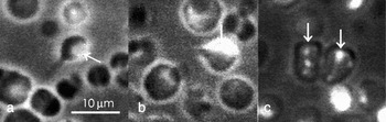
In the euspermatids, fluorescence was typically observed in the cytoplasmic region at the opposite side of the flagellum protrusion (Fig. 6a, b). In contrast, in the paraspermatids, the fluorescence was randomly observed in the cytoplasmic region (Fig. 6c). In many cases, fluorescence was too weak to be detected in the cytoplasmic region of the paraspermatids (data not shown).
Mitochondrion localization in the cultured spermatids of H. gilberti. Germ cells were obtained from the testis at the active spermatogenesis stage and cultured in 1.25× L15 medium containing 10% FBS. At day 8, 10 µM Mitotracker FM was added to medium and incubated for 1 h. Mitochondria in the cytoplasm were fluorescently stained. (a, b) A euspermatid with stained mitochondria localized to one side of the head. (c) Paraspermatids with randomly distributed clusters of mitochondria. Arrows indicate fluorescence from the Mitotracker showing the mitochondrial localization in the germ cells. Bar: 10 µm.

Scanning electron microscopy
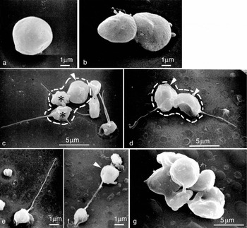
Scanning electron microscopic observation of the developing germ cells showed that most cells were spherical in shape with a diameter of 5–7 µm (Fig. 7a) and had no flagellum on day 0. The cells were not connected to each other by cytoplasmic bridges, probably because the pipetting in preparation disrupted these connections, as seen in previous studies on O. latipes (Saiki et al., Reference Saiki, Tamura, Matsumoto, Katowgi, Watanabe and Onitake1997; Sasaki et al., Reference Sasaki, Watanabe, Takayama-Watanabe, Suzuki, Abe and Onitake2005). On the other hand, many aggregates containing several cells were typically observed after day 4 (Fig. 7b, g). In some cases, the cells were connected to each other by a cytoplasmic bridge, suggesting that these germ cells were proliferating in the culture. Cells with flagella were typically observed after day 8 (Fig. 7c–f). These cells were classified into two types as seen in the morphological observation using the microscope: small spherical cells with a flagellum (Fig. 7c, e, f), which corresponded to euspermatids, and retroflex-shaped large cells (Fig. 7c, d), which corresponded to paraspermatids. Cells with incomplete flagella were often observed among the latter type of cells (Fig. 7d).
Scanning EM observation of cultured germ cells of H. gilberti. Germ cells obtained from the testes at the active spermatogenesis stage were cultured. They were fixed on days 0, 4, 8 and 11. Morphological features of the developing germ cells were observed by the scanning EM. (a) A germ cell with spherical shape. This shape was typically observed at day 0. (b) Asymmetrically divided cells observed on day 4. (c) A cell cluster composed of two eusperm-like cells (asterisks) and one parasperm-like cell (arrowhead) observed on day 8. (d) A cell cluster composed of two parasperm-like cells (arrowheads) observed on day 8. (e) A eusperm-like cell. (f) A eusperm-like cell having a residual body (arrowhead). (g) A cell cluster observed on day 8. Each cell was connected to each other by a long cytoplasmic bridge. Dotted lines in (c, d) indicate a cluster of germ cells. Bars: 1 µm (a, b, e and f) and 5 µm (c, d, g).

BrdU-labelling of germ cells
Since DNA synthesis occurred only in spermatogonia and primary spermatocytes among the spermatogenetic cells, BrdU-labelling in the nuclei of spermatids should reveal that they were differentiated from DNA-synthesizing primary spermatocytes or earlier stages of germ cells (Sasaki et al., Reference Sasaki, Watanabe, Takayama-Watanabe, Suzuki, Abe and Onitake2005). In order to examine when both types of germ cells were differentiated in the cell culture, BrdU was incorporated into the nuclei of the cultured cells on day 0. By day 8, many germ cells showed BrdU in their nuclei (Fig. 4). Among the BrdU-positive cells, germ cells designated as the flagellated euspermatids that possessed a single nucleus and the paraspermatids that possessed two elongated nuclei were observed. This agreed with the results of the morphological observation, indicating that both the euspermatids and the paraspermatids were formed from DNA-duplicating primary spermatocytes in the cell culture.
Discussion
Polymorphism of spermatozoa has been described in many invertebrate species, whereas a few examples of the phenomenon have been observed in vertebrate species. The development of dimorphic sperm in the fish, Hemilepidotus gilberti is therefore a unique and interesting event from the point of view of sperm function and spermatogenesis in vertebrates. The functional role of H. gilberti sperm in sperm competition at fertilization has been clearly explained (Hayakawa et al., Reference Hayakawa, Munehara and Komaru2002b) and the process of spermatogenesis in this species has been investigated in vivo, revealing that the morphological difference between eusperm and parasperm can be first recognized during the second meiotic division (Hayakawa et al., Reference Hayakawa, Komaru and Munehara2002a). However, it remains unknown how the differentiation of the two types of sperm is controlled.
It has been well established that spermatogenesis is generally controlled by hormonal influences and germ cell–somatic cell interaction in teleosts as well as in other vertebrates (reviewed by Chong et al., Reference Chong, Koh and Melamed2005; Sherwood & Adams, Reference Sherwood and Adams2005; Young et al., Reference Young, Kusakabe, Nakamura, Lokman and Goetz2005). Germ cell differentiation synchronously progresses in every cyst under a combined effect of the hormones and signalling molecules from somatic cells. In H. gilberti, both eusperm and parasperm are formed in a single cyst (Hayakawa et al., Reference Hayakawa, Komaru and Munehara2002a; Fig. 1c), indicating that these morphologically different cells differentiate under identical micromilieu in the cyst. Moreover, since they are connected to each other by a cytoplasmic bridge in the developing process (Hayakawa et al., Reference Hayakawa, Komaru and Munehara2002a), some cytoplasmic substances may be shared between the developing euspermatids and paraspermatids. This raises the question of how the eusperm or parasperm development pathway progresses during spermatogenesis in the germ cells.
The present study was the first report of spermatogenesis for developing eusperm and parasperm in H. gilberti in cell culture. The spermatogenesis occurred from the DNA-duplicating primary spermatocytes without contact to Sertoli cells and hormonal influences, although some events in spermiogenesis were incomplete (Figs. 2, 4–6). In in vitro spermatogenesis of vertebrates, from fish to mammals, spermatocytes can undergo meiotic division without keeping contact with any somatic cells or being affected by any hormones (Risley & Eckhardt, Reference Risley and Eckhardt1979; Abe, Reference Abe1981; Saiki et al., Reference Saiki, Tamura, Matsumoto, Katowgi, Watanabe and Onitake1997; Feng et al., Reference Feng, Chen, Dettin, Pera, Herr, Goldberg and Dym2002; Hong et al., Reference Hong, Liu, Zhao, Xu, Wang, Liu, Chen, Deng and Gui2004, Sasaki et al., Reference Sasaki, Watanabe, Takayama-Watanabe, Suzuki, Abe and Onitake2005). Furthermore, fertilizable sperm are formed as the result of in vitro spermatogenesis in O. latipes (Saiki et al., Reference Saiki, Tamura, Matsumoto, Katowgi, Watanabe and Onitake1997; Sasaki et al., Reference Sasaki, Watanabe, Takayama-Watanabe, Suzuki, Abe and Onitake2005). This indicates that meiotic events normally progress under intrinsic control in vertebrate spermatocytes in culture.
The results of the present study clearly indicate that spermatocytes at the initial stage have a potent ability to undergo specific events in meiosis to form either eusperm or parasperm. Thus, the differentiation toward eusperm or parasperm of cottoid fish is an autonomous event in spermatocytes. This scenario – to form distinct types of sperm in a cyst, where an identical micromilleu surrounds the differentiating germ cells – is quite feasable. The fate of each cell type may be determined, possibly, in cultured primary spermatocytes. In order to estimate this alternative, we tried to observe the continuous development of a single germ cell in the culture, but did not obtain a conclusive result because aggregate formation occurred between the germ cells (Fig. 2). These aggregates appeared at the early stage of culture, suggesting that they were formed initially by germ cells dispersed in preparation. Unfortunately, dissociation of the aggregated germ cells appeared to decrease severely the ability of the cells to develop into dimorphic sperm (data not shown). Recently, we succeeded in incorporating BrdU in euspermatids and paraspermatids in the cultured testes of H. gilberti. This system is now available for the further study of dimorphic sperm determination.
In contrast to what occurred in meiosis, spermiogenesis occurred incompletely. As typically, in the testis of H. gilberti, both eusperm and parasperm were formed in a single cyst, some local factor in the cyst may be needed to act in the spermiogenesis of euspermatids and paraspermatids. Germ cells stay in contact with Sertoli cells in the cysts of teleostean testes (Pudney, Reference Pudney1993), and the micromilieu in the cyst has been suggested to be significant for a process of spermiogenesis in newt testes, which also have the cyst structure (Abe, Reference Abe1988). Even in the medaka fish, whose spermiogenesis has been demonstrated fully in cell culture (Saiki et al., Reference Saiki, Tamura, Matsumoto, Katowgi, Watanabe and Onitake1997; Sasaki et al., Reference Sasaki, Watanabe, Takayama-Watanabe, Suzuki, Abe and Onitake2005), this spermiogenesis is highly accelerated compared to natural spermatogenesis when detachment from the Sertoli cells occurs. These facts suggest that contact with Sertoli cells mediates the completion of the spermiogenesis. In the present study, despite nuclear formation, mitochondrion localization and a single moving flagellum being established in developing euspermatids (Figs. 5, 6), and flagella and elongated nuclei being established in developing paraspermatids (Figs. 4, 6), it appeared that the residual bodies in euspermatids and fusion of nuclei in paraspermatids did not occur. Furthermore, flagellum formation was commonly observed in cultured paraspermatids (Fig. 2), whereas many parasperm had no flagellum (Hayakawa et al., Reference Hayakawa, Komaru and Munehara2002a). It is a puzzle how flagellum formation of parasperm is controlled in vivo. It may be necessary for these events to occur under the control of Sertoli cells, in order to form the complete morphology of the eusperm and parasperm of H. gilberti.
Huge diversity exists in the morphology of animal sperm, which assures species-specific fertilization. It is thought that the establishment of different morphologies of the sperm of each species may depend on modification, in addition to the common process of spermatogenesis. Although parasperm of H. gilberti are not fertilizable, they have some morphological characteristics in common with the fertilizable sperm of vertebrates, such as condensed nuclei (Hayakawa et al., Reference Hayakawa, Komaru and Munehara2002a; Hayakawa & Munehara, Reference Hayakawa and Munehara2004; Watanabe & Onitake, Reference Watanabe and Onitake2007). Thus, parasperm formation in cottoid fish is a good model system for investigating the development of distinctive sperm morphologies. This study suggests that a pathway to form sperm dimorphism progresses autonomously from primary spermatocytes in H. gilberti. It is important to form a distinct type of sperm under the same micromilieu in a cyst, although the micromilieu is supposed be involved in progressing some part of spermiogenesis in both euspermatids and paraspermatids. It remains unknown when and how the fate of eusperm or parasperm is determined. Further detailed study of the mechanism for controlling dimorphic sperm formation will help to clarify what modifications of the spermatogenetic process are responsible for producing sperm with different morphologies.
Acknowledgements
We thank Dr Masaaki Morisawa of Yamagata University for his careful reading of the manuscript and many valuable suggestions. We also thank the staff of Usujiri Fisheries Laboratory, Hokkaido University for collection of fish. This study was supported in part by Grants-in-Aids for Scientific Research by the Japan Society for the Promotion of Science (16380125, 17405019).