Introduction
The zona pellucida is an extracellular glycoprotein matrix that surrounds mammalian oocytes and early embryos. As the zona pellucida plays an important role in fertilization, antibodies against it will interrupt sperm–egg recognition in vitro (East et al., Reference East, Mattison and Dean1984). The zona pellucida has therefore been considered as the target for regulating reproduction and fertility.
Mouse is a well characterized model animal. The mouse zona pellucida is made up of three sulphated glycoproteins (ZP1, ZP2, and ZP3). ZP3 is thought to be the primary receptor for spermatozoa and the inducer of acrosomal exocytosis of the spermatozoa (Wassarman, Reference Wassarman1990; Kinloch et al., Reference Kinloch, Mortillo and Stewart1991; Beebe et al., Reference Beebe, Leyton, Burks, Ishikawa, Fuerst, Dean and Saling1992; van Duin et al., Reference van Duin, Polman, De Breet, van Ginneken, Bunschoten, Grootenhuis, Brindle and Aitken1994). After acrosomal exocytosis, spermatozoa expose the inner acrosomal membrane. ZP2 functions as a secondary receptor to bind the inner acrosomal membrane of the acrosome-reacted spermatozoa (Bleil & Wassarman, Reference Bleil and Wassarman1986; Bleil et al., Reference Bleil, Greve and Wassarman1988; Mortillo & Wassarman, Reference Mortillo and Wassarman1991; Hinsch et al, Reference Hinsch, Hagele, Bohle, Schill and Hinsch1998; Tsubamoto et al., Reference Tsubamoto, Hasegawa, Nakata, Naito, Yamasaki and Koyama1999).
Early experiments have shown that, unlike ZP3, ZP2 binds sperm based on the ZP2 polypeptide, rather than on its oligosaccharide side chains (Bleil et al., Reference Bleil, Greve and Wassarman1988; Hinsch et al., Reference Hinsch, Hagele, Bohle, Schill and Hinsch1998; Tsubamoto et al., Reference Tsubamoto, Hasegawa, Nakata, Naito, Yamasaki and Koyama1999). The ZP2 motif used to recognize spermatozoa however remains unclear. Understanding the molecular characteristics and the immunogenicity of the ZP2 is helpful for further research on ZP2 function.
The Zp2 gene is located on chromosome 7. The coding sequence of the Zp2 gene is 2142 bp long, encoding for 713 amino acid residues (Liang et al., Reference Liang, Chamow and Dean1990). Early experiments have shown that there are five monoclonal antibodies that recognized ZP2 (East & Dean, Reference East and Dean1984). Studies with different fragments of the N terminus of mouse ZP2 showed that ZP2 121–140 was immunogenic (Sun et al., Reference Sun, Lou, Dean and Tung1999). Conversely, studies targeted at the C terminus of ZP2 indicated that ZP2P325–A637 elicited a significant immune response (Lea et al., Reference Lea, Widgren and O'Rand2002), and that ZP2 peptide 541–555 was implicated in sperm–egg recognition (Hinsch et al., Reference Hinsch, Hagele, Bohle, Schill and Hinsch1998). Overall, most of the earlier studies were focused on the C or N terminus of ZP2, little investigation has been done to elucidate the role of the medial fragment of the Zp2 polypeptide in sperm–egg interaction. In order to understand whether the medial fragment of ZP2 is involved in sperm–egg interaction, a recombinant peptide ZP2 190–505 was expressed in E. coli and a polyclonal antibody against the recombinant polypeptide was prepared.
Materials and methods
New Zealand white rabbits and Kunming Swiss mice were purchased from the animal experimental centre of Guangdong Academy of Medical Sciences, China for the experiments. During the experiments, animals were handled in accordance with the laboratory animal management regulations of Guangdong province, China.
Anti-Zp2 antibody preparation
In order to prepare the anti-ZP2 antibody, the recombinant polypeptide ZP2 190–505 with a 6His tag at its C terminus (Z2eH) expressed in E. coli Rosetta (DE3) cells was verified by western blotting with a monoclonal anti-6His tag antibody. Then the recombinant Z2eH peptide was separated and emulsified with complete Freund's adjuvant. New Zealand white rabbits were immunized with the emulsified suspensions. Four boosting immunizations were done at 1 week intervals with recombinant Z2eH antigen emulsified in incomplete Freund's adjuvant. One week after the last injection, whole blood was drawn from the rabbit heart following anaesthesia with pentobarbitone and the antiserum was prepared by centrifugation. Antibody purification was carried out using protein A after verification.
The immunospecificity of the antiserum to recombinant ZP2 protein was verified by western blot with the lysate of the z2eh-transformed E. coli cells. The lysates of z2eh-transformed E. coli cells were loaded onto sodium dodecyl sulphate polyacrylamide electrophoresis (SDS-PAGE) gels and then were electronically transferred onto a NC membrane (PALL) in Tris–glycine buffer with 20% methanol. The transferred NC membrane with the two lanes of lysate were cut into two individual strips and then incubated with rabbit anti-Z2eH antiserum or control rabbit serum respectively at the dilution of 1:10,000. Following five washes with phosphate-buffered saline with Tween (PBST), the strips were incubated with horseradish peroxidase (HRP)-conjugated goat anti-rabbit IgG antibody (Boster) at an optimized dilution of 1:3000. Then the bound antibodies were visualized due to the colour reaction of diaminobenzidine (DAB) and H2O2.
Immunohistochemical assay
Two frozen tissue sections (A and B) of mouse ovary were fixed in methanol and treated with 6% H2O2 for 15 min. The samples were blocked in 5% skimmed milk, and then section A was incubated in rabbit anti-Z2eH antibody diluted to 1:500, whereas section B was incubated in control rabbit serum. The sections were next washed in PBST and incubated in the HRP-labelled goat anti-rabbit IgG (Boster) antibody at a dilution of 1:1000. The peroxidase conjugated to the anti-Z2eH antibody was visualized by the DAB colour reaction.
Sperm–zona binding in vitro
Six-week-old female Kunming mice were superovulated with pregnant mare serum gonadotropin (PMSG) and human chorionic gonadotropin (HCG). The cumulus–oocyte cell complexes (COCs) were harvested at 20 h post-HCG injection after cervical vertebra exarticulation and were digested with hyaluronidase (Sigma) to separate the free oocytes. The oocytes were divided into two groups. Group 1 was treated with control rabbit serum. Group 2 oocytes were treated with rabbit anti-Z2eH antibody at a dilution of 1:200 for 30 min.
For the sperm–zona binding assay, the epididymal sperm were separated from caudate epididymides of male Kunming Swiss mouse after cervical vertebra exarticulation and were capacitated in M16 medium in a CO2 in air incubator. The sperm were allowed to swim up into the M16 medium at 37°C in an atmosphere of 5% CO2 in air. The oocytes of different groups were co-cultured with the sperm suspension for the binding assay. After 1 h incubation, the oocytes were washed through serial medium drops to separate the free sperm. Then the sperm-bound eggs were dispersed individually on slides. The bound sperm were detached by dissolving zona pellucida with lactic acid and counted under a microscope. The numbers of the sperm bound on one egg in each group were collected and indicated as mean ± standard deviation (SD). A t-test was performed using SPSS Statistics version 13 software to evaluate the significance of the quantitative difference between the two groups.
Passive immunization of the anti-Z2eH antibody by intraperitoneal injection
Six-week-old female Kunming mice were superovulated with PMSG and HCG. The mice in the antibody group were injected intraperitoneally with 0.1 ml of anti-Z2eH antibody (1 mg/ml) the second day after injection with PMSG. The mice in the control group were injected intraperitoneally with 0.1 ml of control rabbit serum the second day after injection of PMSG. The COCs were harvested at 20 h post-HCG injection and were digested with hyaluronidase to separate the oocytes. Then the nude oocytes were incubated with fluorescein isothiocyanate (FITC)-conjugated goat anti-rabbit IgG (Boster) at an optimized dilution of 1:200 and observed under a fluorescence microscope.
Fertility assay after passive immunization
Six-week-old female Kunming mice were superovulated as above. The mice in the test group were injected intraperitoneally with 0.1 ml of anti-Z2eH antibody the second day after injection with PMSG. The mice in the control group were injected intraperitoneally with 0.1 ml of normal rabbit serum the second day after injection with PMSG. Then all the mice were cohabited with proven fertile male mice at a male:female ratio of 1:1 after HCG administration and any mice with virginal plugs the following morning were raised as pregnant mice. For recovery of 2-cell embryos, the oviducts of the exarticulated pregnant mice were flushed with M2 medium the third day after HCG administration. Then the 2-cell embryos were incubated with FITC-conjugated goat anti-rabbit IgG (Boster) at an optimized dilution of 1:200 and observed under a fluorescence microscope. The other half of the conceived mice group were raised for the fertility test. The pregnancy situation was followed up and newborn pups were counted.
Results
The recombinant Z2eH peptide elicited antibody
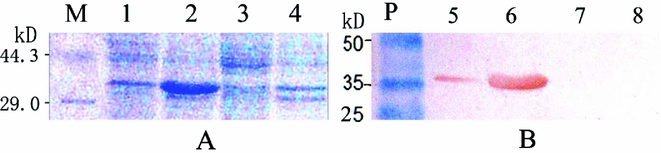
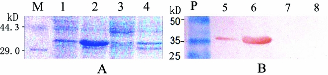
Western blotting with monoclonal antibody against the 6His tag showed that a specific protein with a molecular mass of approximately 36 kDa was detected in the lysate and in the inclusion body of transformed E. coli cells (Fig. 1 B, lanes 5 and 6), but not in the pre-induced E. coli Rosetta cells (Fig. 1 B- 7 , B-8). This result indicates that the fusion protein ‘Z2eH’ was expressed in E. coli Rosetta cells after isopropyl β-d-thiogalactoside (IPTG) induction, and that most of the product formed inclusion bodies (Fig. 1 A- 2 , B-6). Protein quantification showed that the total yield of recombinant protein reached 45 mg in a 1 litre culture after an 8 h induction at 25°C.
SDS-PAGE (A) and western blotting (B) of lysates of the z2eh-transformed E. coli Rosetta cells. Lane M: protein marker. Lane P: pre-stained protein marker. Lanes 1 and 5: the lysate supernatant of the z2eh-transformed Rosetta cells after 8 h IPTG induction. Lanes 2 and 6: urea-dissolved inclusion body of the z2eh-transformed Rosetta cells after IPTG induction. Lanes 3 and 7: the lysate supernatant of the z2eh-transformed E. coli Rosetta cells before induction. Lanes 4 and 8: the dissolved pellet of the z2eh-transformed E. coli Rosetta cells before induction.
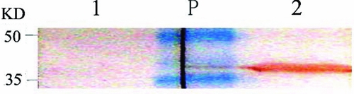
Western blot of the lysate of z2eh-transformed E. coli Rosetta cells. Lane 1: reacted with control rabbit serum. Lane 2: reacted with rabbit anti-Z2eH antiserum. Lane P: pre-stained protein standard. The membrane was split through lane M into two strips for individual antibody incubation and then combined together after staining.
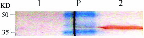
When the two strips of electronically transferred NC membrane were incubated with rabbit anti-Z2eH antiserum and control rabbit serum, respectively, an intense brown band appeared on the lane of the strip incubated with anti-Z2eH antiserum, no band was seen on the strip incubated with the rabbit control serum (Fig. 2). The brown band was a bit more than 35 kDa in weight, based on the pre-stained protein marker, corresponding to the calculated molecular mass of the recombinant Z2eH peptide. This finding indicated that the anti-Z2eH antibody reacted specifically with the recombinant ZP2 peptide.
Anti-Z2eH antibody bound specifically to the native mouse zona pellucida
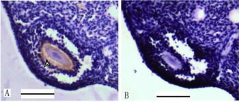
Immunohistochemical assay showed that evident colour reaction occurred in the antrum follicles of section A when incubated with rabbit anti-Z2eH antiserum (Fig. 3 A), whereas no brown particle was seen on the follicles of section B incubated with control rabbit antiserum (Fig. 3 B). The brown particles of section A were distributed on the region typically attributed to the zona pellucida around the oocyte. This result indicated that the rabbit anti-Z2eH antibody reacted specifically with the natural zona pellucida of mouse ovary.
Immunohistochemistry assay of the mouse ovarian sections with rabbit anti-Z2eH antiserum (A) and control rabbit serum (B). The antigen–antibody reaction is shown as the brown particles around the oocyte of (A) (arrowhead). Scale bar: 100 μm.

Antibody-treated oocytes bound sperm as normal
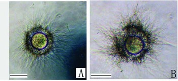
The sperm–egg binding assay showed that anti-Z2eH antibody treatment did not reduce sperm binding (Fig. 4). After the oocytes were co-cultured with sperm suspension, the average number of sperm bound to one oocyte was 382.6 ± 26.2 in the control group, and 400.4 ± 57.0 in the antibody-treated group. Statistical analysis showed that the difference in numbers of sperm bound to the oocytes was not significant between the anti-Z2eH antibody-treated oocytes and the control oocytes (P > 0.05). These results indicated that the anti-Z2eH antibody treatment did not interrupt sperm–egg binding significantly.
Influence of anti-Z2eH antibody on sperm–egg binding. (A) Sperm binding to control oocytes. (B) Sperm binding to anti-Z2eH antibody-treated oocytes. Scale bar: 100 μm.

Anti-Z2eH antibody bound on the zona pellucida of the ovulated oocytes after passive immunization
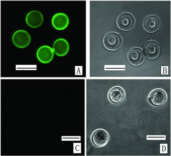
When the mice were given anti-Z2eH antibody intraperitonally 1 day before ovulation, the zona pellucida in the COCs bound the antibody when the oocytes flowed into the oviducts, despite the thick cumulus cells layer (Fig. 5). This finding indicated that antibody in the abdominal cavity diffused and permeated through cumulus cell layer.
The immunofluorescence assay of the zona pellucida of the ovulated oocytes. Fluorescence microscope photograph (A) and phase-contrast microscope photograph (B) of the ovulated oocytes of mice treated with anti-Z2e antibody. Fluorescence microscope photograph (C) and phase-contrast microscope photograph (D) of the ovulated oocytes of control mice administered with rabbit serum. Scale bar: 100 μm.

Binding of antibody to zona pellucida did not interrupt fertilization in vivo
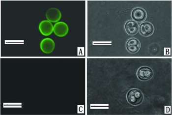
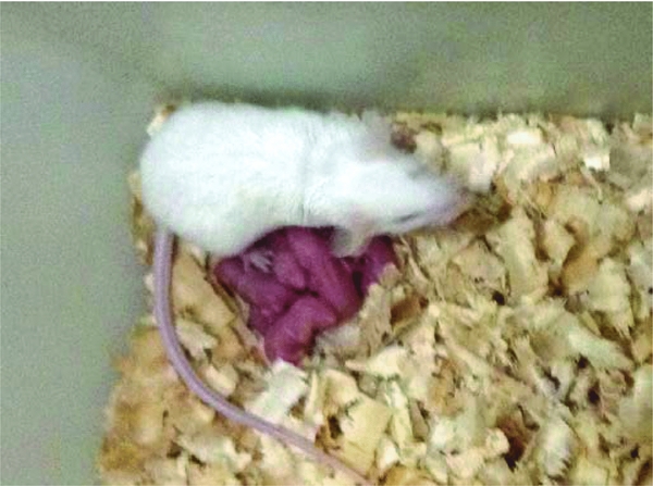
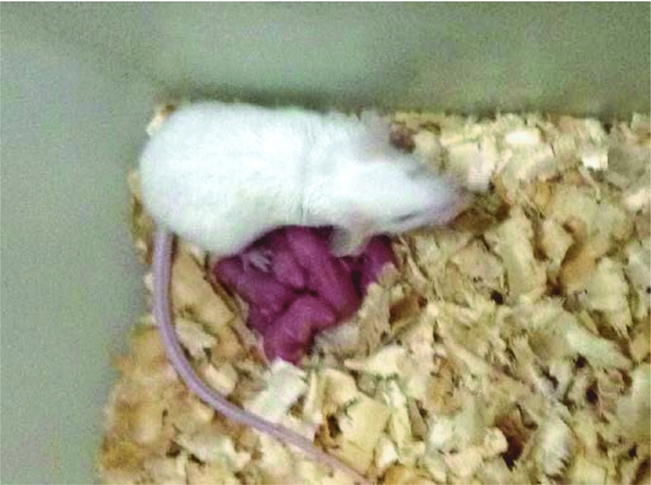
Although antibody bound the zona pellucida of the ovulated oocytes after passive immunization of anti-Z2eH antibody, the oocytes were fertilized by sperm in the oviduct after ovulation and zygotes cleaved into 2-cell embryos as normal (Fig. 6). It is indicated that anti-Z2eH antibody did not stop fertilization in vivo. The conceived mice had a normal pregnancy and gave birth at the regular term (Fig. 7).
Immunofluorescence assay of the 2-cell embryos after passive immunization of anti-Z2eH antibody. Fluorescence microscope photograph (A) and phase-contrast microscope photograph (B) of the 2-cell embryos of mice administered the anti-Z2e antibody. Fluorescence microscope photograph (C) and phase-contrast microscope photograph (D) of the 2-cell embryos of the control mice. Scale bar: 100 μm.

The newborn mouse pups of a passively immunized mouse.
Discussion
The full-length ZP2 is a glycoprotein with 713 amino acid residues. Early experiments reported that five monoclonal antibodies specific to mouse ZP2 were produced following immunization with native zona pellucida glycoproteins (East & Dean, Reference East and Dean1984). At least one epitope of ZP2 was related to sperm–zona recognition (Sun et al., Reference Sun, Lou, Dean and Tung1999; Tsubamoto et al., Reference Tsubamoto, Hasegawa, Nakata, Naito, Yamasaki and Koyama1999).
The synthesized ZP2 peptide 121–140 was reported to elicit IgG antibody that reacted with mouse ovarian ZP and reduced litter sizes (Sun et al., Reference Sun, Lou, Dean and Tung1999). The recombinant N terminal polypeptide 1–206 portion of human ZP2 was also shown to contain a binding site for acrosome-reacted spermatozoa and to play an important role in secondary sperm binding and penetration into the ZP (Tsubamoto, et al., Reference Tsubamoto, Hasegawa, Nakata, Naito, Yamasaki and Koyama1999). However, immunization with recombinant ZP2V35–G200 did not result in a significant immune response that recognized native ZP (Lea et al., Reference Lea, Widgren and O'Rand2002). Other experiments showed that ZP2 peptide 541–555 was implicated in sperm–egg recognition (Hinsch et al., Reference Hinsch, Hagele, Bohle, Schill and Hinsch1998). Therefore the molecular mechanism of the sperm–ZP2 recognition remained confused. Our present experiments showed that the Z2eH polypeptide elicited a strong immune response and the resultant antibody reacted specifically with the natural zona pellucida. Therefore, the central peptide of ZP2 190–505 is immunogenic and should contain ZP-specific epitopes.
When the oocytes were treated with anti-Z2eH antibody, sperm bound actively and stably to the treated zona pellucida. Moreover, fertilization and embryo development were not obstructed, although intraperitoneally injected antibody bound to the ovulated oocytes. Therefore, the central fragment of ZP2 may not contribute to forming the sperm-binding site.
Early research with four recombinant ZP2 fragments, ZP2V35–G200, ZP2V35–L331, ZP2P325–A637 and ZP2V35–A637 showed that antibodies capable of recognizing native zona pellucida occurred only after immunization with ZP2V35–A637 and ZP2P325–A637 Only immunization with ZP2V35–A637, rather than with ZP2P325–A637, was correlated with a reduction in fertility (Lea et al., Reference Lea, Widgren and O'Rand2002). So it seems that the sperm-binding site on ZP2 may not be located simply in a single linear amino acid sequence.
Some experiments showed that the N terminus of ZP2 peptide was verified to bind sperm stably (Baibakov et al., Reference Baibakov, Boggs, Yauger, Baibakov and Dean2012) and sperm–egg recognition depended on the cleavage status of ZP2 (Baibakov et al., Reference Baibakov, Gauthier, Talbot, Rankin and Dean2007; Gahlay et al., Reference Gahlay, Gauthier, Baibakov, Epifano and Dean2010). It is probable that the amino acid residues of the cleavage site form a particular conformation that helps sterically to create a sperm-binding site. The cleavage frame contorts the ZP2 peptide chain and moves the central segment of ZP2 from the sperm-binding site, although ZP2 190–505 is less than 30 amino acid residues apart from the cleavage site. Also ZP2 may bind sperm through the oligosaccharide side chains on the ZP2 polypeptide, rather than on the polypeptide itself.
In conclusion, the central polypeptide of ZP2 190~505 is immunogenic, antibody against the peptide is capable of recognizing native zona pellucida, however it might not be the crucial part needed to form a functional domain to bind sperm.
Financial support
This work was supported in part by the collaborated grant for HK-Macao-TW from the Ministry of Science and Technology of China (2012DFH30060).