Background
Total anomalous pulmonary venous return is a rare congenital anomaly in which the pulmonary veins do not connect to the left atrium. Obstruction of the alternative pathway for the pulmonary venous return will result in marked pulmonary venous congestion, elevated pulmonary pressure, and impairment to right ventricular function. Historically, mortality was high especially in the obstructed cases at over 90%. Reference Duff, Nihill and McNamara1,Reference Louis, Harvey and Menk2 More recently, better operative techniques and improved diagnostic accuracy have increased the survival rates to 93–97%. Reference Shi, Zhu and Chen3 Early recognition and surgical repair will usually result in an optimal outcome, though later anastomotic pulmonary venous obstruction can occur associated with a high mortality. Reference Shi, Zhu and Chen3,Reference Karamlou, Gurofsky and Sukhni4
The development of gastric varices in a newborn with obstructed pulmonary venous return is an extremely rare finding and has the potential for catastrophic gastrointestinal haemorrhage. Reference Elias-Jones and Cordner5,Reference Laurence and Brown6
Case
We present a term newborn male infant, noted to be hypoxic. This prompted an echocardiogram, which was suspicious for total anomalous pulmonary venous return and triggered transfer to our paediatric tertiary centre for diagnosis confirmation and management.
Investigations
An initial chest radiograph demonstrated hyperinflated lungs, bilateral interstitial oedema, and normal cardiac silhouette.
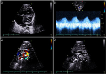
Echocardiography demonstrated the right and left pulmonary veins joining to become a descending common vein, coursing inferior through the diaphragm (Fig 1a–d). Exact location of obstruction could not be defined. An enlarged right ventricle, an atrial septal defect, and patent ductus arteriosus with moderate right-to-left shunting were noted. A small tortuous venous structure was seen coursing across the midline from right to left entering the innominate vein. Obstructed total anomalous pulmonary venous return was diagnosed, suspected to be the mixed type.
(a–d) CRX pre-op.

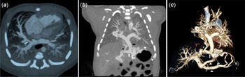
Due to the complicated anatomy, a CT scan was arranged. A sequential, cardiac-gated CT scan was performed using a dual-source scanner. CT images demonstrated infracardiac total anomalous pulmonary venous return with the vertical vein having dual connections to the ductus venosus and a dilated left gastric vein (Fig 2a–c). A focal stenosis was present where the common vein is connected to the ductus venosus. The gastric branch is connected to the patent portal venous system via dilated and tortuous right and left gastric veins, gastroepiploic and short gastric veins.
(a–c) CRX post-op.

All pulmonary veins communicated with the venous confluence. A collateral vein connected the right middle lobe vein with the left brachiocephalic vein (Fig 2). This collateral vein was thought to be a supracardiac mixed connection on echocardiogram (Fig 1).
Management
There was continued clinical deterioration with worsening pulmonary oedema and corrective surgery was undertaken on day 3 of life.
The child is doing well at 1-year old with no other congenital lesion or suspected genetic syndrome. Echocardiography shows good biventricular function and laminar flow across the surgical anastomosis. No episodes of upper gastrointestinal haemorrhage occurred.
Discussion
Total anomalous pulmonary venous return is rare accounting for about <2% of all congenital cardiac anomalies. There are four types described by the Darling classification. The infracardiac (type III) represents 26% of cases with connections to the hepatic veins, inferior vena cava, portal vein, or ductus venosus. Reference Coley7 A significant proportion of the infradiaphragmatic type are associated with other congenital anomalies including cardiac lesions and heterotaxy syndromes. Reference Duff, Nihill and McNamara1,Reference Louis, Harvey and Menk2 There is evidence of an inheritable genetic link. Those with obstructed veins at presentation, 87% of infracardiac type, and other anomalies have poorer outcomes. Reference Louis, Harvey and Menk2,Reference Coley7
The association with gastric varices is rare with only a handful of cases in the literature. The cause of development of gastric varices is unknown, most likely related to the degree of obstruction of the path of alternative pulmonary venous drainage. Chen et al describe two neonatal cases of oesophageal varices with no anastomosis between the anomalous pulmonary vein and portal venous system, which precludes portal hypertension as a prerequisite for the formation of varices. Reference Chen, Chen and Li8,Reference Park, Lee, Ban, Jo and Sung9 Laurence and Brown described infradiaphragmatic common vein draining directly only to the left gastric vein. Reference Laurence and Brown6 Oesophageal varices have been described causing upper gastrointestinal haemorrhage in uncorrected infradiaphragmatic total anomalous pulmonary venous return. Reference Laurence and Brown6
Our patient had two subdiaphragmatic connections: one to the ductus venosus and one to the gastric veins. Given the rapid clinical deterioration, the alternate drainage via the gastric/gastroepiploic venous system was inefficient in decompressing the pulmonary venous hypertension caused by the closing ductus venosus.
Early in embryonic development, the pulmonary and splanchnic venous plexuses are connected via the common cardinal and the umbilico-vitelline venous systems. Reference Kao, Hsieh, Cheng, Chiang and Huang10 In the fourth week of gestation, the lung buds are enmeshed by the pulmonary venous plexus and the splanchnic venous plexus. Reference Kao, Hsieh, Cheng, Chiang and Huang10 In the second month, a common pulmonary vein invagination develops from the posterior wall of the left atrium and subsequently connects with the pulmonary venous plexus as the splanchnic connections involute. Failure of this connection to form can lead to persistence of the splanchnic connections. Reference Kao, Hsieh, Cheng, Chiang and Huang10 This would explain the “common vertical vein” having connections to both the ductus venosus and/or left gastric vein.
The true incidence of total anomalous pulmonary venous return with associated gastrointestinal varices is unknown. Echocardiography is not sensitive for the detection of gastrointestinal varices. The urgency of corrective surgical intervention might not allow for the varices to manifest clinically. The described cases of catastrophic gastrointestinal tract haemorrhage associated with total anomalous pulmonary venous return were mainly in the pre-operative period.
Echocardiography is the current diagnostic modality of choice. However, should there be any question regarding heterotaxy, location, or size of the pulmonary veins, then pre-operative CT angiography of the chest and upper abdomen will provide accurate high-resolution imaging to guide the surgery.
Conclusion
Total anomalous pulmonary venous return is an uncommon congenital cardiovascular anomaly with unusual variations that need to be well documented including associated gastric varices. Early imaging with contrast-enhanced CT should be considered in atypical cases in order to best plan for surgery and offer the best outcome for patients.
Supplementary Material
To view supplementary material for this article, please visit https://doi.org/10.1017/S1047951120003479
Acknowledgements
We greatly appreciate the support and encouragement extended for this brief report by Dr Martin Hosking and Dr Jim Potts.
Financial Support
This research received no specific grant from any funding agency, commercial, or not-for-profit sectors.
Conflicts of Interest
None.
Ethical Standards
No Applicable.