Introduction
Epicardial adipose tissue is a metabolically active fat depot that derives from brown adipose tissue during embryogenesis and is in anatomic and functional proximity to the myocardium and coronary arteries. Epicardial adipose tissue has a protective effect on the heart against hypothermia and has been identified as being predictive of cardiovascular disease in adulthood. Recently, Epicardial adipose tissue has been reported to contribute to early diastolic dysfunction in the heart, similar to changes also found in hypertrophic cardiomyopathy. Reference Monti, Marchetti and Vincentelli1
Gestational diabetes mellitus is a metabolic disease that manifests as insulin resistance during pregnancy and causes many metabolic changes in the fetus. It has been known for many years that asymmetric septal hypertrophy and hypertrophic cardiomyopathy develop in infants of diabetes mothers as a result of insulin and other growth factors that stimulate the heart. Reference Al-Biltagi, El Razaky and El Amrousy2 Even under conditions of good prenatal glycaemic control, fetuses of mothers with gestational diabetes mellitus remain at risk of mild cardiac hypertrophy. Reference Weber, Copel, Reece, Green and Kleinman3 It has been explained that good prenatal glycaemic control, especially in the 3rd trimester, can reduce the frequency of neonatal complications such as macrosomia, but cannot reduce the tendency for cardiac enlargement and septal hypertrophy. Reference Litwin, Sundholm, Rono, Koivusalo, Eriksson and Sarkola4
Epicardial adipose tissue has been reported to increase in insulin resistance and diabetes mellitus patients. It has been shown that the fetuses of pregnant women with diabetes have a higher epicardial fat thickness than the fetuses of pregnant women without diabetes. Reference Yavuz, Akkurt, Yalcin, Karakoc, Varol and Sezik5 However, there are very few publications on the detection of epicardial fat thickness status in infants of diabetes mothers and its association with cardiac hypertrophy. This study aimed to compare epicardial fat thickness and interventricular septum diameter values in infants of diabetes mothers with infants of non-diabetic mothers and evaluate the effect of birth weight on epicardial fat thickness and interventricular septum diameter values in infants of diabetes mothers.
Materials and methods
Study population
Infants of diabetes mothers aged 0-30 days who presented to the paediatric cardiology outpatient clinic between November 2022 and June 2023 were evaluated. Infants with a maternal history of gestational diabetes were included in the study. Those with a maternal history of type 1, type 2 diabetes mellitus, or additional maternal diseases were excluded from the study. Prematurity, low birth weight (< 2500 g), CHD, or a history of comorbidities were also excluded from the study. Healthy infants of non-diabetic mothers aged 0–30 days, whose birth weight was between 2500 g and 4000 g, were included in the study as the control group.
Maternal age, gestational diabetes history, treatment, and diet during pregnancy were evaluated. Demographic data (birth weight, birth length, birth week, and perinatal history) of infants of diabetes mothers were evaluated. Infants of diabetes mothers with a birth weight of ≥ 4000 g were considered macrosomic and those with a birth weight of 2500–4000 g were non-macrosomic.
We classified infants of diabetes mothers into two groups: the infants of mothers with uncontrolled gestational diabetes (mothers who neither use insulin nor apply diet, uncontrolled infants of diabetes mothers) and the infants of mothers with controlled gestational diabetes (mothers who use insulin and apply a diet, controlled infants of diabetes mothers). We compared these two groups.
Echocardiographic measurements
All patients underwent two-dimensional echocardiography, Doppler, color Doppler, and M mode examinations. All measurements were performed using a Vivid 5S (General Electric Medical Systems) echocardiography device.
Left ventricular systolic function measurements were performed in M mode. The diastolic thickness of interventricular septum diameter, the systolic and diastolic diameter of the left ventricle, the diastolic thickness of the posterior wall of the left ventricle, ejection fraction, and shortening fraction measurements were evaluated in M mode in the parasternal long axis position. Aorta, pulmonary artery, mitral, and tricuspid valve diameters were recorded. Interventricular septum diameter > 6 mm was considered asymmetric septal hypertrophy.
Epicardial fat thickness was measured at the end of diastole from the right ventricular free wall in the parasternal long axis position in two-dimensional mode. While the mitral valve, aortic valve, and right and left ventricle were visible, Epicardial fat thickness measurements were performed in a position where a parallel view of the aorta and left atrium could be achieved (most often in an upper costal view) (Figure 1).
Measurement of epicardial fat thickness at the end of diastole from the right ventricular free wall in the parasternal long axis position in two-dimensional mode. EFT = epicardial fat thickness; LA = left atrium; LV = left ventricle; RV = Right ventricle.

Infants of diabetes mothers and the control group (infants of non-diabetic mothers) were compared in terms of demographic data and echocardiography findings. Infants of diabetes mothers were divided into two groups according to their birth weight: macrosomic infants (≥ 4000 g) and non-macrosomic infants (2500–4000 g). These two infants of diabetes mothers groups were compared with each other in terms of demographic and echocardiography findings.
Statistical analysis
The data were analysed using the IBM SPSS Statistics software package (Version 21.0. Armonk, NY: IBM Corp.). Categorical data were expressed as numbers (%) and compared using the Chi-square test. The normality of the data distribution was checked using the Kolmogorov–Smirnov test. Student’s t-test was used to compare unpaired samples and identify the between-group differences. The Mann–Whitney U test was used to compare non-parametric numerical data. Pearson´s correlation test was used to value the correlation between different echocardiographic parameters and epicardial fat thickness. Spearman’s non-parametric correlation test was used to value the correlation between asymmetric septal hypertrophy and epicardial fat thickness. An alpha of 0.05 was applied in all tests.
Results
A total of 78 infants of diabetes mothers and 56 control group (infants of non-diabetic mothers) were evaluated. The ages of the infants of diabetes mothers ranged from 3 to 30 (median: 16) days and the ages of the control group ranged from 1 to 30 (median: 12) days. Forty-two (53.8%) infants of diabetes mothers were girls and 36 were boys. The delivery method of the infants of diabetes mothers was normal vaginal delivery in 34 (44%) and cesarian section delivery in 44 (56%). The mean birth weight of the infants of diabetes mothers was 3600 ± 628 g, and the mean birth length was 50.6 ± 1.5 cm. Thirty-one (35%) infants of diabetes mothers needed incubator care after birth.
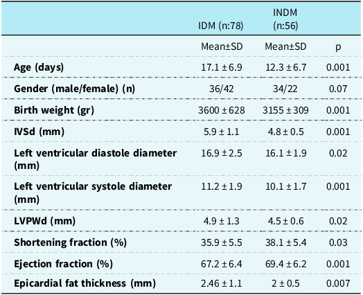
When the epicardial fat thickness and interventricular septum diameter parameters of the infants of diabetes mothers and control group were compared, epicardial fat thickness and interventricular septum diameter measurements were found to be statistically significantly higher in infants of diabetes mothers than in the control group (p = 0.007 and p = 0.001, respectively). Additionally, a statistically significant difference was found between posterior wall of the left ventricle, ejection fraction, and shortening fraction values when infants of diabetes mothers were compared with control group (Table 1). Correlation coefficient values between epicardial fat thickness and interventricular septum diameter in infants of diabetes mothers :0.413
Comparison of infants of mothers with diabetes and infants of mothers without diabetes

IDM = infants of mothers with diabetes; INDM = infants of mothers without diabetes; IVSd = interventricular septum diameter; LVPWd = left ventricle posterior wall diameter.
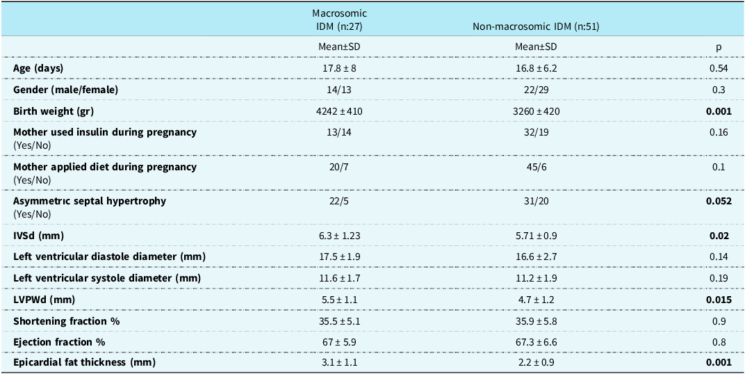
When the birth weights of infants of diabetes mothers were evaluated, it was seen that 27 (35%) infants of diabetes mothers were macrosomic (≥ 4000 g) and 51 (65%) were non-macrosomic (< 4000 g). When macrosomic and non-macrosomic infants of diabetes mothers were compared, epicardial fat thickness, interventricular septum diameter, and posterior wall of the left ventricle measurements were found to be statistically significantly higher in macrosomic infants of diabetes mothers than in non-macrosomic infants of diabetes mothers (p = 0.001, p = 0.02, and p = 0.015, respectively). Asymmetric septal hypertrophy was found to be more common in macrosomic infants of diabetic mothers than in non-macrosomic infants, although not statistically significant (p = 0.052). We also found a positive correlation between epicardial fat thickness and asymmetric septal hypertrophy (Table 2).
Comparison of infants of macrosomic and non-macrosomic of diabetic mothers

IDM = infants of mothers with diabetes; INDM = infants of mothers without diabetes; IVSd = interventricular septum diameter; LVPWd = left ventricle posterior wall diameter.
The mean age of mothers with diabetes was 32.9 ± 5.75 years. The mean number of pregnancies of the mothers with diabetes was 2.9 ± 1.5, and the number of live births was 2.7 ± 1.3. Forty-five (57%) of the mothers with diabetes used insulin during pregnancy, and 65 (83%) dieted during pregnancy. When the maternal age, insulin use, and diet of mothers with diabetes were compared, no significant difference was found between macrosomic and non-macrosomic infants of diabetes mothers regarding maternal age, insulin use, and dieting during pregnancy (Table 2).
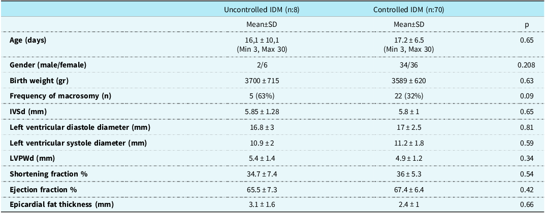
When uncontrolled infants of diabetes mothers and controlled infants of diabetes mothers were compared, although birth weight, interventricular septum diameter, posterior wall of the left ventricle, and epicardial fat thickness values were higher in uncontrolled infants of diabetes mothers cases than in controlled infants of diabetes mothers cases, no statistically significant difference was detected. This may be due to the fewer uncontrolled infants of diabetes mothers than controlled infants of diabetes mothers (Table 3).
Data distribution according to mother’s diabetes control (demographic and echocardiographic)

IDM = infants of mothers with diabetes, INDM = infants of mothers without diabetes, IVSd = interventricular septum diameter, LVPWd = left ventricle posterior wall diameter.
Discussion
Epicardial fat tissue is the visceral fat depot of the heart, anatomically and functionally related to the myocardium, which has a biochemical and thermogenic cardioprotective effect. Reference Iacobellis, Malavazos and Corsi6 Epicardial adipose tissue can locally affect the heart and coronary arteries through the secretion of pro-inflammatory cytokines. Reference Monti, Marchetti and Vincentelli1,Reference Iacobellis, Malavazos and Corsi6 Many studies have reported that epicardial fat thickness is associated with metabolic syndrome and coronary artery disease. Reference Monti, Marchetti and Vincentelli1 It is known that epicardial fat thickness also increases insulin resistance and diabetes mellitus. A study conducted on gestational diabetes mellitus showed that epicardial fat thickness was statistically significantly higher in the fetuses of mothers with gestational diabetes mellitus compared with the control group. Reference Weber, Copel, Reece, Green and Kleinman3,Reference ElSayed, Aleppo and Aroda7
It has been reported that echocardiography evaluation of epicardial fat thickness is a useful method for evaluating cardiovascular risks, especially in children with obesity. Özdemir et al. found that children with obesity had significantly higher epicardial fat thickness compared with lean children. Reference Ozdemir, Hizli, Abaci, Agladioglu and Aksoy8 Avcı et al. reported that carotid intima-media thickness, arterial stiffness, and epicardial fat thickness values were significantly higher in the children of mothers with gestational diabetes mellitus and maternal obesity than in the control group. Reference Avci and Tosun9 In the cross-sectional study of Vela-Huerta et al., epicardial fat thickness was found to be higher in infants of diabetes mothers than in healthy infants. Reference Vela-Huerta, Amador-Licona and Dominguez-Damian10 In our study, we found that the epicardial fat thickness was thicker in infants of diabetes mothers compared with the control group and that the epicardial fat thickness was also thicker in macrosomic infants of diabetes mothers compared with non-macrosomic infants of diabetes mothers.
Many studies reported that epicardial fat tissue affected the structural and functional properties of the heart, such as increased left ventricular mass, abnormal right ventricular geometry, and subclinical diastolic dysfunction. It is reported that the effect of epicardial fat thickness on left ventricular mass increase is independent of coronary artery disease and hypertrophy. Reference Gaborit, Sengenes, Ancel, Jacquier and Dutour-Meyer11 In our study, we found that interventricular septum diameter and posterior wall of the left ventricle measurements were higher in infants of diabetes mothers than in the control group and that interventricular septum diameter and posterior wall of the left ventricle values were also higher in macrosomic infants of diabetes mothers than non-macrosomic infants of diabetes mothers.
In the present study, ejection fraction and shortening fraction were lower in infants of diabetes mothers compared to the control group. We think this is due to the change in ventricular geometry. When we look at the systolic diameters, it provides us with the measurement value of approaching both walls during ventricular systole. This was detected more in infants of diabetes mothers than in the control group. There should be a change in organisation compared to the normal group within thicker walls.
It has been reported that macrosomia, asymmetric septal hypertrophy, hypertrophic cardiomyopathy, and diastolic dysfunction are observed in infants of diabetes mothers as a result of insulin and other growth factors that stimulate the fetal heart. Reference A.Lawlor, Fraser and Lindsay12,Reference Kozák-Bárány, Jokinen, Kero, Tuominen, Rönnemaa and Välimäki13 Asymmetric septal hypertrophy appears to be the most common echocardiographic change found among infants of diabetes mothers. In their study on control group and infants of diabetes mothers, Mohy-Elddin et al showed that macrosomia causes asymmetric septal hypertrophy in both groups. It is known that asymmetric septal hypertrophy is generally benign and often tends to normalise within 6 months postnatally. Reference Rijpert, Breur and Evers14 In our study, we found that there was a significant correlation between epicardial fat thickness and asymmetric septal hypertrophy in infants of diabetes mothers. We also found that asymmetric septal hypertrophy was more common in macrosomic infants of diabetic mothers than in non-macrosomic infants, although it was not statistically significant. This shows us that asymmetric septal hypertrophy is not important for infants of non-macrosomic diabetic mothers. Our study also supports this finding of fetal macrosomia seen in gestational diabetes.
Gestational diabetes has been reported to be the most important cause of neonatal macrosomia. The presence of neonatal macrosomia in infants of diabetes mothers predicts poor glycaemic control of the mother during the gestational period. Additionally, it appears that the increase in epicardial fat thickness parallels fetal–neonatal macrosomia. The lack of a significant increase in epicardial fat thickness and interventricular septum diameter in non-macrosomic infants of diabetes mothers may be indicative of good glycaemic control in non-macrosomic infants of diabetes mothers.
Study limitation
Our study has a small sample size and is a cross-sectional study. We cannot provide information about epicardial fat thickness in long-term follow-up in infants of diabetes mothers. We cannot provide detailed information about the mother’s compliance with diet and insulin treatment because information about the mothers’ diet and insulin use was obtained verbally from the mothers. We cannot make a clear comment about the relationship between pre-gestational obesity and epicardial fat thickness.
Conclusion
In our study, we found epicardial fat thickness, interventricular septum diameter, and posterior wall of the left ventricle values to be significantly higher in infants of diabetes mothers than in control group. We also observed that epicardial fat thickness, interventricular septum diameter, and posterior wall of the left ventricle values were higher in macrosomic infants of diabetes mothers than in non-macrosomic infants of diabetes mothers. This shows that insulin affects epicardial fat thickness just as it has an effect on heart mass in infants of diabetes mothers. Epicardial fat thickness is considered a predictive factor of cardiovascular diseases. For this reason, follow-up of mothers with gestational diabetes mellitus, good glycaemic control, and long-term follow-up of the infants of mothers with diabetes after birth are important.
Acknowledgements
None.
Financial support
This research received no specific grant from any funding agency, commercial, or not-for-profit sectors.
Competing interests
None.
Ethical standards
This study was approved by the Adıyaman University Institutional Review Board (Project No: 2022/7-23).The study protocol was conducted in accordance with the ethical guidelines of the 1975 Helsinki Declaration, as revised in 2008.