There remains much controversy, and perhaps even confusion, regarding those particular anomalies which can justifiably be included within the overarching designation of bronchopulmonary foregut malformations.1 Contributing in part to this confusion are the multiple names given to these malformations in their individual forms. In the past, they have been described in terms of pulmonary sequestration, bronchopulmonary sequestration, expanded sequestration, the spectrum of pulmonary sequestration, pulmonary venolobar syndrome, communicating and non-communicating bronchopulmonary foregut malformations, and malinosculation, with still other terms also being used.2–21 In the light of these various terminologies and classifications, Bush22 has urged that a consistent, uniform, and simplified nomenclature be adopted for congenital malformation of the lung. Others have attempted to provide such unifying approaches based on anatomical and embryological considerations, or aetiological concepts.1, 4–6, 8–10, 13, 14, 17–21 At this juncture, the proposed suggestions for simplification and unification of nomenclature have not been widely adopted. One reason for this, of course, might be that none of the proposed theories is able to explain the origin of these diverse malformations. In this regard, Heithoff et al.,6 concluding their own attempt to provide a unifying aetiological concept, acknowledged the comments and criticisms of Boyden,23–26 who contributed so importantly to our knowledge of congenital pulmonary malformations, and who had reviewed their paper. In the final lines of their paper, they cited Boyden, who had stated “the development of the bronchopulmonary segments and the associated anomalies is so complex that rarely can one hypothesis cover all variations”.6 In this review, we will concentrate on the major vascular components of the anomalies included within the title of bronchopulmonary foregut malformations. Some consider pulmonary arteriovenous malformations as part of the continuum of pulmonary developmental anomalies. Since we have recently described many features of these anomalies in another review published in the journal,27 we will not discuss them further in this one.
Pulmonary sequestration
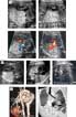
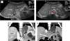
Pulmonary sequestration, a term first used by Pryce2 in 1946, is used to describe a segment of lung that does not have any identifiable connection with the normal tracheobronchial tree, and which receives its arterial supply from one or more systemic arteries, rather than from the right or left pulmonary arteries (Figs 1 and 2). According to Pryce,2 such abnormal pulmonary tissue had previously been said to be ectopic or dislocated. Much has now been written concerning the pathology, the clinical features, the imaging, and the options for treatment of patients with this anomaly. Despite this wealth of literature, there still seems to be confusion as to the best definition. Some use sequestration, as suggested by Pryce,2 in terms of lack of normal tracheobronchial connection, while others use the term in the context of absence of the normal pulmonary arterial supply. In our opinion, it is best if sequestration is used as suggested by Pryce.2 When defined in this fashion, sequestrations are then divided into two types based on the nature of their pleural covering.2–5, 8–11, 13, 17, 21, 28–35 An extralobar sequestration is a mass of pulmonary parenchyma with a distinct pleural covering separating it from the adjacent normal tissues of the lung (Fig. 1).2–5, 8–11, 13, 17, 21, 28–35 In contrast, an intralobar sequestration does not have a separate pleural investment, and the sequestered mass is contiguous with the normal lung, being contained within the normal visceral pleura (Fig. 2).

Figure 1. Extralobar pulmonary sequestration. Reformatted contrast-enhanced CT angiograms in left anterior oblique planes (left-hand and middle panels) and axial plane show a pyramid-shaped, non-functioning, sequestered mass of pulmonary tissue. The mass is supplied by an aberrant systemic artery (asterisk in left-hand panel). It drained to the left atrium through a pulmonary vein (white arrow in middle panel), and to the azygos venous system (black arrow in right-hand panel). It is sharply outlined.

Figure 2. Intralobar pulmonary sequestration. (a) Fetal sonograms obtained at 21 weeks of gestation show a hyperechogenic mass involving the left lower lobe. The heart is displaced to the right and forward. Color Doppler sonograms demonstrate the aberrant systemic arterial branch supplying the echogenic mass and the draining pulmonary vein that connect to the left atrium. (b) Follow-up sonograms at 25 and 36 weeks show spontaneous regression of the mass. At 36 weeks, the heart is only mildly displaced but the left lower lung still is supplied by a systemic artery. (c) Reformatted CT angiograms obtained in neonatal period show a mass of abnormal lung tissue in the left lower lobe that is supplied by a large aberrant systemic artery, and which drained to the left atrium through the dilated left lower pulmonary vein. The abnormal lung contains irregular collections of air and its margin is not clearly delineated.
Pulmonary sequestration is rare, with an incidence ranging from 0.15 percent to 1.8 percent,28, 29, 33 albeit that this figure almost certainly underestimates the true incidence, with some patients, especially those with an intralobar sequestration, being asymptomatic. Some have extended this classification to include as a third pattern those sequestrations associated with bronchopulmonary foregut malformations.1, 4–6, 8–10, 12–18, 21 Thus, seeking to broaden the concept of pulmonary sequestration, Sade et al.4 focussed on the spectrum that included a continuum of developmental anomalies involving all combinations of pulmonary and systemic arterial supply, pulmonary and systemic venous drainage, normal and abnormal pulmonary tissue, gastrointestinal fistula, and deficiencies or accessory formation of the diaphragm. This expanded concept of the spectrum of pulmonary sequestration included all those diverse congenital malformations embraced by the overarching designation of bronchopulmonary foregut malformations, this term having first been used by Gerle and colleagues in 1968.1 It is under this latter term that we have chosen to group together these diverse congenital malformations.
Most consider, therefore, that the spectrum of bronchopulmonary foregut malformations includes:
- Tracheal stenosis
- Bronchogenic cysts
- Bronchopulmonary sequestrations
- Congenital cystic adenomatoid malformations
- Bronchial atresia or stenosis
- Congenital lobar emphysema
The spectrum includes both communicating and non-communicating anomalies.36–40 Thus, the term encompasses a wide variety of anomalies occurring because of abnormal differentiation of the respiratory and alimentary tracts, abnormal separation of the two systems, or abnormal development of arterial blood supply, with the lesions developing perhaps singly, or in combination, during early embryogenesis.1, 6, 12, 14–16, 36–41 Common to all types is the presence of accessory pulmonary tissue that arises from the primitive gastrointestinal tract. In their discussion of the multiple facets of pulmonary sequestration, Bratu et al.17 suggest that, to facilitate management, such malformations should be described according to:
- Types of connection to tracheobronchial tree
- Arrangement of the visceral pleura
- Arterial supply
- Venous drainage
- Nature of communication with the foregut
- Histology
- Mixed or multiple lesions
- Associated malformations
Heithoff et al.,6 whom we have already cited, suggested that a common embryologic pathogenesis leads to a variety of malformations including:
- Intralobar and extralobar sequestration
- Pulmonary sequestration with patent or involuted gastro-oesophageal communication
- Oesophageal or gastric diverticulums
- Oesophageal or bronchogenic duplicated cysts
- Congenital cystic adenomatoid malformations of the lung
Thilenius et al.5 expanded the ongoing dialogue when they emphasized, as had others,42 the association with anomalous pulmonary venous drainage. Several years later, Clements et al.8, 9 reiterated that congenital malformations of the bronchopulmonary airways and their related arterial blood supply represent a complex group of lesions in which abnormalities of lung parenchyma and venous drainage co-exist. They reminded us that the abnormalities of the pulmonary parenchyma included changes such as cysts and foregut inclusions within the sequestrated segments, and that the surrounding pulmonary tissue might exhibit abnormal lobation and hypoplasia. Both Sade et al.,4 and Thilenius et al.,5 had agreed that sequestered areas of lung with normal vasculature should be included within this spectrum. Clements and Warner,8 however, took issue with the use of “sequestration” as the collective term for all bronchopulmonary and vascular anomalies, arguing instead for use of the term “malinosculation”. In advocating this term, they cited Stedman's medical dictionary, which had defined malinosculation as “the establishment of (abnormal) communications by means of small openings or anastomoses, applied especially to the establishment of such communications between already existing blood vessels or other tubular structures that come into contact”. With rare exception,18 nonetheless, their favoured term has failed to become incorporated into the lexicon of these anomalies.
When considering all these discussions, we should note that not only are there distinct differences between the pathology of extralobar and intralobar sequestrations,2–5, 8–11, 13, 17, 21, 28–35, 43 but there are also differences in clinical characteristics. Some of these differences are shown in the various clinical manifestations as seen in children when compared to sequestration in the adult.44–47 An extralobar sequestration may involve a segment, a lobe, or rarely the entire lung.8–11, 28, 29 Amongst patients with an extralobar sequestration, most reports suggest the left hemithorax is more commonly affected than the right, especially with involvement of the left posterior costophrenic groove.2–5, 8–11, 13, 17, 21, 28–35 According to Savic et al.,28 almost four-fifths of extralobar sequestrations are located between the left lower lobe of the lung and the diaphragm, with about one-sixth located below the diaphragm. In those patients with an infradiaphragmatic extralobar sequestration, it is common also to find gastrointestinal abnormalities, diaphragmatic hernia, and congenital cystic adenomatoid malformations (Fig. 3). The arterial supply in about four-fifths of those with extralobar sequestrations comes directly from the descending thoracic aorta or abdominal aorta, with one-sixth receiving blood from another systemic artery, and one-twentieth from the right or left pulmonary artery.28 Uncommon sources of arterial supply are those derived from the brachiocephalic or internal thoracic arteries, amongst others.48–50 The venous drainage from an extralobar sequestration is usually into the systemic circulation, typically the azygos or hemiazygos vein.28 About one-quarter drain into pulmonary veins. Rarely, the drainage of an extralobar sequestration is to the portal vein.51 Dilated subpleural lymphatics are observed in five-sixths of patients with an extralobar sequestration, suggesting congenital pulmonary lymphangiectasia.28 The majority of patients with extralobar sequestration present within the first six months of life.4, 5, 17, 21, 28, 29, 32–34, 44–46 A number of reports have suggested a ratio of males to females of 3 or 4 to 1, although others have not found this male bias. One-quarter of babies with extralobar sequestration present shortly after birth with either feeding difficulties, or respiratory distress, or both.4, 5, 17, 21, 28, 29, 32–34, 44–46 Older infants and children may present with congestive heart failure, mitral regurgitation,52 or respiratory symptoms. There is extensive experience with fetal diagnosis,53–65 including documentation of partial or complete regression during sequential scanning throughout pregnancy (Fig. 2).58–65

Figure 3. Infradiaphragmatic extralobar pulmonary sequestration. (a) Transverse sonograms of the upper abdomen of a 40-day-old baby girl show an echogenic mass in the left upper abdomen along the left adrenal gland. A small arterial branch from the aorta supplies the mass. The differential diagnoses include neuroblastoma, adrenal hemorrhage and pulmonary sequestration. The mass gradually regressed on follow-up examinations. Although it was not confirmed histologically, the mass is consistent with regressing pulmonary sequestration. (b) Contrast-enhanced computerized tomographic images reformatted in oblique coronal plane from another patient show a low-density mass in the medial aspect of the spleen immediately underneath the left diaphragm. It is supplied by an aberrant branch from the aorta and drained by the splenic vein.
The histology of the anomalous systemic artery is well described,28, 29, 31 albeit that both muscular and elastic vessels have been identified. Communications with the foregut are more common in patients with extralobar sequestration when compared to those with an intralobar sequestration, and associated anomalies are similarly frequent in patients with the extralobar sequestration, but rare with the intralobar variant.1, 12, 13, 15, 16, 37, 39, 40 A bronchial trifurcation, as well as a laryngeal cleft, have been described as co-existing with sequestration of the right lower lobe,66 demonstrating the diversity of these malformations in a single patient.
Intralobar sequestration, nonetheless, is far more common than extralobar sequestration.2–5, 8–11, 13, 17, 21, 28–35, 43 Such intralobar sequestration can be detected at any age, but rarely produces symptoms before the age of two years. It has been diagnosed in the asymptomatic older adult found to have an abnormal chest radiograph. Clinical presentation is usually heralded by the presence of recurrent or chronic pneumonia, although some patients may present in heart failure, or with haemoptysis. There is no predilection for gender.28 The intralobar sequestration almost always involves the medial and posterior basal segments of the lower lobes of the lungs, affecting the left lung in about three-fifths of cases.28 Exhaustive reviews have addressed the various sources of arterial supply.28 In more than nine-tenths, the arterial supply is from the descending thoracic aorta or abdominal aorta, although many other arterial sources to the sequestered lobe have been documented, including the subclavian arteries, internal thoracic arteries, and the arteries feeding the chest wall, amongst others.28, 67, 68 Anomalous systemic arterial supply has also been seen from the coronary arterial circulation, which may predispose to myocardial ischaemia,69–74 but these communications are likely acquired, as they are seen predominantly in old age. In about one-sixth of patients, more than one systemic artery supplies the intralobar sequestration.28, 67 Unlike the venous drainage of the extralobar sequestration, the venous drainage of the intralobar sequestration is via the pulmonary veins in more than nineteen-twentieths of patients. For a number of years, suggestions were made that the intralobar sequestration was not a congenital anomaly at all, but likely the result of an acquired inflammatory process.30 This view has largely been abandoned, especially considering that the diagnosis of intralobar sequestration has now been made during fetal life (Fig. 2).57, 58 A significant number of reported cases, nonetheless, could represent acquired lesions. Rarely, extralobar and intralobar sequestrations are identified in the same patient.75
Pulmonary sequestration may be asymptomatic, or it may produce any kind of difficulty in breathing or feeding depending on the spectrum of anomalies with which it is associated. When an extralobar pulmonary sequestration extends to become intrapericardial, it can produce a severe fetal pericardial effusion and fetal hydrops, or it may be recognized as a fetal intrapericardial mass.54–56 Some neonates and infants may experience severe congestive heart failure secondary to the volume-loading from the large systemic artery that supplies the sequestered segment.76 Other fetuses may be found to have a mass within the lungs, such as a congenital cystic adenomatoid malformation. The young infant with an infradiaphragmatic extralobar sequestration may be thought to have a malignant tumour, such as a neuroblastoma. As pointed out by Corbett and Humphrey,33 we should have a high index of suspicion in any child presenting with symptoms such as recurrent chest infection, respiratory distress, or cardiac failure in the absence of obvious congenital cardiac disease. In the older adult, we should remember that an intralobar sequestration may be a rare cause of recurrent bronchitis, pneumonia, or haemoptysis.77 The chest x-ray, ultrasound, Doppler technology, computerized tomography, and magnetic resonance imaging, including resonance angiography, are valuable tools for imaging.78–86 Selective angiography may be required in some cases, especially in those cases considered for embolotherapy.78, 86–90 As pointed out by Manson and Daneman,91 the sonographic and Doppler findings of an abnormal systemic artery and a juxtadiaphragmatic mass, especially in the first few years of life, are strongly suggestive of a pulmonary sequestration (Fig. 3). Yet there are pitfalls in the sonographic diagnosis of pulmonary sequestration, again as discussed by Manson and Daneman.91 Options for treatment vary considerably.28, 29, 32–34, 44–46, 58, 64, 76, 78, 90 Many patients, especially those with small intralobar sequestrations, are asymptomatic and do not require intervention. Some severely symptomatic neonates and young infants may benefit from ligation, or more likely catheter-based occlusion, of the large systemic artery. Other patients with recurrent and severe chest infection may require resection of the sequestered segment or lobe, or rarely pneumonectomy. Suffice it to say that, from the fetus to the adult, therapy will have to be individualized depending on symptoms, the nature of the sequestration, and the presence of any associated bronchopulmonary foregut malformation.
We have made mention earlier of the association of pulmonary sequestration with other pulmonary parenchymal abnormalities. These parenchymal abnormalities include congenital cystic adenomatoid malformation, congenital lobar emphysema, and bronchogenic cyst.8–18, 23, 24, 35, 38, 63 In this regard, there is considerable evidence of the coexistence of sequestration and congenital cystic adenomatoid malformation of the lung.92–104 The congenital cystic adenomatoid malformation, a parenchymal abnormality resulting from an arrest in normal development, is a cystic, intraparenchymal, hamartomatous mass that, when studied histologically, demonstrates an abnormal proliferation of bronchiolar-like airspaces and a lack of normal alveoluses.92–95, 99, 100, 105 The malformation is disorganized in its structure, with different degrees of cystic change. It is rare, with an incidence reported between 1 in 25,000, and 1 in 35,000.92–95, 99, 100, 105 Although the cysts lack normal bronchial supply, they do usually communicate with the tracheobronchial tree. Their blood supply is from the right or left pulmonary arteries. Such malformations have been diagnosed in stillborn infants, fetuses, newborns, and rarely in adults.100–110 The entity can be divided into five types, with distinct levels or stages of tracheobronchial development.92–94, 96 One of the concerns is the potential for malignant transformation, including bronchioloalveolar carcinoma or sarcomatous and blastomatous transformation.107, 111–115 When fetuses are followed serially, however, a significant number of the malformations seemingly resolve partly or completely. Other less fortunate fetuses, in contrast, may develop fetal hydrops, pulmonary hypoplasia, and so on.95–99, 105–115
Some patients with a congenital cystic adenomatoid malformation, nonetheless, can have their arterial supply directly from the aorta, similar to the patient with classic sequestration. This association was well documented by Conran and Stocker,92 who reported 50 cases. In regards to patients with this combination of anomalies, congenital cystic adenomatoid malformations and bronchopulmonary sequestration, both congenital disorders of the lung are classically described as having distinct and separate embryology, pathology, and natural history. With the increasing reports of the coexistence of these two conditions, Cass et al.104 argued that such hybrid cases may have a similar embryologic origin. One fetus has been described as having a bronchogenic cyst, a congenital cystic adenomatoid malformation, and bronchopulmonary sequestration, this combination considered by MacKenzie et al.101 to represent the “missing link”. The constellation of coexisting anomalies continues to expand, with patients exhibiting congenital diaphragmatic hernia, congenital cystic adenomatoid malformation, extralobar sequestration, and laryngotracheoesophageal cleft. Imai and Mark100 have stressed that the congenital cystic adenomatoid malformation is common to various forms of cystic lung disease in children. Most patients with the adenomatoid malformation will require surgical intervention, even those who are asymptomatic.116–120 Occasionally, however, a congenital cystic adenomatoid malformation may be found in association with an extralobar sequestration as independent lesions in the same hemithorax. The congenital cystic adenomatoid malformation and extralobar sequestration can also be found below the diaphragm.121 Cases with duplicated cysts of mixed bronchogenic and/or oesophageal type, together with an extralobar sequestration, are appropriately placed within the spectrum of bronchopulmonary foregut malformations. In patients with an extralobar sequestration and a cystic adenomatoid malformation, diaphragmatic hernia and pulmonary hypoplasia are common, and most will benefit from surgical excision when symptomatic. It is, of course, always necessary to establish the presence or absence of abnormal systemic arterial supply in the patient with a cystic adenomatoid malformation, which can confidently be evaluated by computed tomography, angiography, or magnetic resonance angiography.
Systemic arterial supply to a normal lung
Systemic arterial supply to an otherwise normal lung2, 3 is an uncommon congenital malformation (Fig. 4). Sade et al.,4 and Thilenius et al.,5 included this lesion as a variant of pulmonary sequestration. In distinction to patients with classic pulmonary sequestration as defined by Pryce, 2 however, the bronchial supply to that segment of lung supplied by the anomalous systemic arterial vessels is normal, so we would question such categorisation. Numerous terminologies have been used to describe the normal lung fed by a systemic artery, including amongst others, the arterial type of pulmonary malinosculation, anomalous systemic arterial supply to normal basal segments of the left lower lobe, and systemic arterial supply to the lung without sequestration.7–9, 18, 122–131 The pathological, clinical, imaging, and therapeutic features have been widely addressed. In the majority of the reported cases, there is a bias towards males, and it is the left lung, particularly its basal segment, which is supplied by the anomalous systemic artery or arteries.7–9, 18, 122–131 The anomalous vessel usually originates from the descending thoracic aorta, and has elastic histology.7–9, 18, 122–131 With rare exception, the normal pulmonary artery does not supply that section of lung supplied by the anomalous systemic artery.132 As already emphasized, the bronchial supply is also usually normal.7–9, 18, 122–131 It is this last feature that distinguishes the anomaly from classic pulmonary sequestration. Pulmonary vascular changes in that section of lung supplied by the anomalous systemic artery depend on the age at which the anomaly is detected, and the calibre of the anomalous systemic artery. Occasionally, the anomalous supply may result in congestive heart failure in the neonate and young infant, while it may also be heralded by the presence of a heart murmur in an otherwise asymptomatic patient.133 Symptoms can thus be variable, ranging from mild exertional dyspnoea to severe haemoptyis.134 In defining the pulmonary arterial anatomy, and origin and calibre of the anomalous systemic artery, imaging has classically involved selective pulmonary arteriography and descending thoracic aortography.7, 86–88 More recently, non-invasive imaging, including computerized tomography, magnetic resonance imaging with angiography, and so on, have demonstrated with clarity the pertinent pathology (Fig. 4).124, 127, 130–136 Ligation of the anomalous artery, or catheter-based occlusion without lobectomy, would likely result in infarction because of the inadequacy of collateral vessels between lobes or segments of lung with normal pulmonary arterial supply and the systemic arterialized lobe. Thus, therapy usually involves thoracotomy with ligation of the anomalous systemic artery and lobectomy. An alternative, but less frequently used, approach is to anastomose the anomalous systemic artery to the branches of the right or left pulmonary artery.127

Figure 4. Systemic arterial supply to normal lung. The contrast-enhanced magnetic resonance angiogram reformatted in coronal plane (upper panel) shows a small aberrant systemic artery supplying the left lower lung. Axial computerised tomographic images of the lower lungs (lower panel) shows normal aeration of both lungs and normally patent lower lobar bronchi (arrows). In contrast to the right side, a normal pulmonary arterial branch accompanying the left lower lobar bronchus is missing. Asterisk indicates the descending branch of the right pulmonary artery accompanying the bronchus.
From the embryological observations of Congdon137 on the formation of the arteries feeding the aortic arches in man, and the pathological observations of McCotter138 and others,2–5, 7 it is likely that the anomalous systemic artery represents persistence of a primitive intersegmental artery. These arteries usually involute as the pulmonary arteries derived from the sixth aortic arch connect with the intraparenchymal pulmonary arteries.137, 138 We have published elsewhere139 details of a patient with a non-branching pulmonary trunk (Fig. 5). In this particular child, neither the right nor the left pulmonary arteries originated from the ascending aorta, as has been observed in most of the reported cases in which the pulmonary trunk itself is non-bifurcating.140–143 Rather, the only source of arterial supply to the lungs in our patient was from persistent intersegmental arteries, exactly reminiscent of the direct systemic-to-pulmonary collateral arteries seen in many patients with tetralogy of Fallot and pulmonary atresia.7 In the bigger group of patients with such systemic supply to otherwise normal lungs, it is unclear why there is the predilection for the left lower lobe, yet a similar predilection is observed in patients with extralobar sequestration.

Figure 5. Systemic arterial supply to both lungs with lack of branches from the pulmonary trunk. Selective injections into the arterial trunks show that the pulmonary trunk continues to the descending aorta through the patent arterial duct without giving rise to any branch to the lungs. Both lungs are supplied by the systemic arterial branches (asterisks) arising from the descending aorta.
The anomalous systemic arteries in this condition, and in pulmonary sequestration, as well as the major aortopulmonary collateral arteries in tetralogy of Fallot with pulmonary atresia, almost always enter the lung through the natural opening in the pleural envelop, namely the pulmonary hilum or its inferior ligamentous extension toward the diaphragm, which is called the inferior pulmonary ligament. We have recently been confronted with a case having the systemic arterial supply to the lungs through the transpleural routes, as shown in a separate case reported in this issue.144
Scimitar syndrome
It is Cooper145 to whom most give credit for the earliest description of those anomalies now included under the banner of “scimitar syndrome”. He described the postmortem findings of “imperfect development of the right lung with malposition of the heart” in a 10-month-old girl with right pulmonary hypoplasia, partial pulmonary arterial supply from the aorta, and partial pulmonary venous drainage to the inferior caval vein. A similar report, albeit less complete, was published in the same year by Chassinat.146 It was not until the twentieth century, however, that Park provided a description of similar pathology in the North American literature.147 And it was not until 1956 that Halasz et al.148 first used the word “scimitar” in describing the characteristic appearance on the frontal chest radiograph of the anomalous draining right pulmonary veins, this particular feature now considered an integral component of the hypogenetic right lung, or scimitar, syndrome (Fig. 6). The peculiar curvilinear shadow is likened to the scimitar, a Turkish sword. It was then several years later, in 1960, that Neill et al.149 fully characterized what they then termed the scimitar syndrome, a designation now firmly entrenched in the literature. The syndrome consists of a hypogenetic right lung, frequently a small right pulmonary artery, rightward position of the heart secondary to the small right lung, anomalous drainage of one or more of the right pulmonary veins to the inferior caval vein, and an anomalous systemic artery supplying a portion of the right lung, usually the right lower lobe. This last feature, of course, is also seen in classic pulmonary sequestration, as well as in those patients with systemic arterial supply to otherwise normal lung. The history of the scimitar sign and syndrome has been dutifully recorded.150–152 Other designations have been used for patients with these constellation of findings, including Halasz syndrome, bronchovascular malformations of the lung, caval venous bronchovascular syndrome, dysmorphic right lung, hypogenetic lung, epibronchial right pulmonary arterial syndrome, and the mirror-image lung syndrome.19, 20, 153–159 It was Felson19 who introduced the terminology “pulmonary venolobar syndrome” to encompass such bronchovascular malformations which include the scimitar syndrome. Patients with the scimitar syndrome often demonstrate abnormalities of pulmonary arterial branching, pulmonary lobation, and bronchial supply (Fig. 7).160 It is this last feature, along with the anomalous systemic arterial supply, that raises the spectre of sequestration. We will discuss later those other unusual pulmonary anomalies, the so-called horseshoe lung and crossover lung anomaly, both parenchymal abnormalities found primarily though not exclusively in patients with the scimitar syndrome. Typically the changes seen in the scimitar syndrome are right-sided,145–150, 153–161 although rarely they can be found on the left side,161, 162 and even more rarely, bilaterally.163, 164 What remains to be explained is why the scimitar syndrome involves primarily right-sided pathology, while systemic arterialisation of an apparently otherwise normal lung and extralobar sequestration are predominantly left-sided. The variability and the pathologic spectrum of the scimitar syndrome, nonetheless, have been fully characterized.9, 145–150, 153–156, 159–161 The connection of the anomalously draining right pulmonary veins is usually subdiaphragmatic between the hepatic veins and right atrium, and less commonly supradiaphragmatic. One or all of the right pulmonary veins typically connect to the inferior caval vein.165–169 A few cases have been reported as variants of the syndrome in which the venous drainage of the hypogenetic right lung is through a meandering vein that shows a radiographic shadow similar to that of classic scimitar syndrome, but which connects to the left atrium.170–181 Cukier et al.177 reported a patient with a scimitar sign, systemic arterial supply, but with normal pulmonary venous drainage, asking whether the patient should be considered to have the scimitar syndrome or bronchopulmonary sequestration. It is evident from a number of clinical reports that some patients do not have all the above representative findings now observed in patients with the classic expression of the scimitar syndrome, and these patients have been considered “scimitar variants”, else described as having an “incomplete” scimitar syndrome.176–181, 170–174 We recently encountered a case showing multiple features of scimitar syndrome, but with normal connection of the right pulmonary vein to the left atrium.182 Although this patient did not have aberrant systemic arterial supply to the right lower lung, there was a horseshoe lung, abnormal lobation of the right lung, and abnormal bronchial and pulmonary arterial branching, all of which are often seen in scimitar syndrome. It is also interesting to see that a few cases reported as meandering pulmonary vein, or most cases reported as scimitar variant, had dual connection of the abnormal vein to the inferior caval vein and left atrium.175, 181 With all these common features, the classic scimitar syndrome, and the so-called meandering pulmonary vein and scimitar variant can be considered to be closely related congenital anomalies. Dalith and Neufeld,178 in their tomographic study of anomalous pulmonary venous drainage, have written that the scimitar vein may course through the anterior third of the thorax, or in the mid thorax. As pointed out by Geggel,179 the remainder of the right pulmonary veins may also connect anomalously to a systemic vein or to the right atrium. We have also experienced a few cases in which the basal part of the right lung has its venous drainage to the portal venous tributaries (Fig. 8). We suspect that, in these cases, a part of the right lung base is fused to the hepatic parenchyma through the diaphragmatic defect. The left pulmonary veins typically connect normally to the left atrium, although anomalous connections of the left pulmonary veins, and/or left pulmonary venous stenosis, have been observed as complicating the situation.86, 87 The right pulmonary artery may be nearly normal in calibre, severely hypoplastic, or occasionally absent (Fig. 7).86, 87, 159–162, 183–186 The left pulmonary artery is usually larger than the right, reflecting the preferential flow to the left lung.86, 87, 159–162 There is an occasional exception when left pulmonary arterial stenosis is present, as in the case reported by Platia and Brinker,187 or when there is associated stenosis of the left pulmonary vein. It is common for coexisting cardiac anomalies to be present, including atrial septal defects within the oval fossa, ventricular septal defects, tetralogy of Fallot, and others. Almost two decades ago, Gikonyo et al.188 wrote that, amongst all patients with the scimitar syndrome, additional congenital cardiac malformations are seen in about one-quarter, this number rising to one-third of children, and three-quarters of neonates. Congestive heart failure and pulmonary arterial hypertension are particularly common in symptomatic neonates and young infants.160–162, 183, 189–201 The reasons for these features include pulmonary venous obstruction of both right or left-sided pulmonary veins, left-to-right shunting through the anomalous systemic artery or atrial septal defect, or from other coexisting cardiac lesions, or persistent pulmonary arterial hypertension of the newborn.86, 87, 160–162, 183, 188–202 Rarely, the inferior caval vein may be congenitally stenotic, as in the patient reported by Tumbarello et al.,203 and others,204 this feature also contributing to pulmonary venous obstruction (Fig. 6b). Most of the patients with absence of the right pulmonary artery have some degree of pulmonary arterial hypertension.183–186 The observations about the causes for congestive heart failure have been confirmed by clinical and surgical reports emanating from Toronto and elsewhere.86, 87, 154, 162, 164–166, 188, 189, 191–197 The so-called scimitar sign on the frontal chest radiograph, while characteristically representing the anomalously draining right pulmonary veins, can be mimicked by other structures, including right pulmonary veins meandering through a tortuous course to the left atrium, a large anomalous systemic artery, or anomalous low insertion of the right superior caval vein.156, 182, 205, 206 In at least one patient with a false-positive scimitar sign, azygos continuation of the inferior caval vein was noted, and was likely responsible for the abnormal silhouette. One should also remember that, albeit rarely, the scimitar syndrome may be associated with oesophageal varices.207 The mechanism likely responsible for this unusual complication is the lack of a patent connection of the scimitar vein with the inferior caval vein, but rather with the azygos oesophageal venous complex. The oesophageal varices diminished after re-implantation of the pulmonary vein into the left atrium. Important coronary arterial abnormalities, with pulmonary arterial origin of the left circumflex coronary artery, or origin of the main stem of the left coronary artery from the pulmonary trunk, have been observed in a few patients with the scimitar syndrome.208, 209

Figure 6. Scimitar syndrome. (a) The frontal and lateral chest radiographs show a scimitar vein in the right lower lung and small right lung volume. Notice the indistinctness of the right heart border in frontal view and the retrosternal stripe (arrows) in lateral view. (b) Magnetic resonance angiograms reformatted in oblique coronal planes from a different patient show the scimitar vein having a stenotic connection to the inferior caval vein. A small part of the right lung is drained by a pulmonary vein (asterisk) that connects to the left atrium. A small aberrant systemic arterial branch supplies the right lower lung.

Figure 7. Scimitar syndrome with horseshoe lung and diaphragmatic abnormality. Contrast-enhanced computerized tomographic images reformatted in oblique axial (upper left panel) and oblique coronal (upper right panel) planes show a small scimitar vein draining the right lower lung to the inferior caval vein, hypoplastic right lung, and rightwardly located heart. The right lower lung extends to the left thorax though a space behind the heart, and is in direct contact with the left lung with an intervening fissure. Oblique coronal image through the central airway (lower left panel) shows symmetric branching pattern of the bronchuses. Both the right and left pulmonary arteries (asterisks) are above the main bronchuses. Sagittal image (lower right panel) shows upward displacement of a small part of the liver (arrows), which represents a diaphragmatic abnormality.

Figure 8. Scimitar syndrome with venous drainage partly to portal venous system. Contrast-enhanced computerized tomographic images reformatted in oblique coronal plane (left panel) show a scimitar vein. Axial image (middle panel) shows a venous tributary encircling the solid tissue that is supposed to be the hepatic tissue. This venous tributary connects to the portal venous system shown in the right panel. An aberrant systemic arterial branch supplies the right lower lung and solid tissue along the dome of the liver.
In the context of the scimitar syndrome, we should not be surprised that anomalies of the lung, larynx, tracheobronchial tree, diaphragm and pulmonary veins coexist when we remember that the primordiums of the lungs, larynx, and tracheobronchial tree are all derived from a division of the primitive foregut.6, 11, 12, 24, 37, 41, 95 The developing liver and gut, along with the primitive lung, share the splanchnic vascular plexus. There is also ample evidence of primitive vascular connections between the right and left cardinal venous system and the umbilico-vitelline system.210–214 The persistence and enlargement of these connections form the basis for the anomalies of connection of the pulmonary veins.210–212 The diagnosis of the scimitar syndrome may be expected or suggested by an abnormal chest radiograph, especially when the right lung is hypoplastic with rightward displacement of the heart, and a scimitar-like shadow is seen in the right hemithorax (Fig. 6a). Indeed, features of rightward displacement of the heart and hypoplasia of the right lung lend themselves to the diagnosis of the scimitar syndrome in fetal life, especially in the absence of diaphragmatic hernia.215, 216 Postnatally, imaging modalities include cross-sectional echocardiography, angiography, and more recently magnetic resonance imaging and contrast-enhanced computerized tomography.86–88, 159–162, 175–187, 217–223 Some of these latter non-invasive imaging modalities provide very clear delineation of the pertinent pathology. Because of the propensity for congenital cardiac anomalies and left-sided pulmonary venous anomalies to co-exist in patients with the scimitar syndrome, it is imperative that both sides of the heart be completely imaged.162, 191 We should also remember that the scimitar syndrome may be familial, as in the original report from Neill et al.,149 and then others.224–227 Ashida et al.224 have also reported the scimitar syndrome in sisters, while Tomsick et al.226 reported the scimitar syndrome in three successive generations. More recently, Ruggieri et al.227 reported a brother and sister, products of non-consanguineous parents, who also had multiple cardiac anomalies as well as abnormalities of the craniofacial and central nervous systems. They wondered whether they represented another example of familial scimitar syndrome, or a new syndrome?
Clinical findings in patients with the scimitar syndrome range from the asymptomatic child or adult to the critically ill neonate and young infant. Pulmonary hypertension is particularly common in the critically ill neonate, and the prognosis for this group has historically been poor. Some infants and children will be only modestly symptomatic, with recurrent chest infections and/or wheezing, with a routine chest radiograph suggesting the scimitar diagnosis. Indeed, there is considerable difference in morbidity and mortality in patients diagnosed in infancy when compared to those diagnosed beyond a year of age. Dupuis et al.,185, 191, 199 who have had a long interest in the scimitar syndrome, reported only 4 deaths in their review191 of 122 adults, all of these occurring after surgery. In contrast, 16 of the 25 patients diagnosed in the first year of life died. We194 reported on the surgical experience with 32 patients with the scimitar syndrome, finding 11 of 19 patients diagnosed as infants with features of heart failure, compared to none of the 13 diagnosed beyond infancy. Similarly, pulmonary hypertension was found in 11 of our 19 infants, and in only one of 13 diagnosed at the later age. Rarely, the scimitar syndrome is complicated by pulmonary arteriovenous fistulas.228
The medical, surgical, and catheter-based management of any given patient may vary considerably, with important differences in outcomes, both morbidity and mortality.159–162, 193–201, 229 Therapy has ranged from no intervention for the older asymptomatic adult, to lobectomy or pneumonectomy, this latter operation as a primary or secondary procedure.193, 200, 230, 231 The critically ill infant continues to remain a challenge, because there are potentially numerous issues contributing to the congestive heart failure and pulmonary arterial hypertension in this group. Some babies may be afforded considerable improvement by ligation or catheter-mediated occlusion of the offending large anomalous systemic artery.189, 197 Other patients will require more demanding procedures to relieve pulmonary venous obstruction, with re-routing of the anomalously connected right pulmonary veins, as well as attention to coexisting cardiac lesions.159–162, 193–201, 229 Any number of procedures has been employed to baffle the right pulmonary veins through the atrial septal defect. In the neonate and infant, all too frequently, the baffle becomes obstructive, resulting in severe right-sided pulmonary venous obstruction, albeit abolishing the left-to-right shunt at atrial level. This has necessitated late pneumonectomy. Brown et al.229 have had some success in older children with direct anastomosis of the scimitar vein to the posterior aspect of the left atrium via a right thoracotomy without cardiopulmonary bypass. Calhoun and Mee232 have described yet another approach for the severely symptomatic infant with short, lung-enveloped scimitar veins, an approach that avoids a sharp turn to the baffle that predisposes to obstruction. It is important to clarify the state of the bronchial supply to the involved segment of lung, as this may have an important influence on the type of intervention considered. Indeed, if there is classic sequestration in the setting of the scimitar syndrome, this may be an indication for ligation of the anomalous systemic arterial supply combined with lobectomy. There remains considerable discussion about the merits, if any, of intervening in the minimally symptomatic or asymptomatic patient with normal pulmonary arterial pressures and a ratio of flows of pulmonary-to-systemic blood of less than 2 to 1. If such a patient has an atrial septal defect, it may be difficult to quantitate the relative contributions to the overall left-to-right shunt from that occurring across the atrial defect, the flow through the anomalously connected pulmonary veins, and that from the anomalous systemic artery. Such patients may benefit from catheter-occlusion of the anomalous systemic artery, and closure of the atrial defect with a device. Magnetic resonance imaging with phase-contrast velocity mapping may allow quantification of the total volumes of pulmonary and systemic arterial blood, pulmonary venous drainage, and the volume of flow from the systemic arteries to the right lung. It may also be possible to estimate the amount of right-to-left shunting through the atrial septal defect.
Horseshoe lung and crossover lung segment
As far as we can establish, the first complete description and designation of the so-called horseshoe lung was published by Spencer233 in 1968, although Morestin and Porregaux234 likely described a similar anomaly in 1894, which they characterized as ectopia of the lung.234 The classic lesion is characterized by fusion of the right and left lungs without an intervening pleural fissure (Fig. 7).8, 9, 233, 235–259 The right and left pleural cavities, therefore, communicate through a mediastinal tunnel between the heart anteriorly and the aorta and oesophagus posteriorly, thus creating a passage for the parenchymous pulmonary connection. It has also been shown, in some instances, that the pulmonary tissue extending from one side to the other through the pleural tunnel is in direct contact with the other lung, but with an intervening pleural fissure because the herniated pulmonary tissue keeps its visceral pleural envelop.242, 244 A similar anomaly described by Clements and Warner236 was designated by them the cross-over lung segment. In this anomaly, the right lower pleural cavity extends behind the heart into the left hemithorax. An extension of the right lung occupies the recess, extending into the left chest, but importantly without a mediastinal tunnel connecting the right and left pleural cavities. In the setting of either horseshoe or crossover lung, the isthmus of pulmonary parenchyma extending to the left thorax receives its arterial supply from the right pulmonary artery, and also its bronchial investment from the right bronchial tree (Fig. 9). Typically, then, vessels and bronchuses supplying the isthmus cross the midline, and these features are characteristic findings on angiography, bronchography, and computerized tomographic imaging.237–259 Hyde260 and James261 both queried independently whether the horseshoe lung and crossover lung were the same malformation, albeit referred to by different names. This issue was clarified in correspondence by Warner and Clements,262 who emphasized that the right and left lung parenchyma are not fused in the crossover lung anomaly. There have been numerous reports of the horseshoe lung, and a number of reviews have been published.237–259 What becomes abundantly clear is the striking association between the horseshoe lung and the scimitar syndrome.235, 237–259 Indeed, the majority of patients identified with either horseshoe or crossover lungs is associated with the scimitar syndrome, either in its complete or incomplete expression. This relationship reaches up to four-fifths depending on the specific literature review. The most comprehensive review was published a decade ago by Kelly et al.235 These authors suggest that both horseshoe lung and crossover lung segment are variations in the spectrum of a single malformation of the lung, and they recommend the designation of horseshoe lung to embrace both anomalies, albeit that this view is not universally shared. In their exhaustive review, Kelly et al.235 found an association with the scimitar syndrome in 33 of the 48 cases, and showed that abnormalities of both pulmonary lobation and airways were common. Of the 48 instances of horseshoe lung, the isthmic segment originated from the right lung in 41 patients, one from the right upper lobe, one from the right upper and lower lobes, and 5 from the left lung. They also point out that a rather constant feature is unilateral hypoplasia of the lung from which the isthmus originates.235 Single, or occasionally multiple, systemic arteries supplying the hypoplastic lung arose most frequently from the descending thoracic aorta, but arteries arising from the phrenic arteries, or from the arteries supplying the chest wall, or the abdominal aorta, and/or the coeliac artery, were also observed, a spectrum typical of that seen in the scimitar syndrome without the horseshoe lung malformation. While some reports mention that the isthmus of lung appears histologically normal, this is not always the case.235 As in patients with the scimitar syndrome, anomalous connections of the right pulmonary veins are also common. Indeed, aside from the horseshoe lung malformation itself, there is no specific anomaly other than the association with the scimitar syndrome that would be predictive of a horseshoe or crossover lung. We commented earlier that a few patients have been identified with bilateral pulmonary sequestrations, some of these with a horseshoe lung anomaly.165, 255 Cardiac anomalies were identified in 42 of the 48 patients with horseshoe lung, with rightward location of the heart observed in 34 of 41 patients with right-sided pulmonary isthmic segments. Other malformations included atrial or ventricular septal defects, atrioventricular septal defects, and functionally univentricular hearts. Variations on the theme of a typical horseshoe lung have also been reported, some of these included in the review of Kelly and associates.235 These include the inverted horseshoe,263 a horseshoe lung associated with a pseudo ring-sling,249 and the so-called crossed ectopic left lung with fusion to the right lung.264 It is also of interest that horseshoe lungs have been identified in patients with the VATER and VACTERL syndromes, and in patients with other bronchopulmonary foregut malformations.250, 256 Not all horseshoe lungs, however, are associated with the scimitar syndrome, remembering the patient reported by Lutterman et al.,265 and others cited in Kelly et al.235 The patient reported by Lutterman et al.265 had hypoplasia of the left lung with critical pulmonary venous stenosis. Also not all patients with scimitar syndrome and a horseshoe lung are symptomatic.251, 254 Indeed, it is these wide ranging associations that call for an overarching and uniting theme for these malformations. It is of interest to consider the various appearances of the so-called aberrant left pulmonary artery and its variations with the pulmonary arterial anatomy typical of the horseshoe lung or crossover lung malformations. The abnormal vascular arrangement typical of the aberrant left pulmonary artery was likely first described in 1897 by Glaeveche and Doehle,266 and the designation of vascular sling was probably first introduced in 1958 by Contro et al.267 The typical appearance of the pulmonary arterial sling has since been widely published.268–274 There can be no confusion between the classic horseshoe lung and the typical pulmonary arterial sling. But just as there are unusual variations of the horseshoe lung, so are there atypical pulmonary arterial slings.275, 276 Erickson et al. have published a case they have called a “partial anomalous left pulmonary artery”.276 Figures 1, 2 and 3D from this publication look surprisingly similar to angiograms of typical horseshoe lung. It is also interesting to reflect on the fact that, in at least one case of horseshoe lung, a bridging bronchus was also found, this latter malformation also observed in some patients with a pulmonary arterial sling.277 The bridging bronchus is a rare malformation. In the benchmark case reported by Gonzalez-Crussi et al.,278 a large branch originating from the main stem of the left bronchus bridged the mediastinum to enter the contralateral lung and supply the right middle and lower lobes. This patient also had partially anomalous pulmonary venous connection. A second patient with a bridging bronchus was reported by Starshak et al.279 in 1981. This patient, and the one reported by Gonzalez-Crussi et al.,278 both demonstrated fusion of the right middle and lower lobes of the lung. Other patients with some variation of the topography of the bridging bronchus, along with a left pulmonary arterial sling, have subsequently been reported.277–283 We also note the patient reported by Wheeler et al.,284 who had both a bridging and braided bronchus complicating a horseshoe vascular anomaly. Interestingly, this patient with a thoracic coarctation of the aorta also had anomalous connection of the vein draining the right upper lobe to the junction of the right atrium and the superior caval vein. We are tempted to think there may be a common morphogenetic abnormality in patients with the horseshoe or crossover lung and pulmonary arterial sling. The similarities in vascular topography and associated abnormalities are unlikely to occur by chance alone.182

Figure 9. A pulmonary arteriogram in a patient with scimitar syndrome shows that the right lung segment extending to the left is supplied by a branch (asterisk) arising from the right pulmonary artery.
Symptoms are unlikely related to the horseshoe or crossover lung anomalies themselves, but rather to the usual associated cardiac malformations, usually involving the scimitar complex, and less commonly to vascular compression or bronchial abnormalities. The lack of separate pleural investments implies that a left-sided pneumothorax would likely extend across the midline to the contralateral chest. Thus, it may be important to define through various imaging techniques whether the lungs are fused, and the nature of the pleural investments. The diagnosis of horseshoe lung should be suspected in any patient in whom the chest radiograph either suggests, or is consistent with, the scimitar syndrome. Frank et al.242 have called attention to a finding seen in the chest radiograph that is helpful in making this diagnosis. A fine linear density may be seen in the medial aspect of the base of the left lung, representing the lateral extent of the pulmonary isthmic tissue. In this regard, Figa et al.244 from Toronto proposed a new classification of horseshoe lung based on the nature of the pleural investments and their fissures. We showed that the pleural and bronchial anatomy in such patients is quite variable, and that patients can be classified on the basis of presence or absence and extent of separation of the pleural cavities. The diagnosis of horseshoe lung was initially made by selective right pulmonary arteriography, showing the origin of an inferiorly positioned branch of the right pulmonary artery coursing into the left hemithorax. This invasive imaging has subsequently been complemented by cross sectional echocardiographic imaging, and by computerized tomographic imaging. In the differential diagnosis of typical horseshoe lung, we should consider, as we have already mentioned, the left pulmonary arterial sling and its variants, the bridging bronchus malformation where the left lower lobe bronchus crosses the mediastinum to join the right lung, and so-called accessory right lung.182 In the catalogue of rare bronchial anomalies, we should also include the braided bronchus, an anomaly associated with a bridging bronchus, and the case reported by Wheeler et al. in which their patient was considered to have a bronchopulmonary malinosculation with partially anomalous pulmonary venous drainage.284 The requirement for intervention for the majority of patients with the horseshoe lung is similar to that of patients with the scimitar syndrome. The fusion of lung tissue, however, may make some forms of therapy more difficult, such as lobectomy and so on.
Persistent hepatic venous plexus
Both the scimitar syndrome and persistence of the primitive hepatic venous plexus are uncommon anomalies. There are probably less than 20 reported cases of persistence of the hepatic venous plexus in patients with functionally biventricular hearts recorded in the English literature.284–290 But there are now at least three patients with this unusual combination of anomalies.286, 289, 290 Whether this is just a chance coincidence is still unclear. Persistence of the hepatic venous plexus was first reported by Jolly et al.285 in 1991 in a patient with patency of the arterial duct who also had azygos continuation of the inferior caval vein. While studying a patient with the scimitar syndrome more than two decades ago, we saw an unusually dilated hepatic venous plexus which we originally, and in retrospect, incorrectly attributed to congenital stenosis of the inferior caval vein (Fig. 10). This patient underwent plasty of the stenotic inferior caval vein, as well as repair of the scimitar syndrome, but there was no change in the appearance of the dilated and tortuous hepatic venous plexus on a follow-up angiographic study. Over the next decade, we287, 288 came to appreciate this unusual appearance of a racemose network of a dilated hepatic venous plexus a number of times. One of the consistent features of these eight patients, which we reported in 2000, was underdevelopment of the inferior caval vein with azygos or hemiazygos continuation to the superior caval vein.288 We divided the patients into groups with pulmonary venous problems, which we termed the scimitar group, and those with complex congenital cardiac disease characterized by mixing of systemic and pulmonary venous blood associated with isomerism of the left atrial appendages. All patients in this second group were candidates for the Kawashima type of cavopulmonary connection.291–294 As we pointed out, failure to recognize this hepatic venous abnormality before construction of a circulation as suggested by Kawashima could result in profound systemic desaturation, a situation well described by others.295–300 The 18-month-old patient recently reported by Agnoletti et al.290 also had a scimitar syndrome, but with absence of the right pulmonary artery. In this patient, the inferior caval vein itself was globally hypoplastic, being almost interrupted at its junction with the right atrium. Like our patients, an extensive racemose network of venous channels connected the inferior caval vein to the hepatic veins, which also had a stenotic communication with the right atrium, with hemiazygos connection. We believe that the dilated intrahepatic venous channels represent a primary developmental anomaly, a view shared by Agnoletti et al.,290 as well as by Madan and Moore289 who have also reported the angiographic images of a patient with scimitar syndrome and persistence of the primitive hepatic venous plexus. The benign nature of the hepatic venous plexus in the setting of the scimitar syndrome has been stressed by Agnoletti et al.290 This hepatic venous plexus, nonetheless, cannot be considered benign in those patients considered for a total cavopulmonary connection of the Kawashima type.

Figure 10 Persistent hepatic venous plexus. Angiograms obtained from a 9-year-old female with scimitar syndrome. The venous phase image of right pulmonary arteriography (left-hand panel) shows the scimitar vein having a stenotic connection (black arrow) to the inferior caval vein, which also shows tight stenosis. The drainage of the inferior caval vein and the scimitar vein is through the persistent hepatic venous channels. The largest channel is marked with asterisks.
Conclusions
Bronchopulmonary foregut malformations include a diverse spectrum of congenital anomalies, which are frequently accompanied by a panorama of vascular malformations involving the pulmonary and systemic circulations. Diagnosis of these lesions, previously dependent on imaging by angiography, is now routinely complemented, or indeed replaced, by non-invasive techniques, including computerized tomography and magnetic resonance imaging. Treatment for the specific malformation, and the frequently coexisting vascular pathology, must be individualized both for the fetus and for the patient recognized after birth.
Acknowledgements
Professor Robert H. Anderson is supported by grants from the British Heart Foundation together with the Joseph Levy Foundation.
Research at the Institute of Child Health and Great Ormond Street Hospital for Children NHS Trust benefits from R&D funding received from the NHS Executive.