Introduction
Although over 100 species of Hypogymnia (Nyl.) Nyl. have been reported worldwide, according to the latest statistics from Index Fungorum, only five species have pruina on the upper surface of the lobes (Wei & Jiang Reference Wei and Jiang1980; Chen Reference Chen1994; Wei & Wei Reference Wei and Wei2005). So far, all of these species are endemic to China. Thus, during our studies on the taxonomy of Chinese species of Hypogymnia, the pruinose species group attracted our attention. A new pruinose species collected from Mt. Taibaishan, Shaanxi Province, is described in this paper. The type of Hypogymnia pseudopruinosa X. L. Wei & J. C. Wei (Wei & Wei Reference Wei and Wei2005) was found to be a mixture with H. laccata J. C. Wei & Y. M. Jiang, necessitating the lectotypification of H. pseudopruinosa.
Materials and Methods
Specimens treated here are preserved in the Lichen Section of Herbarium Mycologicum Academiae Sinicae (HMAS-L). A dissecting microscope (ZEISS Stemi SV 11), compound microscope (OPTON III), and scanning electron microscope (SEM, FEI, Quanta 200) were used to study morphology. Colour test reagents (10% aqueous KOH and concentrated alcoholic p-phenylenediamine) and thin-layer chromatography (TLC) were used for the detection of lichen substances (Culberson & Kristinsson Reference Culberson and Kristinsson1970; Culberson Reference Culberson1972).
Taxonomy
Lichen thalli often have a whitish, flour-like surface covering called pruina. It is sometimes treated as a diagnostic character to delimit lichen species (Giralt et al. Reference Giralt, Mayrhofer, van den Boom and Elix2001), although in some genera it is considered to be highly variable and unreliable. To date, six pruinose species of Hypogymnia, H. pruinoidea X. L. Wei & J. C. Wei, sp. nov, H. lijiangensis J. B. Chen, H. pruinosa J. C. Wei & Y. M. Jiang, H. pseudopruinosa X. L. Wei & J. C. Wei, H. subfarinacea X. L. Wei & J. C. Wei, and H. subpruinosa J. B. Chen, all endemic to China, have been found and reported.
Key to the pruinose species of Hypogymnia in China
1 Soredia present ... H. subfarinacea
Soredia absent ... 2
2(1) Medulla PD+ orange-red, containing physodalic acid in medulla ... 3
Medulla PD− ... 4
3(2) Lobes divergent, with black borders; holes mainly on axils, rarely on lower surface ... H. lijiangensis
Lobes crowded, without black borders; holes on lobe tips ... H. subpruinosa
4(2) Pruina layer dense, with a sharp demarcation between pruinose and epruinose regions; containing alectoronic acid in thallus ... H. pruinosa
Pruina layer thin; lacking alectoronic acid in thallus ... 5
5(4) Thallus texture cartilaginous to slightly papery; lobes subdichotomously branched; holes mainly present on lobe tips ... H. pseudopruinosa
Thallus texture cartilaginous; lobes isodichotomously branched; holes present on lobe tips, axils, and lower surfaces ... H. pruinoidea
The Species
Hypogymnia lijiangensis J. B. Chen
Acta Mycologica Sinica 13 (2): 109 (Reference Chen1994); type: China, Yunnan, Lijiang county, 1981, X. Y. Wang, X. Xiao, J. J. Su 6991 (HMAS—holotype).
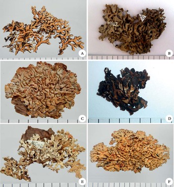
Hypogymnia species. A, H. lijiangensis (holotype); B, H. pruinoidea (holotype); C, of H. pruinosa (holotype); D, H. pseudopruinosa (lectotype); E, H. subfarinacea (holotype); F, H. subpruinosa (holotype). Scale=5 mm increments.

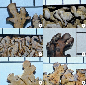
Pruina of Hypogymnia species. A, H. lijiangensis (holotype); B, H. pruinoidea (holotype); C, H. pruinosa (holotype); D, H. pseudopruinosa (lectotype); E, H. subfarinacea (holotype); F, H. subpruinosa (holotype). Scale=5 mm increments.

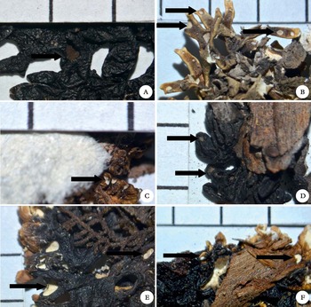
Hypogymnia species, location of holes indicated by arrows. A, H. lijiangensis (holotype); B, H. pruinoidea (holotype); C, H. pruinosa (holotype); D, H. pseudopruinosa (lectotype); E, H. subfarinacea (holotype); F, H. subpruinosa (holotype). Scale=5 mm.

This species is characterized by discrete lobes with black borders, pruina limited to lobe tips, and presence of physodalic acid in the thallus. It is similar to H. subfarinacea X. L. Wei & J. C. Wei and H. subpruinosa J. B. Chen in the location of pruina and chemistry, but clearly differs in morphology. Hypogymnia lijiangensis has separated lobes, holes mainly in the axils, lacks soredia on the upper surface, and has a cartilaginous texture; H. subfarinacea has granular soredia on the upper surface, being easily separated from the other pruinose species of Hypogymnia, cartilaginous texture but slightly papery; and H. subpruinosa has crowded lobes with holes only on the lobe tips.
Chemistry. Thallus cortex K+ yellow; medulla K+ pale reddish brown, PD+ orange-red. TLC: atranorin, physodic acid, physodalic acid, 3-hydroxyphysodic acid, protocetraric acid.
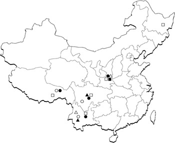
Known distribution. Sichuan, Tibet and Yunnan provinces of China (Fig. 4).
Distribution of the six pruinose species of Hypogymnia. H. lijiangensis (○), H. pruinoidea (▪), H. pruinosa (•), H. pseudopruinosa (Δ), H. subfarinacea (▴), H. subpruinosa (□).

Selected specimens examined. China:Sichuan: Xiangcheng, Mt. Wumingshan, 1983, J. J. Su, H. A. Wen, B. Li 6036 (HMAS-L). Tibet: Riwoqê county, 1976, Y. C. Zong, Y. Z. Liao 256-3 (HMAS-L). Yunnan: Lijiang county, Yulong Snow Mountain, alt. 3060 m, 1981, X. Y. Wang, X. Xiao, J. J. Su 5124 (HMAS-L); Shangri-La county, Mt. Tianbaoshan, alt. 3700 m, 1981, X. Y. Wang, X. Xiao, J. J. Su 5189 (HMAS-L).
Hypogymnia pruinoidea X. L. Wei & J. C. Wei sp. nov.
MycoBank No.: MB 564892
Species characterized by isodichotomously branched lobes, pruinose lobe tips and upper surface, and abundant perforations present on lobe tips, axils, and lower surface.
Type: China, Shaanxi, Mt. Taibaishan, alt. 2800 m, on Abies trunk, 3 August 2005, X. L. Wei 1727 (HMAS—holotype).
Thallus foliose, up to 6 cm wide, texture cartilaginous, loosely adnate, with flat, crowded, mostly isodichotomously branched, hollow lobes 0·5–1·0 mm wide and 0·5–2·0 mm long, apices blunt; upper surface greyish green, rugose, dull, thinly pruinose or pruina limited to the lobe tips, with a sharp demarcation between the pruinose and epruinose region; soredia and isidia lacking; lobules present; lower surface black, pale brown at lobe tips, rugose, dull, perforations present on many lobe tips, axils, and lower surfaces, the holes not rimmed; medulla hollow, both the ceiling and floor of the cavity white to pale dirty brown.
Apothecia not seen.
Pycnidia mostly at lobe tips, immersed to slightly protruding, black, mostly punctiform, aggregated; conidia fusiform, simple, 6·0–7·5×1·0 µm.
Chemistry. Reagent tests: cortex K+ yellow, P−; medulla K+ pale reddish brown, KC+ pale pink, P−. All specimens contain atranorin, physodic and 3-hydroxyphysodic acids, most specimens also contain vittatolic acid.
Etymology. The epithet ‘pruinoidea’ refers to the pruinose lobe tips and upper surface in this species.
Habitat, distribution and substratum. Hypogymnia pruinoidea occurs on bark and wood, usually of Abies in Mt. Taibaishan, Shaanxi province (Fig. 4). The high-elevation mountain habitats are cool and moist, supporting lichen-rich forests, woodlands and subalpine areas. These areas are rich in Hypogymnia species such as H. arcuata Tchaban. & McCune, H. austerodes (Nyl.) Räsänen, H. flavida McCune & Obermayer, H. hypotrypa (Nyl.) Rass., and others.
Remarks. Morphologically the new species resembles H. pseudopruinosa, but differs in having a thallus with a more cartilaginous texture, isodichotomously branched lobes, and abundant perforations present on lobe tips, axils, and lower surface, whereas H. pseudopruinosa is characterized by a thallus with a cartilaginous to slightly papery texture, subdichotomously branched lobes, and perforations mainly present on lobe tips. The new species also resembles H. pruinosa, but differs by perforations sometimes grouped on the lower surface and containing physodic and 3-hydroxyphysodic acids. Hypogymnia pruinosa is characterized by perforations mainly at lobe tips, and contains alectoronic acid.
Specimens examined. China:Shaanxi Province: Mt. Taibaishan, 2800 m, on trunk of Abies tree, 2005, X. L. Wei 1728, 1729; 3136 m, on trunk of Abies tree, 2005, X. L. Wei 1662, 1663, 1664 (HMAS-L).
Hypogymnia pruinosa J. C. Wei & Y. M. Jiang
Acta Phytotax. Sin. 18 (3): 386 (Reference Wei and Jiang1980); type: China, Tibet, Changdu, 1976, Y. C. Zong, Y. Z. Liao 215 (HMAS—holotype; HMAS—isotype).
This species is characterized by almost circular thalli, wide and short lobes, a layer of dense pruina on the upper surface, presence of a sharp demarcation between pruinose and epruinose regions, and in containing alectoronic acid in the thallus.
Chemistry. TLC: atranorin, alectoronic acid.
Distribution. Shaanxi, Sichuan, Tibet, and Yunnan provinces of China (Fig. 4).
Selected specimens examined. China:Shaanxi: Mei county, Mt. Taibaishan, 2005, X. L. Wei 1624 (HMAS-L). Sichuan: Kangding county, Minya Konka, alt. 3600 m, 1982, X. Y. Wang, X. Xiao, B. Li 9438 (HMAS-L). Tibet: Linzhi county, alt. 3011m, 2004, X. L. Wei 1041 (HMAS-L). Yunnan: Lijiang county, Yulong Snow Mountain, alt. 3100 m, 1981, X. Y. Wang, X. Xiao, J. J. Su 4829 (HMAS-L).
Hypogymnia pseudopruinosa X. L. Wei & J. C. Wei
Mycotaxon 94: 155 (Reference Wei and Wei2005); type: China, Yunnan, Dêqên county, 1981, X. L. Wei 7606 (HMAS—lectotype designated here).
Thallus foliose, tightly appressed, texture cartilaginous to slightly papery; lobes subdichotomously branched, 1–2 mm wide, 5 mm long; dense layer of pruina limited to the lobe tips; upper surface greyish green to dark brownish yellow, partly black, rugose; isidia, soredia and lobules lacking; lower surface black, brown near the apices, rugose, and perforations mainly present on the lobe tips; medulla hollow, the ceiling of the cavity white to pale brown, and floor of the cavity white to dark brown.
Upper cortex prosoplectenchymatous, pale yellow, 12·0–14·5 µm thick; algal layer 10·0–14·5 µm thick, consisting of green, subspherical cells of 7·5–9·5 µm diam.; hyphae in medulla colourless, c. 2 µm diam.; lower cortex prosoplectenchymatous, pale yellow, 10 µm thick.
Apothecia rare, 1–3 mm diam., stipitate; disc yellow-brown or red-brown, glossy, concave at first and then slightly plane with entire and thin margin; epithecium brown, 7–9 µm thick; hymenium colourless, 32–36 µm thick; asci clavate, 23·5–25·0×8–14 µm, 8-spored; spores simple, colourless, ellipsoid to nearly spherical, 5·5–7·0×3·5–4·0 µm; paraphyses linear, septate, 2 µm wide, slightly swollen at the tips; hypothecium colourless, 27–36 µm thick.
Pycnidia common, brown to black; conidia weakly bifusiform, c. 2·5 (rarely to 5 µm)×1·0 µm.
Chemistry. Cortex K+ yellow, P−; medulla K+ yellow, P−; containing atranorin and physodic acid.
Etymology. The epithet ‘pseudopruinosa’ refers to the presence of pruina only on the lobe tips.
Distribution. Shaanxi and Yunnan provinces of China (Fig. 4).
Remarks. This species resembles H. macrospora (Zhao Reference Zhao1964; Wei Reference Wei1991) at first sight, but differs by having a dense layer of pruina limited to the tips of the lobes, holes not rimmed, and smaller ascospores. The holotype of H. pseudopruinosa was found to be mixed with H. laccata. Some lobes of the two species overlapped each other, which led to some phenotypic characters of H. laccata being wrongly assigned to H. pseudopruinosa, whereas some diagnostic characters were actually based on both species. The diagnostic characters mistakenly attributed to H. pseudopruinosa include the glossy upper surface and PD+ medulla (physodalic acid), which were actually based on H. laccata. A lectotype is chosen here (ICBN Art. 9.9) from the separated true H. pseudopruinosa portion of the original holotype and isotype. The H. laccata in the mixture has been separated out as specimen no. 7606-1. When this species was published as new, it was based on a single specimen from Yunnan Province but, during our recent research, at least four additional specimens were collected from Shaanxi province.
Selected specimens examined. China:Shaanxi: Mei county, Mt. Taibaishan, 1988, C. H. Ma 074 (HMAS-L); 2011, X. L. Wei w11139, w11143, w11150 (HMAS-L).
Hypogymnia subfarinacea X. L. Wei & J. C. Wei
Mycotaxon 94: 156 (Reference Wei and Wei2005); type: China, Sichuan, Nanping county, Jiuzhai Gou, 1983, X. Y. Wang & X. Xiao 10582 (HMAS—holotype).
This species superficially resembles H. farinacea Zopf, but differs in having more separated lobes, the pruina limited to the lobe tips, and containing physodalic acid; H. farinacea contains atranorin, chloroatranorin, physodic acid, 3-hydroxyphysodic acid, and 2′-O-methylphysodic acid (McCune Reference McCune, Nash, Ryan, Gries and Bungartz2002).
Chemistry. Cortex K+ yellow, P−; medulla K+ yellow, P+ orange-yellow→orange-red. TLC: atranorin, physodic acid, physodalic acid, 3-hydroxyphysodic acid, and protocetraric acids.
Distribution. Sichuan and Yunnan provinces of China (Fig. 4).
Specimens examined. China:Yunnan: Lijiang county, Yulong Snow Mountain, alt. 2900 m, 1981, X. Y. Wang, X. Xiao, J. J. Su 4892 (HMAS-L); ibid., alt. 3100 m, 1981, X. Y. Wang, X. Xiao, J. J. Su 6591 (HMAS-L).
Hypogymnia subpruinosa J. B. Chen
Acta Mycologica Sinica 13 (2): 107 (Reference Chen1994); type: China, Yunnan, Zhongdian county, 1981, X. Y. Wang, X. Xiao, J. J. Su 7094 (HMAS—holotype).
This species is characterized by wide and short lobes with round tips, strongly rugose upper surface, pruina limited near to the lobe tips, and presence of physodalic acid in the thallus. It is similar to H. pruinosa in external appearance, but the latter has pruina all over the upper surface and contains alectoronic acid.
Chemistry. Cortex K+ yellow; medulla K+ pale reddish brown, PD+ orange-red. TLC: atranorin, physodic acid, physodalic acid, 3-hydroxyphysodic acid, and protocetraric acids.
Distribution. Heilongjiang, Shaanxi, Sichuan, Tibet, and Yunnan provinces of China (Fig. 4).
Selected specimens examined. China:Heilongjiang: Dailing, s. n. (HMAS-L). Shaanxi: Mt. Taibaishan, alt. 3280 m, 2005, X. L. Wei 1986 (HMAS-L). Sichuan: Maerkang county, Mt. Mengbishan, alt. 3700 m, X. Y. Wang & X. Xiao 11503 (HMAS-L). Tibet: Linzhi county, alt. 3010 m, X. L. Wei 1036 (HMAS-L). Yunnan: Shangri-La county, Mt. Tianbaoshan, alt. 3800 m, 1981, J. J. Su, X. Xiao, X. Y. Wang 6808 (HMAS-L).
Discussion
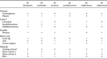
Of the six pruinose species of Hypogymnia, only H. subfarinacea has granular soredia on the upper surface, easily separating it from the other five. Presence or absence of physodalic acid, with the result of the medulla being PD+ orange-red or PD−, respectively, is sometimes a significant diagnostic character to delimit species of Hypogymnia. Two pruinose species H. lijiangensis and H. subpruinosa, have a PD+ medulla. Hypogymnia lijiangensis has separate rather than contiguous lobes with black borders, and holes mainly in the axils, rarely on the lower surface; H. subpruinosa has crowded lobes, without black borders, and holes only on the lobe tips. The three species with a PD− medulla are chemically distinct: alectoronic acid is present in H. pruinosa, but absent in the other two species, H. pseudopruinosa and H.pruinoidea. Hypogymnia pseudopruinosa superficially resembles the epruinose species, H. macrospora, but differs by having a dense layer of pruina limited to the tips of the lobes and smaller ascospores. Compared with H. pruinoidea, the thallus of H. pseudopruinosa has a cartilaginous to papery texture, subdichotomously branched lobes, and perforations mainly present on lobe tips, whereas H. pruinoidea has a thallus with a more cartilaginous texture, isodichotomously branched lobes, and abundant perforations present on lobe tips, axils and lower surface. Table 1 compares six pruinose species of Hypogymnia more clearly, including the main different phenotypic characters, such as thallus texture, lobes, holes, soredia, and medulla PD reactions.
Comparison of phenotypic characters of the six pruinose species of Hypogymnia from China

* Isodichotomous: isodichotomously branched; subdichotomous: subdichotomously branched
† Ceiling colour: white to pale brown in all species; floor colour 1: white to pale brown; floor colour 2: pale to dark brown
Among the six pruinose species of Hypogymnia, all except H. pruinoidea occur in south-western China, and two species, H. pruinosa and H. subpruinosa, extend to north-western and (or) north-eastern China. Hypogymnia pruinoidea is found only in Shaanxi province, north-western China. Located on a low latitude plateau, south-western China arises from south-east Tibet, crosses over west Sichuan, and expands to central and north Yunnan. A combination of complex landforms with advantageous moisture conditions in south-western China results in an extremely abundant biodiversity, with many endemic species of animals and plants. Hypogymnia species are also abundant in south-western China. Over 80% of known Hypogymnia species in China can be found in this area, and about 18% of the species are endemic. South-western China can therefore be regarded as a main centre of speciation for Hypogymnia. Mount Taibaishan is the highest peak of Qinling, Shaanxi province, which is the watershed of Yangtze River and Yellow River. As the highest mountain in the eastern half of China, it has a very complex geography and climate. The marked difference in elevation leads to a vertical distribution of climate, animals, and plants, and many new species of plants endemic to Taibaishan have been reported. Over 30% of known Hypogymnia species in China can be found in this area, including the new endemic species, H. pruinoidea.
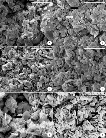
The common character of the six species of Hypogymnia treated in this paper is having pruina. Pruina is a whitish, flour-like surface covering, consisting primarily of superficial deposits, most commonly calcium oxalate (Büdel & Scheidegger Reference Büdel, Scheidegger and Nash1996). There are two kinds of calcium oxalate found in lichen pruina, weddellite and whewellite, and four types of crystal structure have been reported based on SEM study. Types 1–3 belong to the weddellite, and type 4 belongs to the whewellite (Wadsten & Moberg Reference Wadsten and Moberg1985). In our SEM study, three types of crystal structure have been confirmed on the pruina of the six pruinose Hypogymnia species: weddellite type 2, type 3, and whewellite. Hypogymnia pruinoidea has two crystal types, weddellite type 3 and whewellite (Fig. 5A); H. lijiangensis has only one crystal type, whewellite (Fig. 5B); H. pruinosa has only one crystal type, weddellite type 2 (Fig. 5C); H. pseudopruinosa has only one crystal type, whewellite (Fig. 5D); H. subfarinacea has two crystal types, weddellite type 2 and whewellite (Fig. 5E); and H. subpruinosa has two crystal types, weddellite type 3 and whewellite (Fig. 5F). It is postulated that the formation of calcium oxalate on the surface of lichens is caused by a need for the lichen to dispose of an excess of calcium, and there seems to be some relationship between different types of crystal structure and habitat, especially with regard to different conditions of acidity and water content (Wadsten & Moberg Reference Wadsten and Moberg1985). However, the taxonomic significance of different crystal structures of calcium oxalate is not yet clear.
SEM photographs of pruina on Hypogymnia species. A, whewellite in H. lijiangensis (holotype); B, weddellite of crystal type 3 (white arrow) and whewellite (black arrow) in H. pruinoidea (holotype); C, weddellite of crystal type 2 in H. pruinosa (holotype); D, whewellite in H. pseudopruinosa (lectotype); E, weddellite of crystal type 2 (black arrow) and whewellite (white arrow) in H. subfarinacea (holotype); F, weddellite of crystal type 3 (arrow on right) and whewellite (upper arrow) in H. subpruinosa (holotype). Scales: A–D & F=10 µm; E=5 µm.

In conclusion, based on the above mentioned phenotypic characters, presence or absence of soredia and black borders, location of pruina and thallus perforations, and chemistry, all the six pruinose species can be easily separated. All the phenotypic characters used to define the six pruinose species of Hypogymnia are commonly used in the taxonomy of Hypogymnia, while their objectivity and the phylogenetic placement of each species based on them will require molecular methods.
This project was supported by the Main Direction Program of Knowledge Innovation of the Chinese Academy of Sciences (KSCX2-EW-Z-9). We thank Dr Irwin M. Brodo for his careful modification of English grammar in the manuscript. Professor Bruce McCune of Oregon State University is thanked for the significant discussion about Hypogymnia with the first author, and he and one anonymous reviewer are also thanked for their valuable suggestions on the manuscript. Professor Jianbin Chen gave constructive comments on an earlier version of our paper. Mr Chunli Li helped take SEM photograghs of pruina. The authors are also grateful to Ms. Hong Deng for giving considerable assistance during the studies in HMAS-L.