Implications
Glutamate is taken up by special transporters and is oxidized to distribute fuel for the growth of small intestine. l-trans pyrrolidine-2,4-dicarboxylic acid (l-trans-PDC) is one of the inhibitors of the special transporters. Hence, l-trans-PDC solutions were injected into the albumen of fertilized eggs before incubation to evaluate its effects on small intestinal growth of chicks. Small intestinal weights were decreased at hatching and increased on days 7 and 14 after hatching by treatment with l-trans-PDC. The injection of l-trans-PDC may therefore provide a method to regulate the growth of small intestines.
Introduction
Physiological adaption in the small intestine may provide an important contribution to the maintenance of health, development, growth and survival in humans and animals. The small intestine is a well-studied organ that is involved in food digestion and nutrient absorption, the epithelium of which self-renews continuously. The matured intestinal epithelium is covered by a single layer of epithelial cells that are classified as enterocytes, goblet cells, endocrine cells and Paneth cells according to their function, and performs the essential role of digestion and absorption by constantly producing these four types of short-lived cells. Approximately 90% of all epithelial cells are enterocytes that perform absorptive functions (Sheaffer and Kaestner, Reference Sheaffer and Kaestner2012). Hence, the development and growth of the small intestine may be restricted by the functional properties of enterocytes.
Glutamate, which is metabolized during its transcellular journey through enterocytes, acts as the most important contributor to oxidative metabolism in the small intestine mucosa (Wu, Reference Wu1998). More than 98% of the glutamate in the epithelium is converted into α-ketoglutarate to generate ATP through the Krebs cycle pathway (Welbourne and Matthews, Reference Welbourne and Matthews1999). Glutamate supplementation enhances both the total and mucosal growth of the small intestine (Ewtushik et al., Reference Ewtushik, Bertolo and Ball2000). Oral administration of monosodium glutamate increases average daily gain of BW and expression of glutamate transporters in the gastrointestinal tract of young piglets (Zhang et al., Reference Zhang, Yin, Shu, Li, Li, Tan, Wu and Wu2013). In all, five excitatory amino acid transporters (EAAT1 to EAAT5) have been identified in the plasma membrane so far; EAAT2 and EAAT3 are localized in the basolateral and brush border membranes of enterocytes, respectively (Bröer, Reference Bröer2008). EAAT3 plays a more important role in glutamate uptake across the brush border membrane than does the B0 transport system (Speier et al., Reference Speier, Yadgary, Uni and Wong2012). Increased EAAT3 gene expression in the small intestine of broilers changes the small intestine morphology and improves the luminal uptake of nutrients (Barri et al., Reference Barri, Honaker, Sottosanti, Hulet and McElroy2011). Our previous studies showed that EAAT3 gene expression in the small intestine was upregulated with embryonic growth and could be used as a genetic indicator of the small intestinal growth of chick embryos (Li et al., Reference Li, Yan, Zeng, Zhang and Wang2011 and Reference Li, Chen and Wang2013).
l-trans pyrrolidine 2,4-dicarboxylic acid (l-trans-PDC) is a potent competitive inhibitor of EAAT, with approximately equivalent inhibitory activity for each EAAT member, and it has been well studied during excitatory amino acid uptake related to neuronal signaling transduction (Had-Aissouni, Reference Had-Aissouni2011). l-trans-PDC exhibits a Ki value of 4.6 μmol/l for the inhibition of glutamate uptake into the rat brain synaptosome (Dunlop and Butera, Reference Dunlop and Butera2006), whereas no data for inhibition in intestines or embryos are currently available. Although the EAAT protein exists and glutamate may have a transmitter role in the enteric nervous system, it is interesting to explore the effects that the inhibition of EAAT transport activity have on glutamate delivery and small intestinal growth. Therefore, we hypothesized that in vivo l-trans-PDC administration regulates glutamate delivery and small intestinal growth in animals. In contrast to a mammalian fetus, chick embryos mainly obtain nutrients from the yolk sac membrane and the nutrient transporters expressed in the epithelium during the last few days of incubation are important for the absorption of nutrients from the materials that pass through the intestine (Wijtten et al., Reference Wijtten, Langhout and Verstegen2012). In addition, avian embryo development is restricted by the nutrients that are within the eggs in contrast to the development of viviparous mammals (Zeng et al., Reference Zeng, Li, Wang, Zhang, Shu and Luo2011). Thus, the objective of this study was to evaluate the effects of in ovo administration of l-trans-PDC on the EAAT gene expression, amino acid delivery and small intestinal growth of chicks.
Material and methods
Two series of experiments were conducted with hatching eggs. Generally, Experiment I was performed to determine an appropriate feeding dose of l-trans-PDC (Tocris Bioscience, Bristol, UK) and its effects on embryonic development, whereas Experiment II was performed to further explore the effect of in ovo administration of l-trans-PDC on EAAT gene expression, small intestinal growth and amino acid delivery during the embryonic or postembryonic periods. All experimental procedures were approved by the Animal Care Committee of South China Agricultural University (Guangzhou).
Experiment design
In Experiment I, a total of 96 fertilized eggs, which were produced by WENS Yellow Feather Chickens (WYFC; WENS Co., Ltd, Yunfu, China), were used for this study. Eggs with average weights of 39.8±0.2 g were numbered and divided equally into three groups; the egg weights in the three groups were similar and did not show a significant difference (P>0.96). In contrast to brain synaptosome (Dunlop and Butera, Reference Dunlop and Butera2006), higher doses of l-trans-PDC should be injected into the albumen sac owing to the 21-day incubation time and substance turnover in the body. Therefore, 0, 0.075 or 0.225 mg/egg (equal to 0, 12.7 or 38.1 μmol/l of egg volume) of l-trans-PDC, which was diluted by phosphate buffered saline (137 mmol/l NaCl, 2.7 mmol/l KCl, 10 mmol/l Na2HPO4 and 2 mmol/l KH2PO4), was injected into the albumen sac of the eggs before incubation for the Control group (Control), low-dose l-trans-PDC group (L-PDC) and high-dose l-trans-PDC group (H-PDC), respectively. The eggs were quickly incubated at 37.8°C with a relative humidity of 60% and were turned 90° every 2 h until embryonic day 18. The eggs were transferred to the same hatchery for the last 3 days of incubation. The number of hatchlings was recorded at hatching and used to calculate the hatchability, which was measured as the ratio of the number of hatchlings to the number of fertilized eggs.
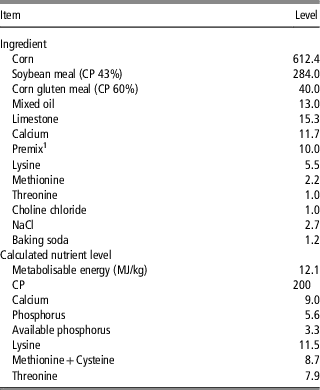
In Experiment II, 200 fertilized eggs with an average weight of 42.3±0.2 g were divided into two groups, the Control group and the H-PDC group, the treatments of which were the same as in Experiment I; the weights of the eggs in the two groups were similar and did not show a significant difference (P>0.98). The eggs were then incubated in the same incubator. A total of 64 hatchlings from either group were divided equally into four replicates of 16 birds each and had ad libitum access to food and water during the experimental period. The ingredient and nutrient analysis of the experimental diet, which met the nutrient requirements of WYFC starters (Wang et al., Reference Wang, Chen, Tan, Zhang, Zhang, Wei and Yan2013a), are described in Table 1. The birds were raised in a room at 30°C to 34°C, with a relative humidity of 55% to 60%.
Ingredient and nutrient analysis (g/kg) of experimental diet
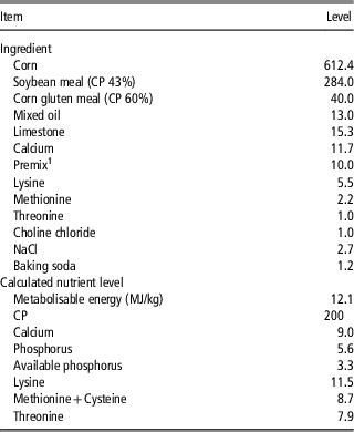
1 Provided (per kg of diet): Cu, 5.00 mg; Fe, 69.00 mg; Zn, 84.00 mg; Mn, 98.6 mg; I, 1.14 mg; Se, 0.30 mg; vitamin A (retinyl acetate), 4.5 mg; vitamin D (cholecalciferol), 0.075 mg; vitamin E (dl-α-tocopheryl acetate), 28.0 mg; vitamin K3 (menadione), 2.1 mg; vitamin B1 (thiamine), 2.4 mg; vitamin B2 (riboflavin), 9 mg; vitamin B6 (pyridoxine), 5.1 mg; vitamin B12 (cyanocobalamin), 0.02 mg; calcium pantothenate, 12 mg; niacin, 48 mg; folic acid, 1.2 mg; biotin, 0.06 mg; roxarsone, 50 mg; salinomycin, 90 mg.
Measurement of body and organ weights
Embryonic development was evaluated by the absolute and relative weights of the body and organs. A total of 10 hatchlings/chicks per group were killed using cervical dislocation and sampled at hatching in Experiment I or at hatching, days 7 and 14 (D7 and D14) in Experiment II. The head, brain, heart, liver, spleen, lungs, kidneys, gizzard and small intestine were isolated and weighed. The gizzard and small intestine were exenterated before they were weighed. The relative weights of the organs were normalized to the BW without yolk. The complete small intestine was divided into the duodenum (from the gizzard to the entry of the bile and pancreatic ducts), jejunum (from the entry of the ducts to Meckel’s diverticulum) and ileum (from Meckel’s diverticulum to the ileocecal junction) in Experiment II.
RNA extraction and real-time RT-PCR
The small intestine (complete in Experiment I and separated segments in Experiment II) was used for RNA extraction and real-time PCR and was processed as previously described (Li et al., Reference Li, Chen and Wang2013). We determined the integrity of the 18S and 28S-ribosomal RNA bands and confirmed the RNA quality by the absence of genomic DNA. The optical density of the RNA was measured using a NanoDrop 2000 UV-Vis spectrophotometer (Thermo, Wilmington, DE, USA) and the 260/280 nm ratios ranged from 1.80 to 2.00. DNA-free RNA (2 μg) was reverse-transcribed to cDNA in a reaction with N10 primers (Sangon, Shanghai, China), M-MLV reverse transcriptase (Promega, Madison, WI, USA), M-MLV buffer and RNase inhibitor (TaKaRa, Shiga, Japan). The abundance of the mRNA was determined by real-time PCR with a Stratagene MxPro 3005 P apparatus (Agilent Technologies, Santa Clara, CA, USA) and the SYBR Green Real-Time PCR Master Mix (TOYOBO, Tokyo, Japan). The real-time PCR primers for EAAT2, EAAT3, glyceraldehyde-3-phosphate dehydrogenase (GAPDH), β-actin, hypoxanthine phosphoribosyltransferase 1 (HPRT1), TATA box binding protein (TBP), ribosomal protein L13 (RPL13) and α-tubulin were the same as described by Li et al. (Reference Li, Yan, Zeng, Zhang and Wang2011 and Reference Li, Chen and Wang2013). The specificity, efficiency and r 2 of each primer were evaluated as previously described (Wang et al., Reference Wang, Jiang, Tan, Zhang, Zhang, Wei and Yan2013b).
Measurement of serum amino acid concentration
Cardiac blood was obtained at hatching and centrifuged at 3500×g for 30 min in Experiment II. The free amino acids in the serum were determined by an amino acid analyzer (Hitachi L-8800; Hitachi, Tokyo, Japan) based on the National Standard Guideline of China for Analysis of Amino Acids (JY/T019-1996) in a room at 24°C with a relative humidity of 67%.
Statistical analysis
The obtained Ct values from real-time RT-PCR were quantified and entered into the geNorm application. Normalized mRNA values of the target genes were calculated as described by Vandesompele et al. (Reference Vandesompele, De Preter, Pattyn, Poppe, Van Roy, De Paepe and Speleman2002). Data were processed by the statistical software package SAS 9.1.3 (SAS Institute Inc., Cary, NC, USA). Variance analysis was performed using the GLM procedure. Differences were considered significant and highly significant for P<0.05 and 0.01, respectively.
Results
Hatchability and the growth of the body and organs
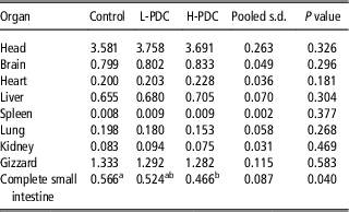
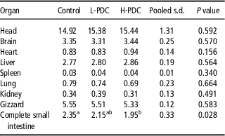
The hatchability of the Control, the L-PDC and the H-PDC groups in Experiment I were 71.43%, 32.00% and 34.62%, respectively. The BWs (both standing and without yolk) did not differ (P>0.05) among the groups in Experiment I at hatching and in Experiment II at hatching, D7 and D14. The absolute and relative weights of most parts, including the head, brain, heart, liver, spleen, lungs, kidneys and gizzard, were not different (P>0.05) among the groups in Experiment I (Tables 2 and 3). Moreover, the absolute and relative weights of the brain and gizzard were not changed (P>0.05) between the Control and the H-PDC groups at hatching, D7 and D14 in Experiment II.
The effect of in ovo administration of l-trans-PDC on absolute weights of the organs in chicks at hatching in Experiment I (g)
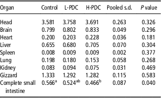
l-trans-PDC=l-trans pyrrolidine-2,4-dicarboxylic acid; Control=Control group; L-PDC=low-dose l-trans-PDC group; H-PDC=high-dose l-trans-PDC group; n=10.
a,bMeans with a significance among groups (P<0.05).
The effect of in ovo administration of l-trans-PDC on relative weights of the organs in chicks at hatching in Experiment I (% of BW without yolk)
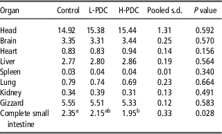
l-trans-PDC=l-trans pyrrolidine-2,4-dicarboxylic acid; Control=Control group; L-PDC=low-dose l-trans-PDC group; H-PDC=high-dose l-trans-PDC group; n=10.
a,bMeans with a high significance among groups (P<0.01).
Growth of the small intestine
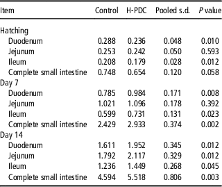
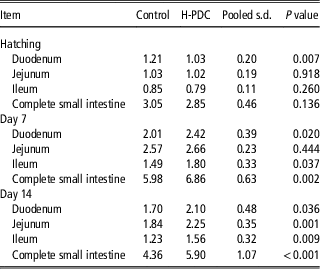
In Experiment I, the weights of the small intestines (absolute and relative) in the Control group were higher (P<0.05) than those in the H-PDC group at hatching (Tables 2 and 3). Similarly, the absolute weights of the duodenum (P<0.01), ileum (P<0.05) and complete small intestine (P=0.058) in the Control group were higher than those in the H-PDC group at hatching (Table 4) in Experiment II. Compared with the Control group, H-PDC group had lower (P<0.01) relative weight of duodenum at hatching (Table 5). Interestingly, the weights (absolute and relative) of the duodenum, ileum and complete small intestine on D7 and D14 and the jejunum on D14 were lower (P<0.05) in the Control group than those in the H-PDC group (Tables 4 and 5).
The effect of in ovo administration of l-trans-PDC on absolute weights of the small intestine in chicks at hatching, days 7 and 14 in Experiment II (g)
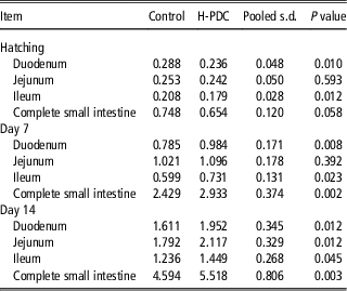
l-trans-PDC=l-trans pyrrolidine-2,4-dicarboxylic acid; Control=Control group; H-PDC=high-dose l-trans-PDC group; n=10.
The effect of in ovo administration of l-trans-PDC on relative weights of the small intestine in chicks at hatching, days 7 and 14 in Experiment II (% of BW without yolk)
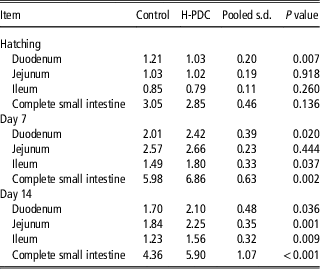
l-trans-PDC=l-trans pyrrolidine-2,4-dicarboxylic acid; Control=Control group; H-PDC=high-dose l-trans-PDC group; n=10.
EAAT gene expression
The average expression stability (M) values of the control genes calculated by geNorm showed that the most stable genes among GAPDH, β-actin, HPRT1, TBP, RPL13 and α-tubulin were β-actin, HPRT1 and TBP in Experiment I and HPRT1, RPL13A and α-tubulin in Experiment II. In addition, the V3/4 values of Experiments I and II were below 0.15. β-actin, HPRT1 and TBP in Experiment I and HPRT1, RPL13A and α-tubulin in Experiment II were utilized as reference genes for normalization.
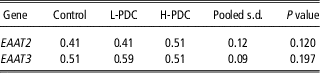
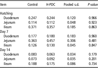
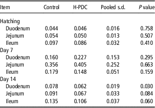
There was no difference (P>0.05) in the gene expression of EAAT2 or EAAT3 among the groups at hatching in Experiment I (Table 6). The results showed that the gene expression of EAAT2 (Table 7) in the duodenum, jejunum and ileum were not different (P>0.05) between the Control and H-PDC groups at hatching, D7 and D14 in Experiment II. EAAT3 gene expression in the duodenum (P<0.05), jejunum (P=0.084) and ileum (P=0.060) on D14 was lower in the H-PDC group than in the Control group (Table 8).
The effect of in ovo administration of l-trans-PDC on gene expression of EAAT2 and EAAT3 in the small intestine of chicks at hatching in Experiment I
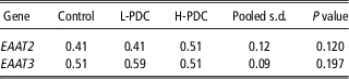
l-trans-PDC=l-trans pyrrolidine-2,4-dicarboxylic acid; EAAT=excitatory amino acids transporter; Control=Control group; L-PDC=low-dose l-trans-PDC group; H-PDC=high-dose l-trans-PDC group; n=10.
The effect of in ovo administration of l-trans-PDC on gene expression of EAAT2 in the small intestinal segments of chicks at hatching, days 7 and 14 in Experiment II
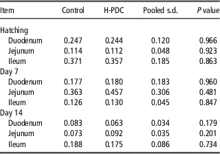
l-trans-PDC=l-trans pyrrolidine-2,4-dicarboxylic acid; Control=Control group; H-PDC=high-dose l-trans-PDC group; n=10.
The effect of in ovo administration of l-trans-PDC on gene expression of EAAT3 in the small intestinal segments of chicks at hatching, days 7 and 14 in Experiment II
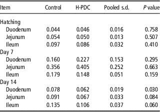
l-trans-PDC=l-trans pyrrolidine-2,4-dicarboxylic acid; EAAT=excitatory amino acids transporter; Control=Control group; H-PDC=high-dose l-trans-PDC group; n=10.
Concentrations of free AAs in the serum
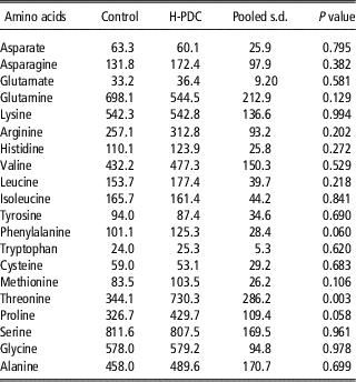
The serum concentrations of free anionic amino acids at hatching were not different (P>0.05) between the Control and H-PDC groups in Experiment II. The concentrations of phenylalanine (P=0.060), proline (P=0.058) and threonine (P=0.003) in the H-PDC group were higher than those in the Control group (Table 9).
The effect of in ovo administration of l-trans-PDC on concentration of the free amino acids in chick serum at hatching in Experiment II (nmol/l)
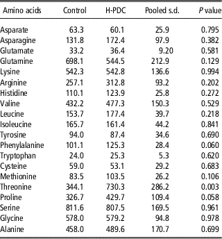
l-trans-PDC=l-trans pyrrolidine-2,4-dicarboxylic acid; Control=Control group; H-PDC=high-dose l-trans-PDC group; n=10.
Discussion
The small intestine has been extensively studied in many organisms owing to its nutrient sensing and absorption, gastrointestinal peptide release and immune response functions. Gastrointestinal dysfunction, such as congenital short bowel syndrome, enterocele and Peutz–Jeghers syndrome, endangers the health, development and survival of the body (Rasoamanana et al., Reference Rasoamanana, Darcel, Fromentin and Tome2012; Wijtten et al., Reference Wijtten, Langhout and Verstegen2012). Quantitative studies demonstrated the regulation of intestinal development by dietary and maternal nutrition, hormones and critical genes. Amino acids, which are the basic constituents of proteins, are signaling molecules that can trigger transportation by enterocytes or the release of hormones by enteroendocrine cells to maintain homeostasis (San Gabriel and Uneyama, Reference San Gabriel and Uneyama2013). Amino acid transporters expressed in the intestinal epithelium connect the amino acid levels in the lumen, epithelium and blood. Furthermore, recent studies confirmed a nutrient-sensing role of special amino acid transporters, which were named transceptors (Hundal and Taylor, Reference Hundal and Taylor2009). Therefore, this study was conducted to regulate small intestinal growth by inhibiting the activity of EAATs. As expected, it was found that in ovo administration of l-trans-PDC selectively regulated the growth of the small intestine but not the body of chicks.
Studies have shown that maternal nutrition restriction during the gestation period decrease the fetal growth of tissues and organs, including the small intestine (Duarte et al., Reference Duarte, Gionbelli, Paulino, Serão, Martins, Tótaro, Neves, Valadares Filho, Dodson and Zhu2013). Bol et al. (Reference Bol, Delattre, Reusens, Raes and Remacle2009) demonstrated that maternal malnutrition at critical time windows resulted not only in the growth retardation of embryos but also in altered postembryonic development because of metabolism programming. Inhibition of glutamate uptake by l-trans-PDC induced significant concentration- and time-dependent neuronal damage (Velasco et al., Reference Velasco, Tapia and Massieu1996). Our results show that the hatchabilities of the L-PDC or H-PDC groups were up to 36% lower than that of the Control group, which might be because of the l-trans-PDC-induced decrease in embryonic survival. In addition, l-trans-PDC administration had no significant effect on the weights of the body and brain among the groups after a 21-day incubation period. Whether l-trans-PDC passes the blood–brain barrier must still be determined. Similarly, local administration of l-trans-PDC triggered no neuronal damage, behavioral changes or alterations in electroencephalographic activity (Montiel et al., Reference Montiel, Camacho, Estrada-Sanchez and Massieu2005) because the oxidative stress that contributes to various forms of cell death is prevented by EAATs, particularly in the cells of the nervous system (Had-Aissouni, Reference Had-Aissouni2011). A novel finding of this study is that in ovo administration of l-trans-PDC induced small intestinal growth retardation during the embryonic period and catch-up growth after hatching. The gene expression of EAAT2 and EAAT3 in the small intestine at hatching and D7 did not change, which might be owing to a recovery after an l-trans-PDC administration that lasted longer than 14 days (Lievens et al., Reference Lievens, Bernal, Forni, Mahy and Goff2000). In addition, the expression of a particular amino acid transporter was immediately regulated by the general control nonrepressed 2 (GCN2) pathway under amino acid deprivation or by target of rapamycin complex 1 (TORC1) under amino acid supplementation in cells (Hundal and Taylor, Reference Hundal and Taylor2009). Whether EAAT3 gene expression increased in H-PDC group on D14 was resulted from GCN2 and/or TORC1 pathways is attractive to further study. Those results showed that the regulation of small intestinal growth by l-trans-PDC was not dependent on EAAT gene expression, although the gene expression of EAAT3 was previously demonstrated to be an indicator of small intestine growth in chick embryos generally (Li et al., Reference Li, Chen and Wang2013).
l-trans-PDC not only competitively inhibited glutamate transport through EAATs but also induced the efflux of endogenous excitatory amino acids by heteroexchange, leading to an increase in the extracellular concentration (Waagepetersen et al., Reference Waagepetersen, Shimamoto and Schousboe2001). It is useful to know whether glutamate delivery is changed by l-trans-PDC treatment in chicks. Therefore, the serum concentrations of amino acids were further studied to evaluate their effects on small intestinal growth owing to their unique physiological functions in intestinal development. Our results showed that the free glutamate concentration in the serum of the H-PDC group did not differ from that in the Control group. As glutamate is the major fuel for the gut, continuously self-renewing enterocytes are extensively involved in the first-pass metabolism of dietary glutamate (Wu, Reference Wu1998). In addition, glutamate is an active molecular participant in transamination, oxidative deamination and amidotransfer (Wu, Reference Wu2009). Therefore, in ovo l-trans-PDC administration complicated glutamate delivery in chicks. It is reasonable to believe that there should be compensation mechanisms to support changes in small intestinal growth. The presence of free phenylalanine, threonine and proline in the serum were particularly notable. The serum concentrations of free phenylalanine, threonine and proline were higher in the H-PDC group than in the Control group of this study.
Oxidation of phenylalanine by phenylalanine hydroxylase, the activity of which was not detectable in enterocytes, was quantitatively negligible in the intestinal mucosa (Wu, Reference Wu2009). The requirement for phenylalanine, which is a precursor of tyrosine, depended on the amount of tyrosine in the diet (Ahmed, Reference Ahmed2009). Phenylalanine or tyrosine were shown to participate in neurological development and function and are precursors for the synthesis of thyroid hormone, which is essential for the embryonic growth and development of avian species (Janz and Bellward, Reference Janz and Bellward1996; Wu, Reference Wu2009). Threonine de novo synthesis was previously shown to not be possible in poultry, which makes it the third most limiting amino acid in the broiler diet (Kidd, Reference Kidd2000). Recently, artificial neural network models based on published data were used to establish the nutritional requirement for threonine in broilers (Mehri, Reference Mehri2012). An adequate level of l-threonine in piglet diet improved the intestinal morphology and gut barrier function by favorably altering the secretion of mucin, which is the major constituent of the mucosal layer that protects against bacterial, chemical and mechanical injures (Wang et al., Reference Wang, Zeng, Mao, Wu and Qiao2010). Proline is a nutritionally essential amino acid for animals and played a key role in the regulation of multiple biochemical and physiological processes in cells (Wu et al., Reference Wu, Bazer, Burghardt, Johnson, Kim, Knabe, Li, Li, McKnight and Satterfield2011). The majority of proline in the small intestine is synthesized via the glutamate/pyrroline 5-carboxylate (P5C) synthase pathway. Neonatal pigs could not synthesize sufficient proline to maintain the plasma concentration owing to the limited expression of P5C synthase in the intestines (Bertolo et al., Reference Bertolo, Brunton, Pencharz and Ball2003). Furthermore, proline was essential during growth and wound repair because proline makes up 25% of collagen and is recently recognized as a potential treatment for intrauterine growth restriction because of its important role in conceptus metabolism, growth and development (Ren et al., Reference Ren, Zou, Ruan, Li, Wang, Peng, Liu, Yin, Li and Hou2013). Collectively, the higher levels of phenylalanine, threonine and proline in the H-PDC group compared with those in the Control group associated with the growth of chick bodies under conditions of intestinal growth retardation.
Conclusion
We are the first to report that in ovo administration of l-trans-PDC induces small intestinal growth retardation during the embryonic period and compensatory growth after hatching. This regulation is not dependent on EAAT gene expression or the serum concentration of anionic amino acids.
Acknowledgments
This work was jointly supported by grants from Special Fund for Agro-scientific Research in the Public Interest (201003011) and Joint Fund of National Natural Science Foundation of China and Guangdong Province Government (U0931004).