INTRODUCTION
Modern Mytilinae, e.g. Mytilus edulis L. 1758, M. galloprovincialis Lamarck 1819, and M. trossulus Gould 1850, is a widespread group of mussels that form abundant populations in the intertidal and upper subtidal zones of cold and temperate waters of the World Ocean. Data on their anatomy, ontogenetic development, geographical distribution and taxonomic position can be found in List (Reference List1902), Pelseneer (Reference Pelseneer1906), Scarlato & Starobogatov (Reference Scarlato, Starobogatov and Vodyanitsky1972), Tanaka (Reference Tanaka1979), Ramorino & Campos (Reference Ramorino and Campos1983) and Kurozumi (Reference Kurozumi and Okutani2000). The taxonomic classifications of Mytilinae, like the Bivalvia as a whole, are based on the definitive characters of the adult specimens while larval and juvenile forms are not generally used in taxonomy (Lutz et al., Reference Lutz, Goodsell, Castagna, Chapman, Newell, Hidu, Mann, Jablonski, Kennedy, Siddall, Goldberg, Beattie, Falmagne, Chestnut and Partridge1982; Cragg, Reference Cragg and Taylor1996). One reason for this is the lack of shared shell features between early ontogenetic and adult stages. Hence, the classifications of larvae and adult forms coexist independently (Rees, Reference Rees1950; Chanley & Andrews, Reference Chanley and Andrews1971; Scarlato & Starobogatov, Reference Scarlato, Starobogatov and Vodyanitsky1972; Kulikova & Kolotukhina, Reference Kulikova and Kolotukhina1989; Scarlato, Reference Scarlato1981). However, the morphological peculiarities of the larval and juvenile stages (commonly size and sculpture) can be used in taxonomy of the adults (Dinamani, Reference Dinamani1973, Reference Dinamani1976; Hayami & Kase, Reference Hayami and Kase1993; Salas & Gofas, Reference Salas and Gofas1997). However, in such an event, they are regarded as distinguishing features assisting to differentiate subspecies, species or genera.
Our data on the morphogenesis of early stages of M. trossulus and Crenomytilus grayanus (Dunker, 1853), along with previous studies of other species of Mytilinae (Evseev et al., Reference Evseev, Semenikhina and Kolotukhina2005; Semenikhina et al., Reference Semenikhina, Kolotukhina and Evseev2008), expand the total number of characters of the subfamily and help to provide more detailed characterizations of its constituent taxa. The results obtained are useful, and yet a problem remains unsolved, especially what is the rank of the early stage features; in other words whether they would be applied to a taxonomic system of the adults as a species or genus, subfamily and family.
The present study deals with the larval–juvenile morphogenesis of two species of Mytilinae—M. trossulus and C. grayanus—which are widespread in the Sea of Japan and its comparison with previously described members of this subfamily. The aim of our work is to the show the importance and role of early stage features for the taxonomy of adult forms, their phylogenetic relationships and ways of macroevolutionary development.
MATERIALS AND METHODS
The material for this study consists of planktonic and benthic samples of larval, juvenile and adult specimens of mytilids that occur in bays and gulfs of the Sea of Japan, as well as samples from mussel aquaculture sites. Planktonic samples were taken with a Juday net and benthic samples were collected with a hand dredge.
In the laboratory, samples of larvae were placed in 70% ethanol and sorted under an MBS-10 binocular microscope. Juvenile forms were extracted by washing the substrate samples through a series of sieves with a minimal mesh size of 250 µm. For light and electron microscopic observations, larvae and juveniles were submerged in a 5% solution of sodium hypochlorite for 1–3 minutes. Thereafter the valves were washed in distilled water, and then placed in glycerol or dehydrated in 96% ethanol. The structure of the hinge system and the surface sculpture of larval and juvenile shells were examined under the Leica DM4500 B light microscope equipped with a Leica DFS300 FX camera and with a Leo 430 scanning electron microscope.
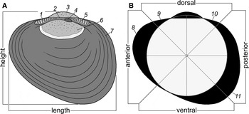
For a taxonomic identification of larvae the previous materials from aquaculture of M. trossulus, M. coruscus Gould 1861, C. grayanus and Septifer keenae Nomura 1936 were used (Kulikova & Kolotukhina, Reference Kulikova and Kolotukhina1989; Semenikhina & Kolotukhina, Reference Semenikhina and Kolotukhina2001). Most terms, including a dimensional scheme and cyclograms (Figure 1) (Evseev & Kolotukhina, Reference Evseev and Kolotukhina2008), were taken from previous works (Rees, Reference Rees1950; Zakhvatkina, Reference Zakhvatkina and Vodyanitsky1972; Booth, Reference Booth1977; Redfearn et al., Reference Redfearn, Chanley and Chanley1986; Fuller & Lutz, Reference Fuller and Lutz1988, Reference Fuller and Lutz1989; Malchus, Reference Malchus2006; Evseev et al., Reference Evseev, Kolotukhina and Semenikhina2007; Evseev & Kolotukhina, Reference Evseev and Kolotukhina2008).
Morphostructures (A) and cyclogram scheme (B) of larval shell of the Mytilinae: 1, anterior provincular teeth; 2, central provincular teeth; 3, umbo; 4, pit of primary (internal) ligament; 5, posterior provincular teeth; 6, primary prodissoconch (PD-I); 7, secondary prodissoconch (PD-II); 8, projection of anterior margin; 9, anterior shoulder; 10, posterior shoulder; 11, enlargement of posteroventral margin.

RESULTS
Mytilus trossulus
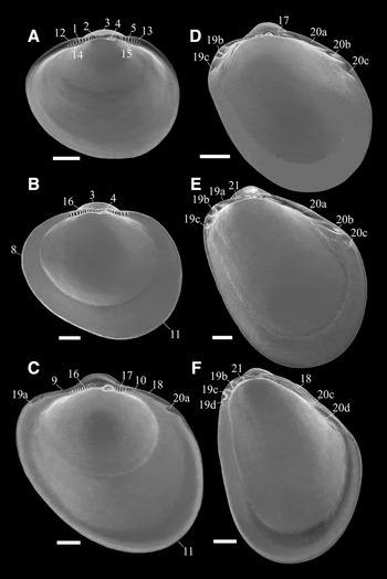
Taxonomically identifiable development starts with a veliconch larva, which is characterized by the formation of an umbo and the secondary prodissoconch (PD-II). The shell of a fully-formed PD-II is 270–290 µm long (Figure 2A). The most important features of larva at this stage are: general outline, hinge plate of the provinculum, and provincular morphostructures, which include anterior, central, and posterior teeth, as well as a primary pit of the internal ligament.
A general view and ontogenetic transformations of larval (A) and juvenile (B–F) shell of the Mytilus trossulus: 12, anterodorsal dilatation of provinculum; 13, posterodorsal dilatation of provinculum; 14, anteroventral dilatation of provinculum; 15, posteroventral dilatation of provinculum; 16, anterior series of postprovincular (juvenile) teeth; 17, posterior series of postprovincular (juvenile) teeth; 18, nymphal layers of the secondary (external) ligament; 19, anterior (dysodont) secondary lateral teeth: a, 1st tooth; b, 2nd tooth; c, 3rd tooth; 20, posterior (dysodont) secondary lateral teeth: a, 1st tooth; b, 2nd tooth; c, 3rd tooth; 21, enlargement of anterodorsal margin. Other numerals are the same as given in Figure 1. Scale bars, A, B, C: 50 µm; D, E: 100 µm; F: 200 µm.
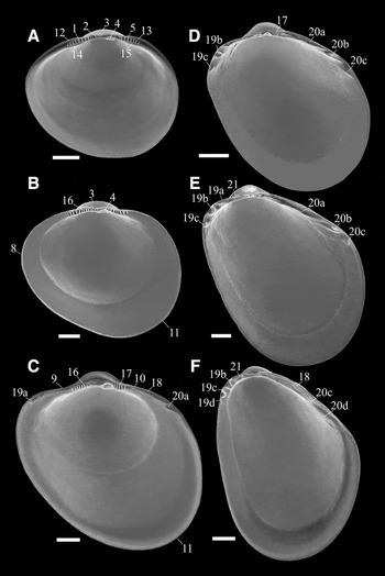
As in most mytilids, the PD-II of M. trossulus is ovate, with a moderately high and symmetrical umbo (3), a broadly pointed anterior end (8), and a broadly rounded posterior one (Figure 2). The anterior margin of the umbo is located above the 6th–7th provincular tooth. The most projecting part of the slightly pointed anterior margin is near the longitudinal midline. The anteroventral shell margin is slightly oblique and the posteroventral margin is regularly rounded, smoothly transitioning into the posterior one. The provinculum about 5–8 µm high bears no more than 15–17 low rectangular central teeth (2), 8–9 higher and wider anterior teeth (1), and 7–8 posterior teeth (5) of the same height and shape. In anterior and posterior parts of the provinculum, two marginal dilatations are arranged dorsally (12, 13) and ventrally (14, 15) to a tooth series. The primary (internal) ligament pit (4) is lens-shaped, and located under the posterior part of central provincular teeth. The ratio of shell height to length is 0.86–0.88.
In an early juvenile shell, at a length of 360–380 µm (Figure 2B), the umbo markedly increases in height, the anterior shell margin acquires a broadly rounded outline, and the posteroventral margin begins to increase in height and length (11). The height of central teeth is up to 10–12 µm. They are an integral part of the anterior juvenile tooth series (16). The length of the hinge plate of the provinculum and the shape of its anterior and posterior teeth, remain unchanged.
A shell about 400–500 µm long (Figure 2C) bears a broad posteroventral enlargement (11), subtriangular umbo and low shoulders (9, 10). The height of its hinge plate, especially in the posterior region, increases to 18–20 µm (17) and the number of posterior teeth is reduced to 5–7. The ligament pit expands mostly ventrally, acquiring a trapezoidal shape. One lateral tooth is formed on both the anterior (19) and posterodorsal (20) shell margins. The first posterior tooth, 30–40 µm long, is located 130–140 µm from the ligament pit. This tooth is inclined at an angle of 120° relative to the juvenile teeth. The first anterior tooth is formed at the same distance from the pit as the first posterior tooth. Behind the postprovincular posterior teeth, a nymph of the external ligament (18) begins to form.
At a length of 600–700 µm (Figure 2D), the shell is oval–elongate ventrally, with a subterminal triangular umbo. Behind the ligament pit are semi-reduced remnants of 4–5 juvenile teeth (17). They appear to be reduced in size because other features are considerably larger. Anterior tooth series of the postprovinculum are not visible. The anterodorsal shell margin bears 3 lateral teeth (19), varying in shape, size, and position relative to one another. The posterodorsal margin also bears 3 lateral teeth (20). However, they are narrower than anterior teeth and are located at a distally increasing angle relative to the postprovincular teeth. The length of teeth decreases from 50–60 to 20–30 µm with increasing distance from the ligament pit. External ligament is up to 120–150 µm long.
A shell of 900–1100 µm in length (Figure 2E) differs from the previous one merely in its more angular posterodorsal margin. The remnants of 4–5 juvenile teeth are still retained behind the ligament pit. However, the anterodorsal shell margin (21) bearing 3–4 lateral teeth begins to expand in front of the umbo. The posterodorsal margin has 3–4 lateral teeth (20), their length decreases towards the umbo from 80–100 to 30–40 µm. The last tooth (20c) is at 500–530 µm away from the ligament pit.
At a shell length of 1500–1700 µm (Figure 2F), the shell is trigonal and elongate ventrally. The widening anterodorsal enlargement (21) extends over the anterior and central parts of the postprovinculum, covering also the base of the umbo. The primary ligament pit and posterior juvenile teeth are overgrown by new shell material. The posterolateral teeth are transformed into narrower, noticeably elongate ridges. The first of them (20c) is about 200 µm long, the last tooth (20d) is about 100 µm long and it is distant 650–700 µm from the umbo base. The number of anterolateral teeth increases to 4. A pseudonymphal layer, the secondary ligament and 2–3 lateral teeth are detectable on shells more than 1500–1700 µm long (e.g. Figure 4A).
Crenomytilus grayanus
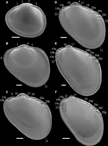
The shell of a competent larva is 260–290 µm long, ovate, with a slightly elongate posteroventral margin and a pointed anterior margin (Figure 3A). The umbo is blunt, low and wide, with its anterior margin above the 4th–5th tooth of the anterior part of the provinculum. The provinculum is 7–8 µm high in its central region, with 17–23 central (2), 8–9 anterior (1) and 7–8 posterior (5) teeth. A wide lens shaped pit of the primary ligament is posterior to the centre of the provinculum, the ventral margin of the pit is straight or slightly oblique anteriorly. The ratio of shell height to length is 0.86–0.88. In optical microscopy, the PD-II has a yellowish-grey hue.

At a shell length of 400–500 µm (Figure 3B), the juvenile shell is elongate–oval, with a broadly rounded anterior (8), wide posterior, elongate posteroventral (11) and almost straight anteroventral margin. The postprovinculum has 8–9 anterior teeth (1) and the same number of the posterior one (5). Teeth of the central plate are elongate, the ligament pit is wide. In optical microscopy (Figure 4B), the shell is semitransparent, with fine comarginal grooves on the external surface, in contrast to the shell at this stage in M. trossulus.
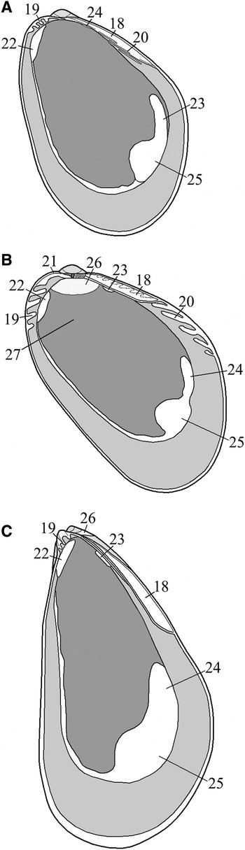
Shell morphostructures of the juvenile Mytilus trossulus (A) and juvenile and adult Crenomytilus grayanus (B, C): 24, anterior pedal–byssal retractor; 25, posterior adductor; 26, nepioconch (juvenile cell); 27, dissoconch (adult shell). Other numerals are the same as given in Figures 1, 2 and 3. Shell length, A, B: 2.5 mm; C: 15 mm.

A shell of between 500 and 700 µm long (Figure 3C) has a conspicuous enlargement of the posteroventral margin. The umbo is subtrigonal in shape, with slightly pointed apex. The postprovinculum is 18–20 µm high and bears 14–16 juvenile teeth of the anterior series and semi-reduced remnants of central provincular teeth separated from juvenile teeth by a marginal ridge of the previous stage. The number of posterior teeth is usually 8–9. Two teardrop-shaped and relatively short lateral teeth appear on both the posterodorsal (20) and anterior (19) shell margins. Of these, the first tooth of the posterodorsal region (20a) reaches 35–45 µm in length and is located 200–240 µm away from the ligament pit. The second tooth (20b) is 60–70 µm long and 310–330 µm distant from the pit. These teeth are about 50 µm apart.
A shell of 850–1000 µm long (Figure 3D) is also oval, elongated ventrally and posteriorly, with a distinct posteroventral enlargement (11). The height of hinge plate is close to 26–30 µm, the primary ligament pit (4) is overgrowing by new shell layers, juvenile teeth are transformed, about 20–25 µm long in the anterior part of the plateau (16) and up to 35–40 µm in the posterior part (17). The number of the anterior teeth usually up to 13–15 and posterior ones up to 7–9. The anterior and posterodorsal margins each have 3–4 lateral teeth up to 70–80 µm long, spaced at about 40–50 µm from each other. The first of posterolateral teeth (20a) is 200–230 µm away from the ligament pit and the last (20c), 400–450 µm away. A nymph of the secondary ligament (18) begins to form behind the postprovinculum on the posterodorsal margin. At this stage, the length of ligament does not exceed 180–200 µm.
The shape of a shell 1100–1400 µm long (Figure 3E) hardly differs from the shape of the previous shell. Juvenile teeth are retained on the postprovinculum, their number is usually up to 10–12 in front of the ligament pit (16) and up to 7–8 behind it (17). Anterior teeth are about 20–25 µm long and posterior, up to 40 µm long. The posterodorsal and anterior shell margins have 5 and 4 lateral teeth respectively. The first tooth of the posterodorsal shell margin (20b) is about 60 µm and is located at 270–290 µm away from the ligament pit, the second (20c) at 360–380 µm, and the last (20e) at 650–670 µm. According to its distance from the pit, the teeth at a distance more than 400-450 µm (20d, e) are ontogenetically new formations of this stage.
A shell 1500–1800 µm long (Figure 3F) is oval–elongate, not differing in its outlines from the previous stage, with trigonal umbo. Juvenile teeth on the hinge plate disappear and the primary ligament pit has been fully overgrown by new shell layers. The anterior shell margin usually has 4 lateral teeth and the posterodorsal margin has 5–6 lateral teeth. The first tooth of the posterior region located 450–470 µm away from the ligament pit corresponds to the fourth tooth (20d) of the tooth series of the previous stage. The last two teeth of this region (20f, g), at 790–810 and 870–890 µm away from the ligament pit, are new formations and occupy the seventh and eighth place in their ontogenetic series.
The general shape of a shell 2500–3000 µm long (Figure 4B) is oval, expanding posteriorly and ventrally. The anterior and central parts of the postprovinculum, as well as the base of the anterior part of the umbo are covered by an expansion (21) of the anterodorsal shell margin. Unlike M. trossulus of the same shell length (Figure 4A), the postprovinculum bears a partially overgrown pit of the primary ligament and 3–5 juvenile teeth. The anterior shell margin usually has 3–4 lateral teeth. The posterior shell margin has 4–5 teardrop-shaped teeth, of which the first is the largest, up to 100–130 µm. The rest of teeth of the posterodorsal region are covered by a pseudonymph of the secondary ligament. The total number of overgrown and functioning lateral teeth is up to 12–14 and the distance from the first tooth of this stage to the ligament pit is 1650–1700 µm. In shells larger than 5–7 mm in length (Figure 4C), the posterior lateral teeth are lacking as a result of cessation in tooth development. The number of anterior teeth, among which the first is the largest, does not usually exceed 3–4. In the ontogenetic series, the final number of posterolateral teeth in C. grayanus is up to 23–25.
DISCUSSION
Morphostructures of larval stage
The competent larvae of Mytilidae provide a good example of the significant role of their features for taxonomic purposes. The most important of these features are: (1) the ratio of shell height/length; (2) width, height and shape of the umbo; (3) outlines of the anterior and posterior margins; (4) number and ratio of anterior, posterior, central and additional provincular teeth; and (5) shape, size, and topology of the primary ligament pit (e.g. Jørgensen, Reference Jørgensen1946; Rees, Reference Rees1950; Chanley & Andrews, Reference Chanley and Andrews1971; Booth, Reference Booth1977; Evseev & Kolotukhina, Reference Evseev and Kolotukhina2008).
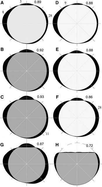
The morphological features of shell make it possible to test if characters are shared or distinctive, when comparable mytilid taxa have been at their larval stages. In that view, the ratio of shell height/length being applied to the M. trossulus–C. grayanus (Figure 5F, E) is not advantageous for distinction between these taxa because their shells are similar. However, the ratio can be highly efficient where compared, for instance, the M. trossulus–M. coruscus (Gould, 1861) (Figure 5C, F) or M. trossulus–M. edulis (Figure 5A, F). The latter pair is a known problem, if the taxa have reached the adult stages. Some details of the umbo M. trossulus (Figure 5F) might be useful distinguishing features as compared with umbos of the M. edulis (Figure 5A), M. galloprovincialis (Figure 5B) and M. coruscus (Figure 5C). The umbos and general shell outlines of M. californianus (Conrad, 1837) (Figure 5D) and M. edulis bear close similarities in shell height and posterior enlargement (28) as opposed to other members of the Mytilinae, but differ from each other in provincular morphostructures (Lutz et al., Reference Lutz, Goodsell, Castagna, Chapman, Newell, Hidu, Mann, Jablonski, Kennedy, Siddall, Goldberg, Beattie, Falmagne, Chestnut and Partridge1982; Fuller & Lutz, Reference Fuller and Lutz1989). It should be also noted that outline and extent of anterior (8), posterior (28) and posteroventral (11) enlargements that might be important distinguishing features if these morphostructures be considered in combination with others, including provincular ones.
Some distinguishing details of larval shell outlines of the Mytilinae: (A) Mytilus edulis, 286 µm (Fuller & Lutz, Reference Fuller and Lutz1989); (B) Mytilus galloprovincialis, 240 µm (Le Pennec & Masson, Reference Le Pennec and Masson1976); (C) Mytilus coruscus, 309 µm, Sea of Japan; (D) Mytilus californianus, 280 µm (Martel et al., Reference Martel, Auffrey, Robles and Honda2000); (E) Crenomytilus grayanus, 277 µm, Sea of Japan; (F) Mytilus trossulus, 307 µm, Sea of Japan; (G) Modiolus kurilensis, 300 µm, Sea of Japan (Evseev & Kolotukhina, Reference Evseev and Kolotukhina2008); (H) Septifer keenae, 180 µm, Sea of Japan (Evseev & Kolotukhina, Reference Evseev and Kolotukhina2008). The numbers to the right of umbo are ratio of the shell height to length. Numerals: 28, enlargement of posterior shell margin. Other numerals are the same as given in Figure 1.

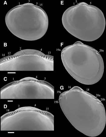
The larval shell of M. galloprovincialis from the Sea of Japan (Figure 6A, B) is similar in its general outline to the shell of M. trossulus. However, it differs by its higher shell with a more flattened umbo, the position and shape of the primary ligament pit (4), and the number of central provincular teeth (2) not exceeding 12–14. Marginal regions of PD-II of this species, specifically ventral, have a specific orange hue. On other hand, our M. galloprovincialis will be different in the shell height, numbers of anterior teeth, and not as wide umbo as M. galloprovincialis from the Mediterranean Sea (Figure 5B) and Black Sea (Zakhvatkina, Reference Zakhvatkina and Vodyanitsky1972). The shell of M. coruscus (Figure 6E) is distinguished by a high and wide umbo with the anterior margin above the 1st–2nd provincular teeth. The provinculum usually has up to 5–6 teeth of the anterior and posterior series but without of the ventral (14, 15) and dorsal (12, 13) dilatations, which are typical of the M. galloprovincialis, M. trossulus (Figure 6C) and C. grayanus (Figure 6D). PD-II of the M. coruscus is light grey.
Some distinguishing details of shell outlines, provincular and postprovincular morphostructures of the Mytilinae: (A, B) Mytilus galloprovincialis, larva, 274 µm, Sea of Japan; (C) Mytilus trossulus, larva, 275 µm, Sea of Japan; (D) Crenomytilus grayanus, larva, 230 µm, Sea of Japan; (E) Mytilus coruscus, juvenile, 388 µm, Sea of Japan; (F) M. coruscus, juvenile, 495 µm, Sea of Japan; (G) M. coruscus, juvenile, 620 µm, Sea of Japan. Numerals are the same as given in Figures 1 and 2. Scale bars, B, C, D: 20 µm.

These morphological peculiarities primarily concerning the general outlines of larval shell, its umbo, number of relatively uniform teeth of the provinculum and topology of the primary ligament pit are discriminating characters of larvae of other taxa of the subfamily as well, among them Mytilaster lineatus (Gmelin, 1790) (Zakhvatkina, Reference Zakhvatkina and Vodyanitsky1972), Choromytilus chorus Molina 1782 and Mytilus chilensis Hupe 1854 (Ramorino & Campos, Reference Ramorino and Campos1983), Ischadium recurvum (Rafinesque, 1820) (Fuller & Lutz, Reference Fuller and Lutz1989), Mytilus californianus Conrad 1837 (Martel et al., Reference Martel, Auffrey, Robles and Honda2000) and Perna viridis (L. 1758) (Hanyu et al., Reference Hanyu, Toyama, Kimura and Sekiguchi2001). An exception may be larvae of Septifer keenae Nomura 1936 (Figure 5H), whose shell, in contrast to that of other taxa of the subfamily, consists of a D-shaped prodissoconch I (PD-I) (Evseev et al., Reference Evseev, Semenikhina and Kolotukhina2004, Reference Evseev, Semenikhina and Kolotukhina2005; Semenikhina et al., Reference Semenikhina, Kolotukhina and Evseev2008).
Morphostructures of juvenile stage
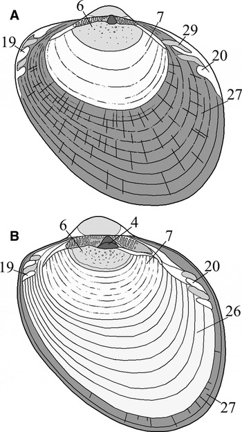
In both M. trosssulus and C. grayanus, in the course of postlarval development the shell elongates posteroventrally, the umbo height and pit size increase, provincular teeth are transformed into lengthened juvenile teeth, lateral teeth are formed on the anterior and posterior shell margins. Morphogenesis terminates with the change in general shape of the shell from rounded to ovate–elongate, its terminal umbo becomes partly covered by an expansion/widening of the anterodorsal margin, the secondary ligament has been formed, the primary ligament pit, juvenile teeth of the postprovinculum and posterolateral teeth of the dissoconch have been reduced. In M. trossulus postlarval shell (Figure 7A), the dissoconch (27), differs from the previous one (7) in its discontinuous radial microstructure, pale violet colour and being demarcated from PD-II by a more or less expressed boundary, which is well-marked on the valve outside. In C. grayanus (Figure 7B), postlarval development begins with an unpigmented and semitransparent nepiconch (26), which is a region of the shell bearing fine regular comarginal grooves on the outside shell surface. Therefore, the dissoconch of C. grayanus, which does not differ in shell colour and microstructure from that of M. trossulus, is formed ontogenetically later, at a shell length exceeding 550–600 µm. Other taxa of the subfamily can be divided into two groups. A C. grayanus type of development is characteristic of Perna viridis, Mytilaster lineatus, Septifer keenae and Ischadium recurvum. The second group, in addition to M. trossulus, includes M. edulis, M. galloprovincialis, M. coruscus, M. chilensis and Choromytilus chorus (Tanaka, Reference Tanaka1979; Ramorino & Campos, Reference Ramorino and Campos1983; Fuller & Lutz, Reference Fuller and Lutz1988; Evseev et al., Reference Evseev, Semenikhina and Kolotukhina2005, Reference Evseev, Kolotukhina and Brykov2006).
Optical microscopy morphostructures of the juveniles and Mytilus trossulus (A) and Crenomytilus grayanus (B) (Evseev et al., Reference Evseev, Kolotukhina and Brykov2006, with some changes): 29, interdental pits. Other numerals are the same as given in Figures 1, 2 and 4. Shell length, A: 495 µm; B: 650 µm.

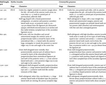
Posterior lateral teeth of M. trossulus and C. grayanus are formed simultaneously with the anterior teeth; however, in the former species, they appear at a shell length of 400 µm and in the latter, at about 600 µm, i.e. at the dissoconch stage in both cases (Table 1). The posterior lateral teeth of M. trossulus are relatively narrow, crest-like, up to 200 µm long. In the course of development, their inclination angle to the postprovincular teeth changes from 104° to 140°, and at a shell length more than 1500 µm when the total number of already formed teeth approaches 3–4, the formations of new teeth ceases. In formation, C. grayanus, the posterolateral teeth are teardrop-shaped and relatively wide, no more than 100–130 µm long. The inclination angle remains unchanged during development, about 110°. The formation of new teeth ends when their total number is up to 23–25 and the shell length exceeds 2500–3000 µm. On their exterior, juvenile shells of this species are densely covered by thin and long hairs of periostracum (Scarlato, Reference Scarlato1960). Hairs on juvenile shells of M. trossulus, like M. edulis (Dixon et al., Reference Dixon, Sole-Cava, Pascoe and Holland1995), are sparse, short, and occur only in the postero-central region of the shell.
Formation of distinguishing features in larval and juvenile shell ontogenetic development of Mytilus trossulus and Crenomytilus grayanus: PD-II, secondary prodissoconch; N, nepioconch; D-I, early juvenile dissoconch; D-II, late juvenile dissoconch.

Anterior and posterior lateral teeth of M. galloprovincialis (Le Pennec & Masson, Reference Le Pennec and Masson1976) and M. edulis (Fuller & Lutz, Reference Fuller and Lutz1989) do not evidently differ from those of M. trossulus. The posterolateral teeth of M. coruscus (Figure 4F, G) are similar in length and inclination angle to teeth of C. grayanus. They appear 180–200 µm distance from the ligament pit at a shell length of about 400 µm, and their formation ceases when the shell is up to 2400–3200 µm long. At the same time, the final number of teeth in the course of development does not exceed 9–10 (Yoshida, Reference Yoshida1953; Tanaka, Reference Tanaka1979). The lateral teeth of M. chinensis, Ch. chorus and M. californianus are also of the dysodont-type; however, details of their morphogenesis are little known, and specific peculiarities have been inadequately studied.
Among the rest of the taxa of the subfamily, I. recurvum deserves particular attention. Its anterodorsal shell margin bears 1–2 secondary lateral teeth of the dysodont-type that are formed at the stage of dissoconch when its length probably reaches 1800–2000 µm (Fuller & Lutz, Reference Fuller and Lutz1989). By their topology, shape, length (500–600 µm) and inclination angle (125–128°), these lateral teeth can be homologous to lateral teeth of Mytilidae. However, as regards to the postprovinculum height, the shape, position, and size of the primary ligament pit, as well as transformations of postprovincular teeth during juvenile development, I. recurvum is not closer to taxa of the Mytilinae (Scarlato & Starobogatov, Reference Scarlato and Starobogatov1984).
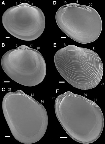
During the ontogenetic development of Septifer keenae (Figure 8D–F), no teeth similar to teeth of M. trossulus and C. grayanus were found (Evseev et al., Reference Evseev, Semenikhina and Kolotukhina2004, Reference Evseev, Semenikhina and Kolotukhina2005). Instead of dysodont-type secondary teeth, the posterodorsal margin of their shells bears primary lateral teeth of the nepioconch (30) and secondary lateral teeth of the dissoconch (32). These primary lateral teeth are known as juvenile of the Modiolus modiolus (Fuller & Lutz, Reference Fuller and Lutz1989), M. philippinarum, (Ozawa & Sekiguchi, Reference Ozawa and Sekiguchi2002) M. kurilensis (Figure 8B, C), and others. However, in some taxa of a paedomorphic development, for example, Modiolus phaseolinus and M. margaritaceus, these type of teeth are inherent at adult stage. The primary and secondary lateral teeth non dysodont-type besides the Septiferinae, can be found in Perninae (Siddall, Reference Siddall1980), Musculinae (Tanaka, Reference Tanaka1979), Brachidontinae (Campos & Ramorino, Reference Campos and Ramorino1980) and other Mytilidae. These data give grounds to consider that we are dealing here with taxa of the modioloid line, in contrast to the taxa of Mytilinae. However, in context of juvenile features, the Mytilinae is not a compact group of taxa. The C. grayanus, I. recurvum and possibly M. californianus might be separated as forms with nepioconch. Other species, M. coruscus and probably M. californianus have need for a new generic name when larval features are also considered.
A general view and morphostructures of out-group mytilids, the larval–juvenile Modiolus kurilensis (A–C) and juvenile Septifer keenae (D–F): 30, primary lateral teeth of nepioconch; 31, comarginal ribs of nepioconch; 32, secondary lateral teeth of dissoconch. Other numerals are the same as given in Figures 1, 2 and 4. Scale bars, A, D: 30 µm; B, E: 50 µm; C, F: 100 µm.

Thus, both larval and juvenile stages of Mytilinae exhibit features, which can be considered as characters of taxa of different levels. The most important characters of a species and generic rank are: (a) shell shape; (b) size characteristics of PD-I and PD-II; (c) shape of the umbo and position of its anterior margin relative to the provincular teeth of the first series; and (d) colour hues of the central and marginal parts of PD-II. However, despite some differences, larvae of Mytilinae usually have the same general morphology due to shared morphostructures: PD-I and PD-II, provinculum, provincular teeth, primary ligament pit.
Characters of the specific or generic rank of juvenile stages can be: (a) peculiarities of posterior lateral teeth of the dysodont type teeth (shape, length, total number, possibly inclination angle); (b) details of structure and sculpture of the dissoconch; and (c) topology and configuration of adductors and retractors.
Our study suggests that the peculiarities of larval and juvenile morphostructures can further extend the general list of taxonomic characters and bridge the gaps in the characterization of the subfamily Mytilinae, in the solution of problems of morphological identification of adult forms of Mytilus spp. and phylogenetic relationships of Mytilinae.
ACKNOWLEDGEMENTS
We are greatly indebted to Dr K.A. Lutaenko and Dr O.Ya. Semenikhina for their helpful comments on the manuscript. Our cordial thanks are due to Professor Nicolaus Malchus for his help, important comments and fruitful discussion about a large part of the manuscript. This research was supported by a grant 08-04-00929 from the Russian Academy of Sciences.