INTRODUCTION
Polychaetes exhibit diversity in reproductive traits that are unique among metazoans (Wilson, Reference Wilson1991). These reproductive diversities are due to the physiological and morphological constraints imposed by the environment in which they live (Giangrande, Reference Giangrande1997; Brommer, Reference Brommer2000). One reproductive strategy found in many invertebrates and a few vertebrates is the encapsulation of eggs and developing larvae within gelatinous or firm leathery capsules (Thorson, Reference Thorson1946). Egg deposition within gelatinous masses is the least common strategy among polychaetes with only 13.6% of the total polychaetes displaying this strategy (Wilson, Reference Wilson1991; Giangrande, Reference Giangrande1997).
Marphysa gravelyi (Southern, Reference Southern1921), a Eunicid polychaete found in the brackish waters of Pulicat Lake, India exhibits continuous synchronous breeding throughout the year, encapsulating eggs within a gelatinous mass where indirect development takes place (Aiyar, Reference Aiyar1931). The jelly masses are pear shaped with a broad distal end that remains attached to the burrow of the adult female via a narrow stalk. The fertilized eggs present within the jelly mass undergo development involving four larval stages: prototrochophore (Stage I), early metatrochophore (Stage II), late metatrochophore (Stage III) and nectochaeta (Stage IV). At the end of the nectochaetal stage the jelly mass disintegrates and the larva commences a creeping behaviour, settling to the bottom (Malathi et al., Reference Malathi, Sunder Raj and Mercy Bai2011). Though the larval stages and the protective function of the jelly mass of M. gravelyi have been discussed by Aiyar (Reference Aiyar1931) and Malathi et al. (Reference Malathi, Sunder Raj and Mercy Bai2011), there have been no studies on the biochemical composition of the gelatinous masses of M. gravelyi. Chapman (Reference Chapman1965) investigated the carbohydrate, protein, lipid and calcium content of cocoons of the polychaete Scoloplos armiger in relation to the function of the egg system. The present study aims to understand the physical and biological function of the jelly masses of M. gravelyi by morphometric, histochemical, biochemical and antimicrobial analyses.
MATERIALS AND METHODS
Sample collection
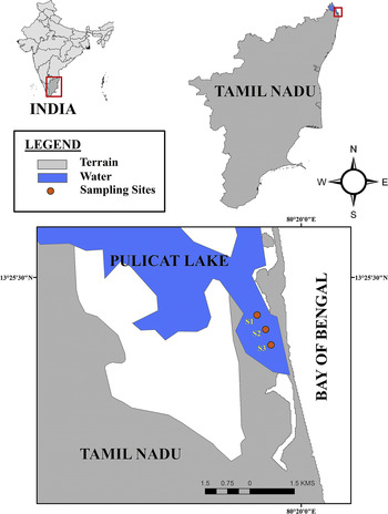
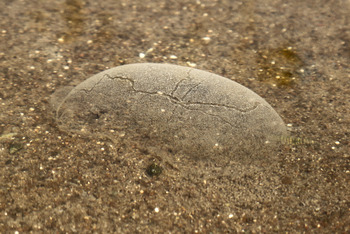
The jelly masses of M. gravelyi are found abundantly in Pulicat Lake (Figure 1) located 60 km north of Chennai, on the east coast of south India (13°.24′49.40″–13°.66′N and 80°19′12.48″–80°.25′E). The jelly masses and their stalks were collected by scooping the mud around them, to a depth of 15 cm (Figure 2). The developmental stage of larvae within the jelly masses was noted by examination under a microscope in order to determine the age of the jelly mass. Jelly masses containing Stage I prototrochophore and Stage IV nectochaeta larva were selected for the present study.
Location and collection site of jelly masses of Marphysa gravelyi in Pulicat Lake, Chennai, India.

Jelly mass of Marphysa gravelyi in situ.

Morphometric studies
The morphometric characters of a total of 30 jelly masses were analysed. For the measurement of length and width, the jelly masses were spread out in a white tray and measured using Vernier calipers to the nearest 0.2 mm. The egg mass volume was measured by recording (to the nearest 0.1 cm3) the displacement of seawater upon total immersion of the jelly mass. The number of larvae in 1 ml of the jelly mass was manually counted using a dissection microscope. Measurements were log transformed for statistical analysis. Variables were analysed by Pearson's correlation tests to determine the relationship between volume of the jelly mass and number of larvae. Regression analysis was used to evaluate the relationship between parameters. All analyses were performed using statistical software GraphPad Prism 7.02.
Histochemical studies
The Stage I and IV jelly masses were fixed in Carnoy's fixative (Humason, Reference Humason1979) for 6 h and washed in three changes of alcohol for a total of 6 h prior to histochemical analysis. The fixed jelly masses were stained with Sudan black B for lipids (Chiffelle & Putt, Reference Chiffelle and Putt1951); Periodic Acid – Schiff Reaction (PAS) for carbohydrates (Lillie & Fullmer, Reference Lillie and Fullmer1976); fast green staining for mucus (Gurr, Reference Gurr1956); alcian blue staining at pH 2.5 for acid mucopolysaccharides (Mowry, Reference Mowry1956); Mayer's mucicarmine (Southgate, Reference Southgate1927) and metachromasia with toluidine blue for mucin (Lillie & Fullmer, Reference Lillie and Fullmer1976).
Biochemical analysis
SAMPLE PREPARATION
The jelly masses were gently washed in 0.2 µm Filtered Sea Water (FSW) to remove surface debris and centrifuged at 20,000 g for 20 min at 5°C. The process was repeated until a clean, larvae free jelly mass was obtained and the pellet containing larvae and debris was discarded.
SOLUBILITY OF THE JELLY MASS
The solubility was tested by suspending 30 mg jelly mass in 1 ml of the following solvents – water, 2.5 M sodium hydroxide (NaOH), 0.6 M sodium bicarbonate (NaHCO3), 1.5 M hydrochloric acid (HCl), dilute sulphuric acid, acetone and ethanol.
BIOCHEMICAL ASSAY
Moisture content (Woodcock & Benkendorff, Reference Woodcock and Benkendorff2008), total carbohydrate (Roe & Dailey, Reference Roe and Dailey1966), glucose (Racker, Reference Racker1946), total protein (Bradford, Reference Bradford1976), total lipid (Folch et al., Reference Folch, Lees and Sloane Stanley1957; Barnes & Blackstock, Reference Barnes and Blackstock1973), HDL (Izzo et al., Reference Izzo, Grub and Murador1981), LDL (Nakamura et al., Reference Nakamura, Taniguti, Yamamoto, Hino and Manabe1997), triglycerides (Sugiura et al., Reference Sugiura, Oikawa, Hirano, Maeda, Yoshimura, Sugiyama and Kuratsu1977) and calcium (Schwarzenbach, Reference Schwarzenbach1955; Biggs & Moorehead, Reference Biggs and Moorehead1974) were determined in Stage I and IV jelly masses.
Antimicrobial assay
PREPARATION OF EXTRACT
To screen for potential antimicrobial properties, Stage I and IV jelly masses were prepared as previously mentioned and extracted with 10 volumes (w/v) of 70% methanol in a shaker at 37°C for 24 h. The extracts were centrifuged at 12,000 g for 10 min at 4°C and filtered solvents removed by rotary evaporation under vacuum. The dried extracts were weighed and suspended in dimethyl sulphoxide (DMSO) for antimicrobial studies by disc diffusion method.
ANTIBACTERIAL ACTIVITY OF JELLY MASS EXTRACT
The bacterial pathogens Escherichia coli (ATCC 10536), Staphylococcus aureus (ATCC 11632), Pseudomonas aeruginosa (ATCC 10145) were grown and maintained in LB agar/broth at 37°C. Vibrio parahaemolyticus (ATCC 17802), Vibrio alginolyticus (ATCC 11632), Vibrio vulnificus (MTCC 1145) and Vibrio harveyi (MTCC 3438) were maintained in Zobell Marine Agar/Broth at 37°C. Exponentially growing bacteria were spread plated and discs with 20 µl of extract (at final concentrations of 1000, 500, 250, 125 and 62.5 µg); 20 µl DMSO (negative control) and 10 µl (10 µg streptomycin – positive control) were placed on Mueller Hinton agar plates and incubated for 24 h at 37°C. The plates were examined for zones of inhibition and the diameters of which were measured in millimetres, using Vernier calipers.
ANTIFUNGAL ACTIVITY OF JELLY MASS EXTRACT
The fungal pathogens Candida albicans (MTCC 186), Candida tropicalis (MTCC 184) and Candida glabrata (MTCC 3019) were grown and maintained in Potato Dextrose Agar/Broth at 37°C. Exponentially growing fungi were spread plated and discs with 20 µl of extract (at final concentration of 1000, 500, 250, 125 and 62.5 µg); 20 µl DMSO (negative control) and 20 µl (20 µg ketoconazole – positive control) were placed on Potato Dextrose Agar plates and incubated for 24–48 h at 37°C. The plates were examined for zones of inhibition and the diameters of these were measured in millimetres, using Vernier calipers.
RESULTS
Morphometrics
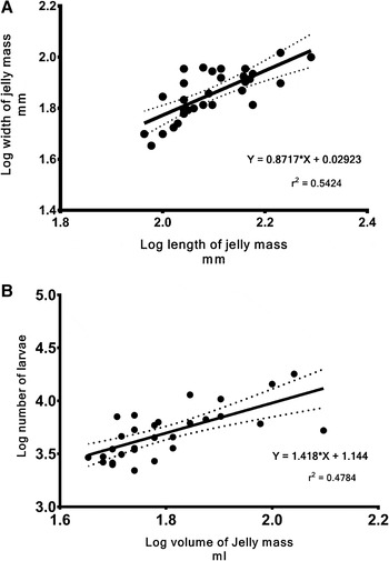
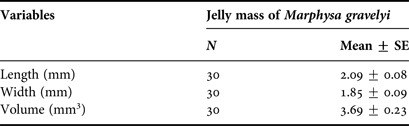
The mean length, width and volume of the jelly masses were 2.09, 1.85 and 3.69 mm3 respectively (Table 1). Significant positive correlations between the length and width of the jelly mass (F = 33.19, P < 0.0001, r = 0.7365) (Figure 3A), and between the volume of the jelly mass and the number of larvae (F = 26.68, P < 0.0001, r = 0.6916) were observed (Figure 3B).
Relationship between (A) length and width of the jelly mass; (B) volume of jelly mass and the number of larvae, calculated for the total sampled specimen.

Jelly mass measurements in Marphysa gravelyi.

N, Number; SE, standard error.
Histochemical observation
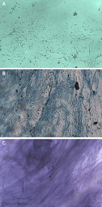
The jelly masses appeared non-homogeneous and fibrous. Histochemical staining of the Stage I jelly mass showed a strong positive reaction for PAS, alcian blue and metachromatic dyes indicating that the jelly mass was acidophilic. Staining the Stage I jelly mass by standard PAS technique indicated the presence of glycogen, mucopolysaccharides such as glycoprotein and glycolipid. Alcian blue staining followed by PAS indicated the presence of carbohydrates with 1,2 glycol and acidic groups. Staining with Sudan black B indicated the presence of lipids. The Stage IV jelly mass showed a mild reaction for carbohydrates, lipids and glycolipids. Staining studies with fast green showed the jelly mass to be mucous (Figure 4A).
Histochemical analysis of jelly mass of Marphysa gravelyi with (A) fast green staining indicates the fibrous jelly mass is primarily mucous; (B) alcian blue indicates the presence of acid mucopolysaccharide; (C) toluidine blue staining for metachromacy indicates the presence of mucins.

ACID MUCOPOLYSACCHARIDES
The Stage I jelly mass showed intense reaction to β-metachromasia and alcianophilia with toluidine blue and alcian blue respectively. Alcian blue staining indicated the carbohydrate to be acid mucopolysaccharides (Figure 4B) and subsequent staining with Mayer's mucicarmine indicated the presence of mucin. Further staining with toluidine blue for metachromasia changed the tissue colouration from blue to a reddish violet hue confirming the presence of mucins (Figure 4C).
Biochemical studies
SOLUBILITY OF THE JELLY MASS
The jelly mass did not dissolve in water but dissolved in NaOH, showing the presence of acidic groups like carboxylic acid or phenols. The jelly mass further solubilized with formation of bubbles on treatment with 0.6 M NaHCO3. The jelly mass was mildly soluble in acetone but did not dissolve in 1.5 M HCl, dilute sulphuric acid and ethanol.
BIOCHEMICAL ASSAY
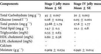
The biochemical study of the jelly mass for carbohydrates showed that Stage I jelly masses have a total carbohydrate content of 45 mg g−1 and 0.08 mmol l−1 glucose and Stage IV jelly masses have a total carbohydrate content of 20 mg g−1 and 0.05 mmol l−1 glucose. The total protein content was 33.68 mg g−1 for Stage I jelly masses, while the protein content in the jelly masses with Stage IV larvae was 27.6 mg g−1. The Stage I jelly mass of M. gravelyi consisted of ~14.7 mg g−1 lipid, 0.55 mg % triglycerides and 0.60 mg % HDL, while the Stage IV jelly mass consisted of 10.1 mg g−1 lipid, triglycerides, although HDL were not detected. LDL and calcium were absent in both Stage I and IV jelly masses. The biochemical study indicates a discernible depletion of organic content in Stage IV jelly masses (Table 2).
Biochemical analysis of Stage I and Stage IV jelly mass of Marphysa gravelyi.

SE, standard error.
Antibacterial activity
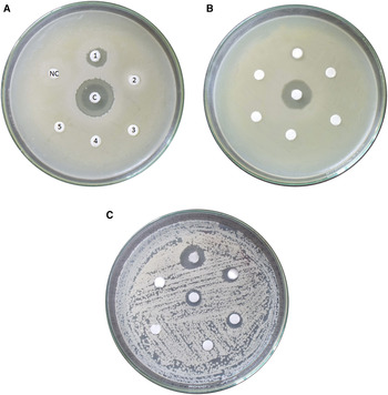
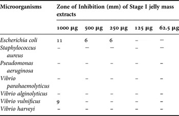
The Stage I jelly mass extract of M. gravelyi showed inhibition at 250 µg against E. coli (Figure 5A) and at 1000 µg against V. vulnificus (Figure 5B). No activity was observed against other bacterial pathogens (Table 3). The Stage IV jelly mass exhibited no antibacterial activity against any of the test organisms.
Antimicrobial activity of jelly mass extracts as observed on (A) Escherichia coli; (B) Vibrio vulnificus; (C) Candida albicans. C – Positive control; NC – Negative control; 1–5 concentration range of 1000, 500, 250, 125, 62.5 µg respectively.

Antibacterial activity in extracts from Stage I jelly mass at different concentrations (diameter of zone of inhibition in mm).

–, Indicates no antibacterial activity. No activity was observed for Stage IV jelly mass (Data not represented).
Antifungal activity
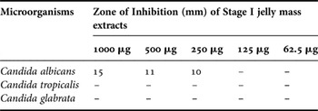
The extract from the Stage I jelly mass exhibited significant inhibitory activity against C. albicans at 250 µg and above (Figure 5C), while no inhibitory activity was seen against C. tropicalis and C. glabrata (Table 4). The Stage IV jelly mass exhibited no antifungal activity against any of the test organisms.
Antifungal activity in extracts of Stage I jelly mass at different concentrations (diameter of zone of inhibition in mm).

–, Indicates no antifungal activity. No activity was observed for Stage IV jelly mass (data not presented).
DISCUSSION
The jelly masses of M. gravelyi were transparent, gelatinous balloons. The newly laid jelly masses were covered by a thin, soft, sticky transparent layer, a characteristic that is lost with gradual adherence of mud and accumulation of algal growth. The body of the jelly mass had a thin watery central core surrounded by an outer thick gelatinous region containing individual fertilized eggs visible as diffusely distributed black specks. The development of the embryo within the gelatinous masses was highly synchronized as each jelly mass represented a single stage of development. The early stages of the egg mass contained only fertilized eggs fully laden with yolk and developing larvae. Unfertilized eggs were not observed within the jelly masses. Slow accumulation of different phytoplanktons and meiofaunal organisms like copepods, nematodes and protozoans were found in the core of jelly masses along with the Stage IV nectochaeta. Statistical studies showed a significant correlation between the parameters chosen. The results indicate that the volume of the jelly mass can be used as a measure for the number of larvae present within.
Microscopic examination of the jelly mass of M. gravelyi showed it to be fibrous. However, this fibrous appearance is due to the local condensation of the jelly and not due to the presence of clearly defined fibres, as observed by Chapman (Reference Chapman1965). The biochemical study of the jelly masses of M. gravelyi demonstrated the presence of proteins, lipids and carbohydrates. The histochemical study revealed the jelly masses to be predominantly mucous, composed primarily of water. Staining with PAS and sudan black indicated the presence of carbohydrates with 1–2 glycol groups like polysaccharides, acid mucopolysaccharides, glycoproteins and glycolipids. Further the solubility study showed that the jelly masses were soluble in NaOH and NaHCO3 indicating the presence of strong acid like carboxylic acid. Acid mucopolysaccharides are polyanions containing sulphate esters and carboxylic acid residues. Subsequently, staining with alcian blue, a polyvalent cationic dye that binds to polyanions, indicates the presence of acid mucopolysaccharide (Scott & Dorling, Reference Scott and Dorling1965). Further staining with toluidine blue and mucicarmine, confirms the presence of mucin of acid mucopolysaccharide, a highly glycosylated mucoprotein consisting of ~80% carbohydrates primarily N-acetylgalactosamine, N-acetylglucosamine and glycogen.
Mucins exhibit the tendency to aggregate and form gels. The property of mucins to bind large amounts of water to a very small amount of organic matter contributes to the viscous and elastic gel-like property (Chapman, Reference Chapman1965) of the jelly mass of M. gravelyi. The glycoprotein mucins exhibit electrostatic, hydrophobic and H-bonding interactions, which cause other substances to adhere to them. This property, known as mucoadhesivity, could explain the accumulation of mud and algal growth on the surface of the jelly mass (Harding et al., Reference Harding, Davis, Deacon and Fiebrig1999). The loss of jelly structure and depletion of organic content present in the Stage IV jelly mass as compared with the early stages may be due to grazing of the meiofaunal organisms seen within the jelly mass.
Antimicrobial studies indicate that Stage I jelly masses have mild antibacterial and antifungal property while no activity was detected in stage IV. Since marine invertebrates are constantly exposed to a high microbial load in seawater and sediments, the antimicrobial property observed could be due to the natural population of microorganisms living in the jelly mass (Benkendorff et al., Reference Benkendorff, Davis and Bremner2001). Another interesting possibility is that there is a parental transfer of antimicrobial secretions into the jelly mass. Previous studies on the immune response of polychaeta have shown that they have the potential for cellular and humoral immune responses (Cuvillier-Hot et al., Reference Cuvillier-Hot, Boidin-Wichlacz and Tasiemski2014).
It is interesting to note that the only species of polychaete producing egg masses in the intertidal waters of Pulicat Lake is M. gravelyi. Although there is no evidence that the laying of eggs in jelly mass confers any thermal or osmotic advantage, the insoluble nature of jelly mass presents several advantages including protection against physical stress, predators and bacterial attack (Pechenik, Reference Pechenik1978, Reference Pechenik1983) and offers nutritional benefits. The jelly material of M. gravelyi performs the important task of protection, aggregation, preventing desiccation and dispersion of larvae.
ACKNOWLEDGEMENTS
The authors thank Mr M. Karunanidhi of Estuarine research station, Madras Christian College for his help with polychaete collection; Dr S. Karuthapandian, Department of Biotechnology, Alagappa University, Karaikudi for his help with microbiological studies; the Principal, Queen Mary's College for providing the required facilities and the anonymous referees for the comments that substantially improved this manuscript.
FINANCIAL SUPPORT
This research received no specific grant from any funding agency, commercial or not-for-profit sectors.