Introduction
Cricothyroid cannulation is part of the armamentarium in management of the difficult airway.Reference Benumof1 It has traditionally been used to establish an emergency airway in cases of acute airway obstruction, and in the ‘can't intubate, can't ventilate’ scenario.Reference Benumof and Scheller2 There is little published literature on its use in a more elective setting. In our unit, cricothyroid cannulation has been increasingly used in those patients undergoing head and neck surgery in whom a ‘difficult airway’ is anticipated. In this situation, cricothyroid cannulation is used to establish a temporary airway, until a definitive airway (i.e. endotracheal tube or tracheostomy) can be secured. The procedure thereby provides a route for jet ventilation if airway obstruction occurs during induction of anaesthesia or awake fibre-optic intubation. In addition, the cannula may be left in place as a prophylactic measure if airway obstruction is considered a post-operative risk (‘dangerous extubation’). Cricothyroid cannulation is not performed routinely, but only in select, difficult airway cases.
The aim of this study was to review the use of cricothyroid cannulation in patients undergoing head and neck surgery, including its indications, technique and complications. We advocate it as a useful adjunct in the management of a difficult airway; in some situations, a tracheostomy can be avoided.
Materials and methods
Written approval was obtained from the Hospital Trust to conduct the study.
The details of all patients undergoing cricothyroid cannulation were prospectively recorded over a two-year period. Inclusion criteria were patients having surgery primarily for diagnosis and treatment of head and neck neoplasm (benign and malignant), or for complications related to this. We recorded the patients' demographic data, diagnosis, surgical procedure, indication for cricothyroid cannulation and complications. The indication for cannulation was classified as difficult intubation, dangerous extubation or both. Case notes were also reviewed to record the disease stage and to identify any late complications related to cricothyroid cannulation.
An anaesthetist discussed the reasons for using the technique, and the procedure itself, with the patient. Verbal consent was obtained. Equipment was prepared, including the following: Ravussin 13 gauge cannula, 5 ml syringe containing 2 mls 4 per cent lignocaine for transtracheal local anaesthesia, 2 ml syringe containing local anaesthetic (LA) for skin infiltration, sterile pack, and a Manujet ventilator (VBM Medizintechnik GmbH, Sulz, Germany) to provide jet ventilation.
If the airway was considered to be potentially at risk during induction of anaesthesia, then the cricothyroid cannula was sited under LA prior to this, with the patient awake. However, if the primary concern was potential airway compromise after surgery, then the cannula was sited with the patient anaesthetised, breathing spontaneously.
All cannulae were sited through the cricothyroid membrane. Confirmation of tracheal placement involved the aspiration of free gas through the needle and cannula during insertion, the presence of exhaled carbon dioxide on a capnograph, and the demonstration of chest expansion denoting successful transtracheal jet ventilation. The cannula was secured using dressings or sutures; on occasion, it was necessary to trim the wings of the cannula to maximise surgical access. In the majority of cases, oxygenation and ventilation of the patient was to be carried out via a definitive airway device, such as an endotracheal tube or a tracheostomy. Only in a minority of cases was sustained transtracheal jet ventilation performed. In all cases, exhalation via a patent larynx and upper airway was confirmed to be possible. Patients receiving prolonged transtracheal jet ventilation were paralysed using neuromuscular blocking agents.
In all cases, an alternative airway approach was also considered for use if cricothyroid cannulation was unsuccessful or resulted in a complication. These alternatives included awake tracheostomy and awake fibre-optic intubation.
Results and analysis
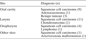
A total of 39 cricothyroid cannulae were inserted into 32 patients undergoing head and neck surgery, over a two-year period. Five patients underwent cricothyroid cannula insertion on two separate occasions, and one patient underwent three separate cannulations for surgery related to osteoradionecrosis of the mandible. Of the 32 patients, 25 had a primary diagnosis of squamous cell carcinoma, and 19 of these had advanced stage disease (tumour–node–metastasis stage III or IV). The most common disease sites were the oral cavity and larynx (Table I).
Patients' primary diagnosis

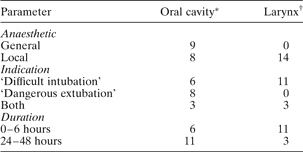
Of the 39 cricothyroid cannulations performed, 21 were performed for anticipated difficult intubations, 10 for anticipated dangerous extubations and eight for both these indications. Most patients with laryngeal cancer underwent cannulation for anticipated difficult intubation, whereas anticipated dangerous extubation was a more common indication in patients with oral cavity tumours (Table II).
Data for cricothyroid cannulations in patients with oral cavity and laryngeal tumours

* n = 17;
† n = 14.
The indication for cricothyroid cannulation is a factor in determining whether the procedure is performed with the patient awake or under general anaesthesia (GA), as well as in determining the duration the cannula is left in place post-operatively. Twenty-seven of the 39 cannulations were performed with the patient awake; this included all patients with laryngeal tumours, in whom a difficult intubation was anticipated. In these patients, the ability to perform transtracheal jet ventilation was confirmed prior to proceeding to securing a definitive airway, either awake or under GA. Of the 27 cannulations performed on awake patients, 21 were for anticipated difficult intubation alone, and all of these cannulae were removed within six hours of insertion. This included seven patients who received a tracheostomy once GA had been established. Conversely, nine of the 12 cannulations performed under GA were for anticipated dangerous extubation in patients with oral cavity tumours. In these patients, the development of acute airway compromise post-operatively, secondary to oedema and/or haematoma, was considered to be a risk. All of these cannulae remained in place for at least 24 hours post-operatively (Table II), in order to allow access for emergency transtracheal jet ventilation in the scenario of failed oxygenation and ventilation via the upper airway.
In eight patients, tracheostomy was performed as part of the planned operative procedure; in all these cases, it could be sited under general rather than local anaesthetic. Seven of these patients had advanced laryngeal squamous cell carcinoma and one had recurrent ameloblastoma of the mandible.
There were six cases in which tracheostomy was possibly avoided. All patients were identified as being at risk of post-operative airway obstruction, and had prophylactic cricothyroid cannulae in situ for a minimum of 24 hours, rather than a tracheostomy. No adverse post-operative airway events occurred in these patients.
One patient did require a tracheostomy in the immediate post-operative period. This patient had a recurrent T0 N2c squamous cell carcinoma of the oropharynx and developed signs of acute airway obstruction immediately post-extubation, following a right radical neck dissection. He had been identified as being at risk and so had a cricothyroid cannula in place. Transtracheal jet ventilation was given prior to reinduction of anaesthesia, allowing confirmation that ventilation was possible. At direct laryngoscopy, the patient had a very oedematous supraglottis. Transtracheal jet ventilation was continued and he was able to be re-intubated despite difficult laryngoscopy. This patient went on to have a tracheostomy and was transferred to the intensive care unit, where he made a good recovery.
No significant complications of cricothyroid cannulation were identified in this study group. In addition, the procedure did not have to be abandoned in any patient due to difficulty in cannula placement.
Discussion
We have found cricothyroid cannulation to be a useful technique in the management of those patients with head and neck neoplasms in whom a potentially difficult airway is anticipated. It may be used as a temporary measure until a definitive airway is established, as ‘prophylaxis’ for post-operative airway obstruction, or for both indications. Generally, when a difficult intubation is anticipated, cricothyroid cannulation tends to be performed with the patient awake, prior to induction of anaesthesia, and the cannula is kept in situ in the short term (2–6 hours). When performed as a precaution for post-operative airway management (dangerous extubation), cricothyroid cannulation can usually be performed safely under GA, and the cannula tends to be left in situ for 24 hours or longer.
There are few articles describing the use of this technique in the management of patients with head and neck cancer. The concept of prophylactic transtracheal catheterisation was reported by Carden et al. in 1976,Reference Carden, Becker and Hamood3 and Miller et al. subsequently reported four cases in 1982.Reference Miller, Lovino, Fine and Klain4 Transtracheal jet ventilation has been used extensively during elective endolaryngeal surgery, without major complication.Reference Monnier, Ravussin, Savary and Freeman5–Reference Russell, Maguire and Jones8 Gerig et al. recently reported the use of prophylactic transtracheal catheterisation in 11 patients with anticipated difficult airway, including five patients with head and neck cancer;Reference Gerig, Schnider and Heidegger9 primary tracheostomy was avoided in all 11 patients. Standley and Smith described a case of tracheal catheterisation and jet ventilation in a patient with failed intubation due to advanced laryngeal cancer; this approach allowed ventilation until a formal tracheostomy could be performed.Reference Standley and Smith10 In this situation, we would advocate placing the cricothyroid cannula prior to any attempt at intubation, in order to avoid the ‘can't intubate, can't ventilate’ scenario. In this way, the cannula is placed in a controlled manner with the patient awake, and jet ventilation may be easily commenced if there are difficulties with intubation.
One of the advantages of cricothyroid cannulation is that if tracheostomy is required, it may be performed safely under GA rather than LA. This has obvious benefits for both the surgical team and the patient. We believe a tracheostomy was avoided in six of our patients, based on the degree of airway obstruction present and our experience in managing such patients prior to the introduction of cricothyroid cannulation.
A potential criticism of this approach to airway management is that cricothyroid cannulation is unnecessary as a tracheostomy could be performed under LA or GA to provide a secure airway. Although tracheostomy is the ‘gold standard’ in management of upper airway obstruction in patients with advanced head and neck cancer, it is not without risk. Emergency tracheostomy carries a significant morbidity and mortality rate, and complications include haemorrhage, airway obstruction, decannulation, pneumothorax and recurrent laryngeal nerve injury.Reference Higgins and Punthakee11
• Cricothyroid cannulation is a valuable airway management procedure in patients undergoing head and neck surgery
• Tracheostomy may be avoided
• There is a low risk of complication
• Careful patient selection is required
• Cricothyroid cannulation should not replace tracheostomy where the latter is clearly indicated
Although there are also complications of cricothyroid cannulation, we did not experience any in our series. The reported complications can be divided into insertional difficulties (e.g. failure to cannulate the trachea, kinking of the cannula and damage to adjacent structures)Reference Benumof and Scheller2, Reference McGill, Clinton and Ruiz12 and complications relating to jet ventilation (e.g. surgical emphysema and pneumothorax).Reference Monnier, Ravussin, Savary and Freeman5, Reference Smith, Babinski, Klain and Pfaeffle13 The barotraumatic complications relate to sustained transtracheal jet ventilation and not to the insertion of the cannula itself, and so in many of our cases were not a significant risk. No specific late complications of cricothyroid cannulation have been reported.
We believe cricothyroid cannulation to be a safe, reliable technique that is a useful adjunct in the management of patients with an anticipated difficult airway, both prior to the induction of anaesthesia and after extubation. In some cases, performing cricothyroid cannulation allowed us to avoid a tracheostomy. There were no significant complications associated with the procedure.
Further study is needed to confirm the usefulness and safety profile of this procedure in patients with advanced head and neck cancer.
Sound judgement is required in patient selection, and cricothyroid cannulation should not take the place of tracheostomy where the latter is clearly indicated.
Acknowledgements
Mr P McLoughlin, Oral and Maxillofacial Surgeon, and Mr R E Mountain, ENT Surgeon, Ninewells Hospital, Dundee, Scotland, UK.