Introduction
Tinnitus refers to a diverse set of phenomena, all of which share the property of the perception of a sound in the absence of an external sound. Some tinnitus can be evoked by stimuli such as gaze, light touch, active finger movement, or strong contractions and/or compressions of the neck and jaw muscles.Reference Cullington1, Reference Levine and Hamilton2
We report a case of unilateral tinnitus evoked by rapid head rotation. This case is unique because the patient's tinnitus was not produced by somatosensory activation but rather was due to abnormal inner ear fluid movement caused by dehiscence of the superior semicircular canal.
Case report
A 50-year-old man sought medical attention for momentary right ear ‘cricket’ tinnitus occurring whenever he turned his head quickly to either side. This symptom had begun a year earlier, following several minutes of belt-sanding. He also noted autophony in his right ear and sound distortion – specifically, tapping his teeth together was perceived as sounding like ‘a metal hammer on ceramic tile’. He denied any hearing loss or vestibular symptoms.
The patient's ENT examination was unremarkable. Right periauricular auscultation was negative even when the patient's tinnitus was induced with rapid head turns. The patient's right ear cricket tinnitus was pitch-matched to 4 kHz. Somatic testing elicited no right ear cricket tinnitus, but intense left sternocleidomastoid muscle contraction provoked faint, high-pitched left ear tinnitus. The use of Frenzel lenses resulted in no spontaneous, head-shaking or gaze-holding nystagmus. Nystagmus was not provoked by the use of a noise box or the Valsalva manoeuvre. The patient's right ear cricket tinnitus could be evoked by yaw head rotations to either side, as well as by head rotations in the plane of his right superior semicircular canal but not in the plane of his left superior semicircular canal. Head velocity measurements revealed that right ear cricket tinnitus was heard only when the yaw head velocity exceeded 600°/second (Figure 1). Saccades to visual targets and yaw-axis optokinetic nystagmus did not provoke right ear cricket tinnitus. However, this tinnitus was provoked by head rotations even when the head and neck moved en bloc with the trunk. The right ear cricket tinnitus was not affected by suppression or enhancement of eye movements during head rotations, which were produced by fixating upon a visual target that either moved with the patient's head (cancelling the vestibulo-ocular reflex) or was earth-fixed (visually augmenting the vestibulo-ocular reflex).
The patient's angular head velocity measurements when his right ear ‘cricket’ tinnitus commenced. An Ascension miniBird position sensor (sampling at 100 Hz) secured to his head measured angular head velocity during head rotation. The arrow at about 4.4 seconds of the yaw velocity trace indicates the first instance when the patient's right ear cricket tinnitus was heard (angular head velocity >600°/second). Deg/sec = degrees per second

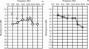
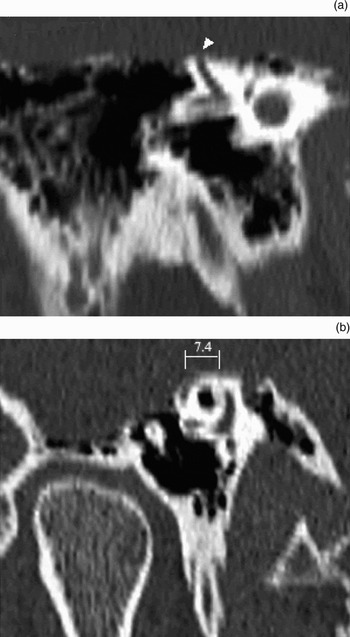
Audiography demonstrated a right conductive hearing loss of 35 dB at 250 Hz and 15 dB at 500 and 1000 Hz, in addition to a symmetrical, mild, high frequency sensorineural hearing loss (Figure 2). The results of speech hearing testing are given in Table I; speech discrimination scores were within the normal range in both ears. The patient's tympanograms were normal, and ipsilateral and contralateral acoustic reflexes were present bilaterally. Temporal bone computed tomography revealed a 7.4 mm defect in the right superior canal wall (Figure 3). Laser Doppler vibrometry measurements of the patient's umbo velocity were consistent with right superior semicircular canal dehiscence. Specifically, given the degree of conductive hearing loss, the right umbo velocity was differentiable from other middle-ear pathologies that could cause a conductive hearing loss.Reference Rosowski, Nakajima and Merchant3
The patient's audiogram for the (a) right and (b) left ear. High frequency sensorineural hearing loss was shown for both ears. The right ear also demonstrated a conductive hearing loss, with a maximum air–bone gap of 35 dB at 250 Hz and also gaps of 15 dB at 500 and 1000 Hz. [O and X air-conduction threshold; < and > = bone-conduction threshold [ and ] = bone conduction threshold with contralateral masking]

Reformatted computed tomography images in planes (a) parallel (Stenver view) and (b) perpendicular (Poschl view) to the patient's right superior semicircular canal, showing an approximately 7.4 mm defect of the bony wall overlying the superior semicircular canal (arrowhead).

Speech hearing test results

R = right; L = left
All studies were performed with the approval of the Human Studies Committee of the Massachusetts Eye and Ear Infirmary.
Discussion
Most tinnitus evoked by head movements has a somatosensory basis.Reference Levine and Hamilton2 However, somatosensory activation did not evoke our patient's right ear cricket tinnitus, although it did evoke transient faint left ear tinnitus, as occurs in about 60 per cent of people. An association between our patient's right ear cricket tinnitus and his superior semicircular canal dehiscence was suggested by tinnitus localisation to the superior semicircular canal dehiscent ear and elicitation by rapid head rotation (>600°/second) in the plane of his dehiscent semicircular canal and yaw, but not in the plane of his left superior semicircular canal. Since some patients with superior semicircular canal dehiscence have heightened sensitivity to bone-conducted sounds (such as those produced by eye or neck movements), this patient's tinnitus could potentially be produced by (1) the vestibulo-ocular reflex elicited by head rotation, or (2) sounds produced by motion of the head on the neck.Reference Minor4 Eye motion was ruled out, since the patient's right ear cricket tinnitus was not elicited by isolated saccadic or optokinetic eye movements, which exceed 600°/second, or changed by suppression or augmentation of eye movements with head rotation. Because the patient's right ear cricket tinnitus was elicited whether or not motion of the cervical vertebrae and associated soft tissues was minimised by en bloc rotation of the body, this tinnitus did not appear to originate from neck sounds.
We therefore conclude that our patient's right ear cricket tinnitus was caused by enhanced inner ear fluid flow, due to his right ear's ‘third window’.
In patients with benign paroxysmal positional vertigo (BPPV), the presence of an otolith may alter the endolymphatic flow of the affected posterior semicircular canal. In contrast, the existence of a bony defect of the superior semicircular canal (a third window) might enhance endolymphatic flow and possibly produce tinnitus. Thus, movement of the head in the same plane evokes vertigo in BPPV patients and tinnitus in patients with superior semicircular canal dehiscence.
• This paper reports a case of head rotation evoked tinnitus due to superior semicircular canal dehiscence
• Head rotation with a velocity of over 600°/second in the plane of the superior semicircular canal induced a cricket-like sound at the right ear; the diagnosis was also supported by (a) high resolution temporal bone computed tomography changes, and (b) umbo velocity
• This case suggests that superior semicircular canal dehiscence may present with head rotation evoked tinnitus, without vestibular symptoms; the mechanism of such tinnitus may be enhanced inner ear fluid flow, due to a ‘third window’
Conclusion
In addition to pulse-synchronous or continuous tinnitus, head rotation induced tinnitus can be a symptom of superior semicircular canal dehiscence, and may be its only presenting symptom.Reference Brantberg, Bergenius and Mendel5 Head rotation tinnitus probably occurs because the dehiscence results in alterations in labyrinthine fluid flow.