Introduction
Non-communicable diseases, such as cardiovascular disease (CVD), type II diabetes mellitus (T2DM), cancer and chronic respiratory diseases, are responsible for a large proportion of early deaths worldwide. 1 Although certain lifestyle choices in combination with genetic predisposition are recognized to markedly increase the risk of developing these diseases,Reference Yang, Kelly and He 2 the physiological and nutritional state of the mother during gestation and lactation also have a significant role in determining future health of the offspring.Reference Kunz and King 3 , Reference Warner and Ozanne 4
It was highly controversial when Barker proposed the concept of ‘Fetal origins of adult disease’ in 1990,Reference Barker 5 but epidemiological studies revealed a clear relationship between maternal undernutrition during pregnancy and postnatal exposure to nutrient-rich environment, with an increased prevalence of CVD and T2DM in adult male offspring.Reference Barker 6 , Reference Hales, Barker and Clarc 7 These observations lead to the formation of thrifty phenotype hypothesisReference Hales and Barker 8 that has been widely accepted in the present time. In addition to epidemiological observations, animal models have repeatedly shown that intrauterine growth restriction imposed by maternal protein or caloric restriction during pregnancy leads to low birth weight and development of CVD, T2DM and obesity in the offspring.Reference Bhasin, van Nas and Martin 9 , Reference Vickers, Breier, Cutfield, Hofman and Gluckman 10
Although the evidence appears strong and is consistent when it comes to effects caused by maternal nutrition during pregnancy, the effects of maternal nutrition before pregnancy on the future offspring are far less studied. Although indistinct, epidemiological evidence indicates an association of preconceptional consumption of protein and vegetables with reduced risk for low birth weightReference Cuco, Arija and Iranzo 11 , Reference Weisman, Misra and Hillemeier 12 and consecutively development of T2DM and CVD.Reference Barker, Winter, Osmond, Margetts and Simmonds 13 , Reference Harder, Rodekamp, Schellong, Dudenhausen and Plagemann 14 Animal studies supplement these findings by demonstrating that maternal caloric and protein restriction exclusively before pregnancy leads to accelerated postnatal growth in ewes and affects offspring vascular function in ewes and mice,Reference Torrens, Snelling and Chau 15 , Reference Watkins, Wilkins and Cunningham 16 as well as increases male offspring adiposity at young adulthood in mice.Reference Mortensen, Wang, Malte, Raubenheimer and Mayntz 17
Here we study how reduced maternal protein intake before conception affects offspring early body mass and their insulin-mediated glucose metabolism. As the offspring are not directly exposed to protein restriction during development, we expect the effects to operate through maternal body composition and/or physiological state, and we therefore examine maternal food intake, body mass, organ size and adiposity before conception to investigate whether any of these factors account for changes induced in the offspring.
Materials and Methods
Animals and housing
All experiments were performed in accordance with the principles and guidelines of Danish legislation on animal welfare, permit number 2006/561–1257. Danish Inspectorate for Animal Experiments specifically approved this study.
Eight-week-old virgin C57BL/6JBom mice (53 females and 36 males) were obtained from Taconic (Lille Skensved, Denmark) and caged individually in type 2 Macrolon cages (length: 23 cm; width: 17 cm; height: 14 cm) at 12:12 h light:dark regime, 23°C and 60% humidity. Food and water was available ad libitum at all times, except for the overnight starvation before Glucose Tolerance Test (GTT), when food was removed for 14 h.
Experimental diets
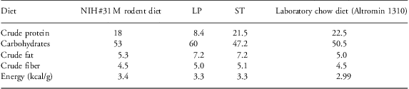
Four dry pellet diets were used in the experiment (composition is described in Table 1). NIH #31 M rodent diet was purchased from Taconic (Denmark) and two custom-made isocaloric experimental diets – Low protein (LP) and Standard (ST) – were purchased from Brogaarden/Altromin (Denmark). Both diets were similar in composition apart from the crude protein and carbohydrate content (Table 1). Laboratory chow diet (Altromin 1310) for rats and mice (Brogaarden/Altromin, Denmark) was used as a standard laboratory chow diet.
Composition of the diets used before and during the experiment

LP, low protein; ST, standard diet.
Values presented as g per 100 g dry mass.
Experimental protocol
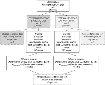
Before the start of the experiment, all 53 female mice were allowed to acclimate to the facilities for 2 weeks; during this period, they were fed the same diet as they received at the breeding facilities (NIH #31 M rodent diet). Subsequently, they were weighed and divided into two groups that received either an LP (n = 27) or an ST (n = 26) diet for the next 9–10 weeks. Following that, nine mice from each treatment were subjected to a GTT, allowed to recover from this test for 5 days and sacrificed by cervical dislocation. The remaining mice (n = 35, n ST = 17; n LP = 18) were placed in cages, with an 8–9-week-old male for every female, for 48 h. From this point onwards, all animals received laboratory chow diet (Altromin 1310). After mating, the male was removed, but the female remained in the individual cage until offspring were born and weaned onto laboratory chow diet (Altromin 1310) at the age of 23 days and caged individually. Laboratory chow diet (Altromin 1310) was used after mating and thereafter to ensure that both groups of female mice were exposed to a change in the diet close to the time of conception. In order to minimize handling stress upon the animals, neither maternal nor offspring body masses were measured at birth. Owing to the low litter numbers, litter sizes were not adjusted. At the age of 10 weeks, the offspring were subjected to GTT, allowed to recover from it for 5 days and sacrificed by cervical dislocation (Fig. 1).
Experimental protocol used during the study.

Food intake measurements
Maternal food intake was measured weekly using a spillage collecting system, for example,Reference Sorensen, Mayntz, Raubenheimer and Simpson 18 on individually caged animals. Animals received a preweighed portion of food, large enough to ensure that they had access to food at all times. After 1 week, the food remaining in the feeding tray and spillage collector was collected and stored at −25°C for later measurements. Before the measurements, the collected food was defrosted and dried at 60°C for 24 h, and weighed to the nearest 0.001 g. A standard curve for conversion of wet mass to dry mass was created for each feed separately.
Offspring food intake was measured on individually housed animals once a week after weaning. As all offspring received laboratory chow diet (Altromin 1310), spillage of food was expected to be similar between groups, and therefore only the difference of given and leftover food in the feeding tray was measured.
GTTs
GTTs were performed after a 14-h overnight fast. Upon measuring fasting blood glucose, 10% glucose solution was injected intraperitoneally (2 g/kg), and blood was collected from an incision at the tip of the tail for determination of glucose concentration at 15, 30, 60 and 120 min. All blood glucose concentration measurements were performed using Bayer Contour® glucometer and Bayer Contour® test strips.
Insulin levels
Blood samples for insulin measurements were collected after the 14 h starvation preceding the GTT and 2 h after glucose injection during GTT. Blood was collected from an incision at the tip of the tail into a heparinized hematoctit tube. Trunk blood was collected after sacrifice to determine non-fasting insulin concentration. All collected blood samples were heparinized and spun, and plasma was collected and frozen at −80°C for later analysis. Plasma insulin concentration was measured using Ultra Sensitive Mouse Insulin ELISA kit according to manufacturer's instructions.
Organ size measurements
Five days after GTT, animals were weighed to a precision of 0.1 g and sacrificed by cervical dislocation and decapitation. Kidneys, liver and heart were dissected, blotted on a filter paper in a standardized manner and weighed. The gastrointestinal tract was dissected free to determine the length from the pyloric sphincter to the rectum and the length of the small intestine with precision to 1 mm. Stomach, cecum and colon were separated and flushed with 0.9% saline, dried at 60°C for 72 h and weighed. The small intestine was blotted on filter paper and wet mass taken. The mucosal layer was separated from a 4–6-cm-long piece of the small intestine (as described by Jensen et al. Reference Jensen, Elnif, Burrin and Sangild 19 ). The proportion of mucosal layer to the layer of muscular tissue of the small intestine was determined after drying both layers separately for 72 h at 60°C. The rest of the small intestine was used for another experiment, and therefore wet mass to dry mass relationship of the 4–6-cm-long piece was used to calculate the dry mass of the whole small intestine. All weight measurements were recorded with precision to 0.1 mg.
Data analysis
Data are presented as means (±s.e.) or as least square means (±s.e.), and the differences between treatment groups (ST v. LP) were tested within the same sex by Student's t-test. For comparison of GTT results, the area under the GTT curve [Area Under the Curve (AUC)] was calculated. The fasting blood glucose concentration was compared between ST and LP groups within the same gender by Student's t-test, and if the values were not significantly different – which was always the case – baseline was calculated (fasting glucose value × 120 min) and subtracted from the total AUC.
Analysis of Covariance (ANCOVA) was used to test the effects of body mass and dietary treatment on organ size of females and the effects of maternal body mass and diet before conception on offspring early body mass. Maternal diet and offspring body mass were used as covariates when offspring organ size was measured. ANCOVA was used to test the effect of maternal body mass before conception on mean body mass of male or female offspring per litter at weaning. Maternal diet and litter size were included as covariates initially, but were removed from the model if not significant.
Litter size was always included in the covariate model initially, but was removed if nonsignificant, which was always the case.
Offspring growth and food intake were compared using repeated measures Multivariate Analysis of Variance (MANOVA) in both genders separately.
Homogeneity of variances was tested using Levene's test (α > 0.05), and if the variances proved to be unequal data were compared using Welch Analysis of Variance (ANOVA).
All data analysis was conducted using JMP 8 and Sigma Plot 11.0.
Results
Food intake and body mass changes of mothers
After 9 weeks of feeding on experimental diets, the total food consumption of females averaged 149 ± 2 g (dry mass) per animal in both groups, and there was no significant difference between LP and ST treatments (t-test, DF = 51, t = 0.08, P = 0.94). On average, after 9 weeks of feeding dams had consumed a total of 493 ± 5 kcal per animal and this result was not affected by the dietary treatment (t-test, DF = 51, t = 0.08, P = 0.94). However, owing to differences in the protein content of experimental diets, over 9 weeks of feeding females on the LP diet ingested a total of 12.5 ± 0.4 g crude protein, whereas females on the ST diet consumed 2.6 times more protein (32.1 ± 0.4 g; t-test, DF = 51, t = 39.64, P < 0.0001).
At the end of the feeding period, we observed significant differences in female body mass. Dams exposed to the LP diet had lower body mass and weighed 21.7 ± 0.3 g, whereas ST females weighed 22.5 ± 0.3 g (t-test, DF = 51, t = 2.1, P = 0.04).
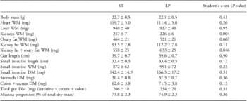
We found no significant differences in the body mass of dams exposed to experimental diets and used for GTT and organ size measurements (Table 2).
Organ sizes of female mice that had been fed the ST or the LP diet for a period of 9 weeks

ST, standard diet; LP, low-protein diet; WM, wet mass; DM, dry mass.
All values presented as mean ± s.e. (n = 9 in each group) and compared by Student's t-test.
GTT and non-fasting plasma insulin concentration of females fed on experimental diets
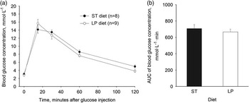
We found no significant differences in GTT results (Fig. 2; t-test, DF = 15, t = −0.71, P = 0.49) between females fed the LP diet and those fed the ST diet for 9 weeks. However, we observed a nearly significant reduction in the non-fasting plasma insulin concentration of females fed the LP diet (0.33 ± 0.07 μg/l; n = 8) compared with the non-fasting insulin levels of females fed the ST diet (0.66 ± 0.14 μg/l; n = 8; t-test, DF = 14, t = 2.06, P = 0.06).
Intraperitoneal glucose tolerance test (GTT) results. (a) Plasma glucose concentrations during GTT and (b) calculated Area Under the Curve (AUC). GTT was performed on female mice fed either the standard (ST; n = 9) or the low-protein (LP; n = 9) diet for a period of 9 weeks (mean ± s.e.). AUC values were compared using Student's t-test.

Organ sizes of females fed experimental diets
Females fed the ST diet had significantly larger kidneys than females on the LP diet (Table 2). The difference remained significant when corrected for body mass (least square means ± s.e., ST = 260 ± 5, LP = 230 ± 5, ANCOVA, body mass F 1 = 35.46, P = 0.0001, diet: F 1 = 22.09, P = 0.0003). Fat deposits were significantly larger in females fed the LP diet (Table 2), even after correcting for total body mass (least square means ± s.e., ST = 551 ± 23, LP = 640 ± 23, ANCOVA, body mass F 1 = 4.4, P = 0.053, diet: F 1 = 7.63, P = 0.01). Visceral organ masses varied with body mass, but neither the absolute (Table 2) nor the body mass-corrected organ masses proved to be significantly different between the two treatments.
Mating success
From the 35 females that were mated (LP: n = 18, ST: n = 17), 10 females delivered offspring (LP: n = 6, ST: n = 4). The mean litter size in the LP group was 6 ± 7 (range 3–8 pups) and in the ST group 5.5 ± 0.9 (range 4–7 pups). The mean body mass of LP females who delivered offspring was 18.8 ± 0.5 g before and 21.4 ± 0.6 g after 9 weeks of feeding on the LP diet (n = 6). The mean body mass of ST diet females who delivered offspring was 20.0 ± 0.6 g before and 22.0 ± 0.8 after the dietary treatment (n = 4). In total, 51 pups were born: LP females delivered 31 pups (females: n = 23, males: n = 8) and ST females delivered 20 pups (females: n = 13, males: n = 7).
Offspring growth and food intake
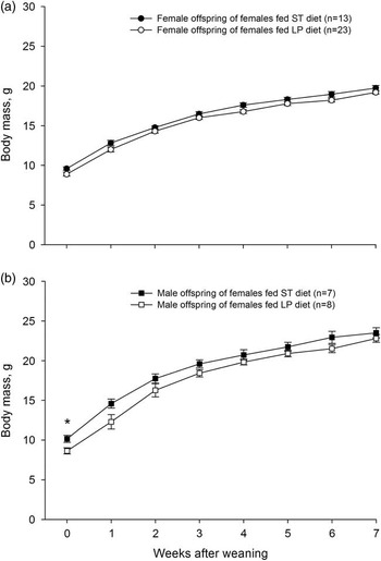
Male offspring born to mothers exposed to the LP diet before conception had significantly lower body mass at weaning (8.64 ± 0.4 g) than male offspring from the ST group (10.16 ± 0.4 g; t-test, DF = 13, t = 2.59, P = 0.02). A similar tendency was observed for female offspring, but it was only marginally significant. LP female offspring weighed 8.88 ± 0.2 g and ST female offspring had a body mass of 9.59 ± 0.6 g (t-test, DF = 34, t = 1.91, P = 0.065).
A positive significant relationship was observed between the average male offspring body mass per litter at weaning and maternal body mass before conception (ANCOVA, maternal body mass before conception: F 1 = 6.25, P = 0.04). A similar but nonsignificant tendency was observed for female offspring (ANCOVA, maternal body mass before conception: F 1 = 4.56, P = 0.06). These differences in offspring body mass disappeared within 1 week after weaning (Fig. 3). The differences in body mass at weaning were not affected by litter size for either males (ANCOVA, litter size: F 1 = 0.007, P = 0.94) or females (ANCOVA, litter size: F 1 = 0.16, P = 0.69).
Offspring body mass after weaning. Female (a) and male (b) offspring born to mothers fed the standard (ST) or the low-protein (LP) diet for 10 weeks before conception. Litter number, ST: n = 4, LP: n = 6. Body masses were compared by Student's t-test within the same gender for separate time points. *Denotes a significant difference between the maternal dietary treatments, P < 0.05.

Over the entire female offspring growth period, maternal diet and offspring age had significant effects on body mass; however, there was no interaction between the two (Fig. 3a; repeated measures MANOVA, maternal diet: F 1, 33 = 4.44, P = 0.04; offspring age: F 7, 27 = 254.6, P < 0.0001; maternal diet × time: F 7, 27 = 0.95, P = 0.48). Thus, growth curves did not have a significantly different pattern. Only offspring age had a significant effect on male offspring body mass, indicating that growth in body mass did occur during the experiment (Fig. 3b; repeated measures MANOVA, maternal diet: F 1, 13 = 3.23, P = 0.1; offspring age: F 7, 7 = 0.72, P < 0.0001; maternal diet × offspring age: F 7, 7 = 0.72, P = 0.66).
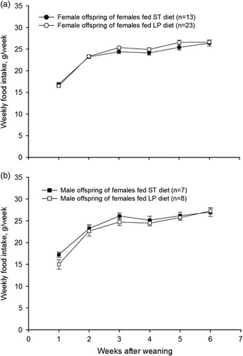
After the 6-week-long post-weaning period, female offspring total food consumption averaged at 197.1 ± 4 g per individual in each group and there was no significant difference between ST and LP treatments (t-test, DF = 34, t = 0.117, P = 0.91). For the same period of time, male offspring total food intake averaged at 193.7 ± 5 g per individual in both ST and LP groups (t-test, DF = 14, t = 0.21, P = 0.84; Fig. 4).
Offspring food intake after weaning. Female (a) and male (b) offspring born to mothers fed the standard (ST) or the low-protein (LP) diet for 10 weeks before conception. Litter number, ST: n = 4, LP: n = 6.

Offspring GTT and insulin
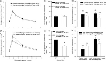
GTT performed on adult offspring revealed that LP male offspring were more glucose tolerant than ST male offspring (Fig. 5d and 5e, DF = 11, t-test, t = 2.2, P = 0.047), but this was not observed for female offspring (Fig. 5a and 5b, t-test, DF = 33, t = −0.06, P = 0.948). However, LP female offspring had a significantly lower blood insulin concentration than ST female offspring 2 h after glucose challenge (Fig. 5c, t-test, DF = 15, t = 2.4, P = 0.03). Such differences were not observed for male offspring (Fig. 5f, t-test, DF = 7, t = 1.7, P = 0.13). No differences were observed in male and female offspring fasting (Fig. 5c and 5f, male offspring, t-test, DF = 7, t = −0.93, P = 0.38; female offspring, t-test, DF = 26, t = 0.94, P = 0.35) and non-fasting insulin concentrations (male offspring, DF = 12, t-test, t = −0.04, P = 0.97; female offspring, t-test, DF = 30, t = 1.02, P = 0.32).
Offspring glucose tolerance test (GTT) results and plasma insulin concentration. Blood glucose concentrations and Area Under the Curve (AUC) during GTT in female (a, b, respectively) and male (d, e, respectively) offspring born to mothers fed either the standard (ST) or the low-protein (LP) diet for 10 weeks before conception. Female (c) and male (f) offspring plasma insulin concentration after fasting and 2 h after glucose injection (2g/kg) during GTT. In (c) and (f ), numbers in brackets indicate number of samples tested. Litter number, ST: n = 4, LP: n = 6. AUC and plasma insulin concentrations were compared by Student's t-test within the same gender. *Denotes a significant difference between the maternal dietary treatments, P < 0.05.

Offspring organ size
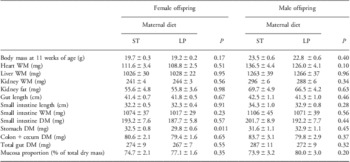
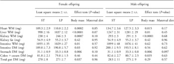
We found no differences in offspring absolute and body mass-corrected visceral organ size (Tables 3 and 4). Stomach dry mass was significantly lower in LP female group (Table 3), but this difference disappeared when stomach dry mass was corrected for wet body mass (Table 4). Litter size had no significant effect on any of the measured values.
Organ size of female and male offspring born to mothers fed either the ST or the LP diet for 10 weeks before conception

ST, standard diet; LP, low-protein diet; WM, wet mass; DM, dry mass.
All values presented as mean ± s.e. (female offspring, ST: n = 13, LP: n = 23; male offspring, ST: n = 7, LP: n = 8; litter number, ST: n = 4, LP: n = 6) and compared by Student's t-test.
Body mass-corrected organ size of female and male offspring born to mothers who were fed either the ST or the LP diet for 10 weeks before conception

ST, standard diet; LP, low-protein diet; WM, wet mass; DM, dry mass.
All values presented as least square mean ± s.e. (female offspring, ST: n = 13, LP: n = 23; male offspring, ST: n = 7, LP: n = 8; litter number, ST: n = 4, LP: n = 6) of measures adjusted for wet body mass. In all analysis, litter size was used as a covariate first, but as it did not have a significant effect it was removed from the final model.
Discussion
Our study shows that protein restriction in the diet before conception may lower body mass at weaning, followed by catch-up growth and increased insulin sensitivity at early adulthood in mice. These effects are similar to those previously described for protein restriction during pregnancy and shown to be an indicator for increased risk of developing T2DM, CVD and obesity in humans and animals.Reference Barker 6 , Reference Hales, Barker and Clarc 7 , Reference Bhasin, van Nas and Martin 9 , Reference Baird, Fisher and Lucas 20 – Reference Zambrano, Bautista and Deas 27 It is therefore possible that LP diet before pregnancy imposes similar types of risks for the development of lifestyle diseases in the offspring as those described for malnutrition during pregnancy.
Offspring subjected to intrauterine growth restriction are often observed to display rapid postnatal growth if exposed to a nutrient-rich environment after birth.Reference Bieswal, Ahn and Reusens 21 , Reference Chen, Martin-Gronert, Tarry-Adkins and Ozanne 28 It is not clear whether catch-up growth is a risk factor that acts in addition to protein restriction when it comes to development of T2DM, CVD and obesity. However, in rodents, it appears to reduce offspring lifespan.Reference Chen, Martin-Gronert, Tarry-Adkins and Ozanne 28 – Reference Jennings, Ozanne, Dorling and Hales 30 In our study, male offspring from the LP group had lower body mass at weaning than those from the ST group, but these differences disappeared within a few weeks after weaning (Fig. 3). This implies that the offspring have gone through a period of fast catch-up growth. Such catch-up growth has been shown in humans and several different animal species,Reference Shepherd, Crowther, Desai, Hales and Ozanne 24 , Reference De Blasio, Gatford, McMillen, Robinson and Owens 31 , Reference Dulloo, Jacquet, Seydoux and Montani 32 including mice,Reference Cottrell, Martin-Gronert and Fernandez-Twinn 33 and is associated with enhanced insulin sensitivity.Reference Morrison, Duffield, Muhlhausler, Gentili and McMillen 34 This pattern corresponds well with our observed results of low male offspring early body mass, catch-up growth and enhanced insulin sensitivity in both sexes (Fig. 5) and indicates the presence of an event cascade similar to that induced by maternal gestational malnutrition.
In other animal models, similar offspring responses have been found. For example, rat offspring from mothers exposed to the LP diet during pregnancy also have increased insulin sensitivity at early adulthood (6–10 weeks), but this effect disappears with age (15 months), when offspring develop insulin resistance and glucose intolerance.Reference Hales, Desai, Ozanne and Crowther 22 – Reference Shepherd, Crowther, Desai, Hales and Ozanne 24 As our findings from manipulating preconception nutrition resemble the cascade of events caused by malnutrition during pregnancy, it is possible that offspring born from mothers that received the LP diet before pregnancy are at the same increased risk of developing insulin resistance later in life. In a previous study with the same mouse strain and the same experimental protocol as in the current experiment, it was found that LP male offspring have increased adiposity as early as 46 days of age.Reference Mortensen, Wang, Malte, Raubenheimer and Mayntz 17 The two studies in combination indicate that maternal protein restriction before pregnancy affects offspring body mass, body composition and glucose metabolism and can therefore modify offspring risk of developing lifestyle-acquired diseases.
Although animal models using the LP diet after conception are rather well studied in terms of produced offspring phenotypes, the mechanisms underlying these effects are less clear.Reference Rosario, Jansson and Kanai 35 Some of the effects of LP diet exposure during pregnancy have been attributed to insufficient amount of amino acid supply to the embryo during development;Reference Bhasin, van Nas and Martin 9 however, our study and previous experimentsReference Mortensen, Wang, Malte, Raubenheimer and Mayntz 17 have showed that similar effects can be induced by preconceptional protein restriction. This indicates that factors other than maternal diet during pregnancy can be involved in fetal programming. Unfortunately, our study design does not allow us to fully elucidate whether or not the risks are imposed through maternal body composition or physiological state, rather than by direct effects of amino acid insufficiency during development. Although studies exposing animals to protein restriction during pregnancy mostly ascribe the effects obtained to reduced amino acid supply,Reference Bhasin, van Nas and Martin 9 there is evidence that maternal body mass index, body composition, hypertension and diabetes among other factors can all have an impact on offspring health.Reference de Bernabe, Soriano and Albaladejo 36 , Reference Ehrenberg, Mercer and Catalano 37
Studies in human population show that maternal body mass index before pregnancy and weight gain during pregnancy are good predictors of offspring birth weight.Reference Weisman, Misra and Hillemeier 12 , Reference Ehrenberg, Dierker, Milluzzi and Mercer 38 Although we did not measure maternal weight gain during pregnancy to avoid stress to the pregnant females,Reference Decatanzaro and Macniven 39 we observed a significant relationship between maternal body mass at conception and male offspring body mass at weaning and a similar, nearly significant tendency for female offspring. It is likely that low body mass at weaning among LP offspring can be explained, at least partly, by low maternal body mass at conception, but these effects remain to be investigated.
Studies on rodents pertaining to maternal obesity induced by a high-fat diet show that obesity at conception programs offspring obesity and insulin resistance at adulthood but do not necessarily affect birth weight.Reference Shankar, Harrell and Liu 40 Our results showed that females fed the LP diet had significantly larger fat deposits at the time of conception than did females fed the ST diet (Table 2). It is difficult to discriminate whether increased adiposity in mothers fed the LP diet had an effect on offspring early body mass, but as suggested by previous studies it could have influenced their glucose metabolism.Reference Howie, Sloboda, Kamal and Vickers 41
Another aspect of offspring programming is maternal insulin-mediated glucose metabolism. Elevated maternal blood glucose differentially affects birth weight: whereas mild elevation above normoglycemia produces macrosomic offspring, highly elevated glucose levels induce fetal growth restriction.Reference Oh, Gelardi and Cha 42 GTT on females at the end of preconceptional feeding period showed no differences in fasting glucose values or glucose tolerance (Fig. 2). However, non-fasting blood insulin concentration was nearly significantly lower in LP-fed dams, a known result of LP feeding.Reference Jansson, Pettersson and Haafiz 43 , Reference Kwong, Wild, Roberts, Willis and Fleming 44 As exposure of preimplantation murine embryos to insulin increases fetal growth rate in vitro,Reference Kaye and Gardner 45 it is possible that lowered maternal insulin levels in the LP group at the time of conception have restricted offspring growth.
Whereas our study mainly addresses the physiological changes of the mother that can affect offspring development and phenotype, other studies have shown that similar effects in offspring can be induced without apparent changes in maternal phenotype. Thus, female mice of the MF-1 strain fed an LP diet for a very short period of time close to conception (±3.5 days, which includes the period of oocyte maturation and preimplantation) produced offspring that at 21 and 52 weeks of age had elevated systolic blood pressureReference Watkins, Wilkins and Cunningham 16 , Reference Watkins, Lucas, Wilkins, Cagampang and Fleming 46 and attenuated endothelium-dependent vasodilation in mesenteric arteries at the age of 22 weeksReference Watkins, Wilkins and Cunningham 16 compared with control offspring whose mothers were supplied with sufficient protein. As such short feeding periods may be expected to have only minor effects on the body composition of the mothers, compared with the 10-week period of feeding in our experiment, the measured alterations in the MF-1 offspring could very well have been caused by epigenetic changes in the mothers eggs or in the young zygote. It is therefore of high importance to investigate such epigenetic effects in the future studies on the presented animal model of preconceptional nutrition.
Other studies have found that maternal LP nutrition during pregnancy can program offspring food intake and preferences,Reference Bellinger, Lilley and Langley-Evans 47 which can potentially explain differences in offspring growth, development of obesity, etc. In our study, we measured offspring food intake throughout the experiment and found no differences. Thus, we find no evidence that differences observed in offspring phenotypes were caused by differences in their feeding behavior after weaning.
Fertility rates observed in the study were rather low. Such low conception rates may be explained by the use of suboptimal mating protocol. Estrous cycle frequency in mice is 4–5 days, but the females were only caged with the males for 2 days, which may explain the low rate of conception. For the future studies, females and males should be housed together for a longer period (4–5 days). The study would have had greater statistical power if the litter numbers had been higher; however, the data presented were tested using appropriate statistical tests, and the effects of maternal preconceptional treatment are evident. It is, however, possible that because of low litter numbers some of the effects could not have not been detected, and therefore future studies need to be carried out, where another mating protocol is used and higher litter numbers are obtained.
As suggested by previous epidemiological studies, maternal preconceptional nutrition and physiological state can have an impact on the future health of the offspring in humans.Reference Cuco, Arija and Iranzo 11 , Reference Weisman, Misra and Hillemeier 12 Our studies using an animal model of preconceptional nutritionReference Mortensen, Wang, Malte, Raubenheimer and Mayntz 17 confirm the importance of these effects. It is therefore important to investigate the effects of maternal preconceptional state further, as, once established, they can provide effective grounds for intervention and prevention of disease development in future generations of human populations.
Overall, our findings show that maternal protein restriction before pregnancy induces gender-specific effects in the offspring, namely, reduced early body mass, male offspring catch-up growth and enhanced insulin sensitivity at early adulthood in male and female offspring. These effects closely resemble the effects of maternal protein restriction during pregnancy on offspring physiology and may therefore imply an increased risk for the offspring to develop obesity, type II diabetes and CVD in later life.
The physiological mechanisms inducing these changes are less clear and remain to be investigated.
Acknowledgments
D.M. and T.W. were funded by the grants from Danish Research Council. G.W. was funded by Danish Medical Research Council, grant no. 271-08-0768. We thank Heidi Meldgaard Jensen and Rasmus Buchanan for their assistance during experiments.