INTRODUCTION
Tropical shallow coral reefs are under serious threat due to natural catastrophes such as bleaching events, hurricanes and anthropogenic activity that leads to serious damage of coral colonies (Woodley et al., Reference Woodley, Chornesky, Clifford, Jackson, Kaufinan, Knowlton, Lang, Pearson, Porter, Rooney, Rylaarsdam, Tunnicliffe, Wahle, Wulff, Curtis, Dallmeyer, Jupp, Kodhl, Neigel and Sides1981; Connell, Reference Connell1997; Marshall, Reference Marshall2000; Bruckner & Bruckner, Reference Bruckner and Bruckner2001). Hall (Reference Hall1997, Reference Hall2001) divided coral damages into four major categories: (i) partial tissue loss (grazing, predation); (ii) tissue mortality (diseases, bleaching events, sediment stress and pollution of the environment); (iii) superficial tissue and skeleton loss (predation by excavating fish, natural disturbances such as turbidity, sand and rubble abrasion and human activity); and (iv) substantial tissue and skeleton loss (breakage due to storms and human activity).
The regenerative ability of damaged colonies have been experimentally studied in the Caribbean (e.g. Bak & Steward-Van Es, Reference Bak and Steward-Van Es1980; Meesters & Bak, Reference Meesters and Bak1995), in the Red Sea (Rinkevich & Loya, Reference Rinkevich, Loya and Spanier1989), in the East China Sea (Kobayashi, Reference Kobayashi1984; Titlyanov et al., Reference Titlyanov, Titlyanova, Yakovleva, Nakano and Bhagooli2005), in Western Australia (Ward, Reference Ward1995) and on the Great Barrier Reef, Australia (Hall, Reference Hall1997, Reference Hall2001). It was shown in the field as well as in laboratory experiments that the regenerative ability of injuries depends on the location of the injuries within a colony, their shape, size and type (Hall, Reference Hall1997; Titlyanov et al., Reference Titlyanov, Titlyanova, Yakovleva, Nakano and Bhagooli2005). Some studies were dedicated to the influence of external factors (light, sedimentation) on lesion healing (Meesters et al., Reference Meesters, Bos and Gast1993; Titlyanov et al., Reference Titlyanov, Titlyanova, Yakovleva, Nakano and Bhagooli2005).
Previous studies demonstrated that epilithic algae rapidly colonized dead and/or injured areas of coral tissue (uncovered skeleton with live tissue), and endolithic algae penetrated the skeleton (Diaz-Pulido & McCook, Reference Diaz-Pulido and McCook2002). After algal settlement a competitive struggle ensues among coral polyps and algae for spatial acquisition and resources (McCook et al., Reference McCook, Jompa and Diaz-Pulido2001; Titlyanov et al., Reference Titlyanov, Titlyanova, Yakovleva, Nakano and Bhagooli2005). Who will become the competitive dominant depends greatly on the physiological status of the competing organisms and on the influence of external factors (e.g. light intensity, the presence of herbivorous fish and probably temperature) (Meesters et al., Reference Meesters, Bos and Gast1993; Hall, Reference Hall1997; Titlyanov et al., Reference Titlyanov, Titlyanova, Yakovleva, Nakano and Bhagooli2005).
This study was conducted to elucidate the winners and main mechanisms of coral–algal competition for newly formed substratum (dead area of lesion) on mechanically damaged Porites lutea colonies at reef localities of different light.
It was hypothetically supposed that the highest abilities of acclimation of red algae (especially calcareous ones) to extremely low light in comparison with that of coral polyps (Littler & Littler, 1968; Titlyanov & Titlyanova, Reference Titlyanov and Titlyanova2002) promote to overgrow coral polyps and become the winners in competition for the substratum. Experiments on mechanically damaged colonies of the massive coral Porites lutea at reef localities of different light were conducted to confirm the supposition. Colour light microscope and scanning electron microscope photographs at sites of direct contact of coral polyps and epilithic algae were analysed. Overgrowing of one of the competitors by the other was determined as a final victory for the substratum.
MATERIALS AND METHODS
Six colonies of the hermatypic massive coral Porites lutea Edwards & Haime, 1860 weighting 0.5–1 kg each were collected at a depth of 1–2.5 m from the fringing reef of Sesoko Island (Okinawa, Japan, 26°38′N 127°52′E) and were placed into an outdoor aquarium for one day. In the aquarium, coral colonies were cut into lower and upper portions with a hand-saw. The upper portions of the colonies (equally exposed to light) were used in the experiment.
Injuries were inflicted with the removal of a superficial layer of tissue and skeleton (2–3 mm) from a colony (chisel injury). The area of such injury amounted to 4–9 cm2. Three chisel injuries were inflicted to each colony. The colonies with the inflicted injuries were fixed with cement to six blocks. These blocks were fixed to the bottom in the shade at a depth of 2–2.5 m under light intensity of about 20–30% of incident photosynthetic active radiation (PAR0) and at the same depth at extremely shaded sites under light intensity of 2–5% PAR0.
Light intensity was measured at the water surface in the field and near the upper portions of the coral fragments with a Li-Cor (Lincoln, NB, USA) radiation sensor (Model LI-192 CB). The measurements were performed three times a day: at 0900–1000, 1300–1400 and 1700–1800 hours. Relative light intensity near the coral was calculated from these measurements and recorded as per cent of incident surface PAR0.
After six months of growth and environmental acclimation (from February to August), coral colonies were removed from the experimental site and photographed (Olympus C5050). Prior areas of inflicted injury, found in contact with algal turf communities, were excised with a delicate hand-saw. Excised fragments were subsequently fixed in a mixture of 2.5% glutaraldehyde, 0.1 M sodium cacodylic phosphate buffer and 0.1 M sucrose (pH 7.4) for a minimum of 3 hours at room temperature (Hayat, Reference Hayatt2000). The samples were freeze-dried (Hitachi ES-2030), and then sputter coated with gold (Hitachi E-101 ion sputter). The species were examined with a Hitachi 2460-N scanning electron microscope. For the first time, we demonstrate photography of coral tissue and epilithic algae in direct contact.
RESULTS AND DISCUSSION
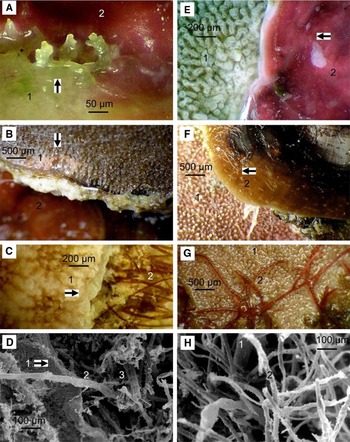
Analysis of photographs of the injuries showed that polyps were able to regenerate under maintenance at moderate and low light in the range of light intensities from 2 to 30% PAR0 (Figure 1A–D). The injuries (except completely recovered) were occupied by algae. In total, 40 algal species were found. Corallophila apiculata, Parviphycus tenuissimus (=Gelidiella pannosa), Polysiphonia sp., Peyssonnelia conchicola, Jania capillacea, Gelidiopsis intricata, Anotrichium tenue (Rhodophyta), Sphacelaria novae-hollandiae, Lobophora variegata (Phaeophyta) were common on the injuries. In moderate light, three of the nine injuries recovered completely. Three of them were partially occupied with algal turf communities (Figure 1A–C), two injuries were covered with red algal crusts (Figure 1A) and one injury with L. variegata (Figure 1B). In low light (2–5% PAR0), all nine injuries partially recovered from injury. Four corals were overgrown by red crust algae (Figure 1E), three were overgrown by algal turf (Figure 1G, H) and two by L. variegata (Figure 1F).
Coral–algal competition for substratum on mechanically damaged colonies of the coral Porites lutea in moderate (20–30% PAR0) and low (2–5% PAR0) light. (A) 20–30% PAR0, the expansion of coral tissue on the red crust alga Peyssonnelia conchicola. In all parts of the figure, arrows indicate directions of the expansion of the objects; 1, coral; 2, algae; (B) 20–30% PAR0, coral expansion on the brown alga Lobophora variegata; (C) 20–30% PAR0, coral expansion on algal turf; (D) scanning electron microscopy photograph, 20–30% PAR0, encounter of coral live tissue with algal turf; 1, live tissue; 2, the red alga Parviphycus tenuissimus; 3, the red alga Polysiphonia sp.; (E) 2–5% PAR0, expansion of the red crust alga Peyssonnelia conchicola on the coral polyps; (F) 2–5% PAR0, expansion of the brown alga L. variegata on coral surface; (G) 2–5% PAR0, expansion of the red alga Corallophila apiculata on coral live tissue; (H) scanning electron microscopy photograph, 2–5% PAR0, the red alga Parviphycus tenuissimus on live coral tissue.

Under the moderate light (20–30% PAR0) corals overgrew algal thalli with thin, transparent, azooxanthellate tissue (50–200 µm wide; Figure 1A–D). Expanding coral tissue produced skeletal crystals (Figure 1A) that enclosed algal thalli occurring in direct contact with coral tissues (Figure 1C). The brown alga, L. variegata, can overgrow coral polyps (Done, Reference Done1992; Jompa & McCook, Reference Jompa and McCook2002), however, under moderate light (20–30% PAR0), we demonstrated that the coral, Porites lutea, overgrew L. variegata (Figure 1B). We also observed the release of mucus by P. lutea when competing with L. variegata.
Furthermore, in low light (2–5% PAR0), Peyssonnelia conchicola (Figure 1E), L. variegata (Figure 1F), C. apiculata (Figure 1G) and turf algae (e.g. Polysiphonia sp. and Parviphycus tenuissimus) (Figure 1H) were the competitive dominant in substratum acquisition when interacting with Porites lutea (Figure 1E–H) and live tissue was not able to enclose the algae in a skeletal matrix (Figure 1G,H) as occurred under favourable light conditions (Titlyanov et al., Reference Titlyanov, Titlyanova, Yakovleva, Nakano and Bhagooli2005).
The presented photographs show that P. lutea polyps overgrow algal settlers on injured colonies under moderate light (20–30% PAR0), while under low light (2–5% PAR0) coral polyps were overgrown by the brown alga L. variegata and some red algae.
In our opinion, the main reason for the differences is unequal acclimative capacities of algae and corals to low light. It was shown that the majority of scleractinian coral species in the South China Sea dwell at a light range from 1 to 80% PAR0 (Titlyanov & Latypov, Reference Titlyanov and Latypov1991) while light limit of red coralline algae is lower at 0.1% PAR0 (Littler & Littler, Reference Littler, Littler, Lembi and Waaland1988).
Colour photography of coral polyps and algae in direct contact and analysis of algal species composition (Titlyanov et al., Reference Titlyanov, Titlyanova and Chapman2008) illustrate that in moderate light (20–30% PAR0) coral polyps were able to overgrow all algal species occupying injured areas excluding some toxic for corals cyanobacteria such as Lyngbya majuscula, L. semiplena and L. bouillonii) (Titlyanov et al., Reference Titlyanov, Titlyanova, Yakovleva, Nakano and Bhagooli2005, Reference Titlyanov, Titlyanova, Yakovleva and Sergeeva2007).
Under low light (to 5% PAR0 and lower), corals partially lose their zooxanthellae to the detriment of autotrophy function and vitality (Titlyanov & Titlyanova, Reference Titlyanov and Titlyanova2002). Under low light, algae become a physical insurmountable obstacle for coral tissue expansion on substratum. Such algae as Peyssonnelia conchicola, Corallophila apiculata, Centroceras clavulatum, Anotrichium tenue, (Rhodophyta), Lobophora variegata, Sphacelaria novae-hollandiae and S. tribuloides (Phaeophyta) overgrew coral polyps becoming the winners in the competition for the substratum.
ACKNOWLEDGEMENTS
The authors are grateful to the referees for their comments that improved this paper and to all staff at the Sesoko Station (Ryukyu University) for the use of facilities, technical help, hospitality and convenience of research work.