Although the Fontan operation has improved survival of patients with univentricular hearts, these patients are at a lifelong risk for complications related to the Fontan physiology.Reference Cetta, Feldt and O’Leary 1 , Reference Gentles, Mayer and Gauvreau 2 In recent years, it has become clear that the Fontan circulation affects most of the organs in the human body, with low-cardiac output and chronically elevated systemic venous pressure likely being the substrates for the development of long-term adverse effects.Reference Gewillig and Goldberg 3 – Reference Rychik, Goldberg and Rand 6 Therefore, patient management has increasingly focussed on the identification of modifiable risk factors in order to optimise Fontan haemodynamics, prevent deterioration of organ function, and develop new therapeutic strategies.Reference Avitabile, Leonard and Zemel 7 – Reference Thacker, Patel, Dodds, Goldberg, Semeao and Rychik 10
Vitamin D deficiency might represent such a modifiable risk factor.Reference Avitabile, Leonard and Zemel 7 , Reference Chin, Stephens, Goldmuntz and Leonard 11 – Reference Kim, Park, Song, Lee and Shim 13 Emerging evidence suggests important additional physiological actions of vitamin D and its metabolites beyond maintaining bone health.Reference Holick, Binkley and Bischoff-Ferrari 14 – Reference Norman and Powell 16 Vitamin D deficiency may adversely influence fundamental processes such as cell proliferation and differentiation, apoptosis, cell adhesion, membrane transport, and matrix homoeostasis in various non-skeletal tissues.Reference Holick, Binkley and Bischoff-Ferrari 14 – Reference Norman and Powell 16 A sub-optimal vitamin D status is not uncommon among healthy and chronically ill children, and it has emerged as an important public health problem in the general population.Reference Holick, Binkley and Bischoff-Ferrari 14 – Reference McNally, Sampson, Matheson, Hutton and Little 22 Indeed, a low serum 25-hydroxyvitamin D level has been associated with worse clinical outcome in various clinical settings, including cardiovascular disease.Reference Beigelman, Zeiger and Mauger 17 , Reference Kelly, Brooks, Dougherty, Carlow and Zemel 19 – Reference McNally, Sampson, Matheson, Hutton and Little 22 Thus, assessing vitamin D status in Fontan patients may have clinical implications as well. Data on vitamin D status in Fontan patients are limited, and potential risk factors for vitamin D deficiency have not been fully elucidated.Reference Avitabile, Leonard and Zemel 7 , Reference Chin, Stephens, Goldmuntz and Leonard 11 Furthermore, recent studies have raised concerns regarding bone mineral density, growth, and whole-body lean mass in patients following the Fontan operation.Reference Avitabile, Leonard and Zemel 7 , Reference Goldberg, Dodds and Avitabile 12
After two of our Fontan patients with protein-losing enteropathy experienced a pathological fracture of the femur, we routinely measured vitamin D status in Fontan patients who regularly visited our outpatient clinic, as well as examined the prevalence of and the potential risk factors for decreased serum 25-hydroxyvitamin D levels.
Methods
Study group
A retrospective chart review was performed of Fontan patients routinely followed-up at our institution who had been screened for vitamin D status by obtaining serum 25-hydroxyvitamin D levels. Medical records were searched for cardiac diagnosis, age at Fontan operation, type of Fontan – extra-cardiac conduit or lateral tunnel – gender, presence of protein-losing enteropathy, echocardiographic findings, medications, pacemaker implantation, season of screening, and anthropometric measures. Gender-specific Z-scores (SD scores) were calculated for height, weight, and body mass index (kg/m2) relative to age using paediatric reference values obtained by the German Health Interview and Examination Survey for Children and Adolescents.Reference Schaffrath Rosario, Schienkiewitz and Neuhauser 23 , Reference Schaffrath Rosario, Kurth, Stolzenberg, Ellert and Neuhauser 24 Z-scores for patients aged 18 years and older were calculated using the adult references obtained by the same national health institution (Robert Koch Institution, Berlin).Reference Bergmann and Mensink 25
If available, data on vitamin D and calcium supplementation, duration of treatment, therapeutic effect on laboratory parameters, and occurrence of adverse effects were collected. Patients were diagnosed as having protein-losing enteropathy if clinical manifestations of hypoproteinaemia were present, such as peripheral oedema, abdominal distention, abdominal pain, diarrhoea, and pleural or pericardial effusion, combined with a decreased serum albumin level <3.0 g/dl, and elevation of the stool α-1 antitrypsin concentration, as previously described.Reference Thacker, Patel, Dodds, Goldberg, Semeao and Rychik 10 , Reference John, Johnson, Khan, Driscoll, Warnes and Cetta 26 Other causes of hypoproteinaemia were ruled out. Skin type was classified using the Fitzpatrick Skin Phototype Classification.Reference Huibers, Visser, Deckers, van Schoor, van Furth and Wolf 18 In brief, skin type was divided into three groups, depending on its pigmentation: type 1 (white), type 2 (coloured), and type 3 (black). Patients receiving calcium or vitamin D supplementation at the time of their vitamin D status screening, or having chronic kidney disease, were excluded from this study.
Laboratory measurements
Serum levels of albumin, total protein, calcium, phosphorus, total alkaline phosphatase, and white blood cell count were measured using standard techniques. Corrected calcium level was calculated from the serum calcium and serum albumin levels (0.8×(serum albumin)+serum calcium)).Reference Goldberg, Dodds and Avitabile 12 From the white blood cell count, the blood neutrophil-to-lymphocyte ratio, a marker of systemic inflammation, was calculated.Reference Uthamalingam, Patvardhan and Subramanian 27 Lymphocytopenia was defined as a relative lymphocyte count of <20%. The normal range for lymphocytes in our laboratory is 20–50%.
The serum 25-hydroxyvitamin D and intact parathyroid hormone concentrations were measured using a competitive electrochemiluminescence immunoassay (Elecsys; Roche Diagnostics GmbH, Mannheim, Germany). The established normal range for serum parathyroid hormone levels for this assay was 12–72 ng/L.
Definition of vitamin D status
Vitamin D deficiency was defined as a serum 25-hydroxyvitamin D level of <20 ng/ml, and vitamin D insufficiency was defined as a serum 25-hydroxyvitamin D level of 20–30 ng/ml.Reference Avitabile, Leonard and Zemel 7 , Reference Holick, Binkley and Bischoff-Ferrari 14 Patients with a serum 25-hydroxyvitamin D levels of<10 ng/ml were diagnosed as having severe vitamin D deficiency, as previously described.Reference Holick, Binkley and Bischoff-Ferrari 14 , Reference Lee, O’Keefe, Bell, Hensrud and Holick 20 Vitamin D sufficiency was defined as a serum 25-hydroxyvitamin D level of >30 ng/ml.Reference Holick, Binkley and Bischoff-Ferrari 14 , Reference Kelly, Brooks, Dougherty, Carlow and Zemel 19 Circulating parathyroid hormone levels of >72 ng/L were considered elevated.
Statistical analysis
Patient characteristics and laboratory variables were summarised using descriptive statistical methods and expressed as mean±SD or median (range). Normality of measurements was tested using the Shapiro–Wilk test. Clinical and laboratory measurements were all normally distributed. Differences between groups in continuous and categorical variables were assessed using Student’s t-test or the χ2 test, respectively; two-sided tests were used throughout. Pearson’s correlation testing was performed to investigate associations between serum 25-hydroxyvitamin D levels and other parameters. The effect of vitamin D supplementation on calcium homoeostasis was analysed using one-way analysis of variance with a design for repeated measures; p values<0.05 were considered statistically significant. SPSS Version 20 (SPSS, Chicago, IL, United States of America) was used to perform all the statistical analyses.
Results
Study group
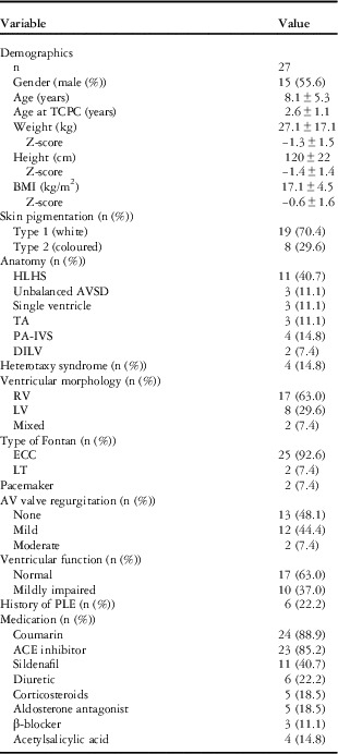
A total of 28 Fontan patients who had been screened for vitamin D deficiency were identified; one patient with associated chronic kidney disease was excluded. Therefore, 27 Fontan patients – 55.6% male, mean age 8.1±5.3 years, median age 7.0 years (2.0–22.0 years) – were included in our study. Protein-losing enteropathy was present in 6/27 patients (22.2%). The clinical and demographic characteristics of the study group are outlined in Table 1.
Clinical characteristics of Fontan patients.

ACE=angiotensin-converting enzyme; AV=atrioventricular; AVSD=atrioventricular septal defect; BMI=body mass index; DILV=double-inlet left ventricle; ECC=extra-cardiac conduit; HLHS=hypoplastic left heart syndrome; LT=lateral tunnel; LV=left ventricle; PA-IVS=pulmonary atresia with intact ventricular septum; PLE=protein-losing enteropathy; RV=right ventricle; TA=tricuspid atresia; TCPC=total cavopulmonary connection
Data are expressed as frequencies (percentage)
Vitamin D deficiency in Fontan
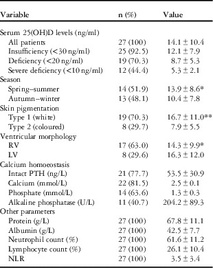
The results of the laboratory measurements are summarised in Table 2. The mean serum 25-hydroxyvitamin D level was 14.1±10.4 ng/ml. Vitamin D deficiency was prevalent in our Fontan cohort, with 70.3% (19/27 patients) having a serum 25-hydroxyvitamin D value of <20 ng/ml and 44.4% (12/27 patients) having severe vitamin D deficiency. Sufficient serum 25-hydroxyvitamin D values were found in only two patients (7.4%). Significantly lower mean serum 25-hydroxyvitamin D levels were seen in patients with skin type 2 (p=0.04). Secondary hyperparathyroidism was seen in 5/21 (23.8%) patients.
Laboratory measurements.

LV=left ventricle; NLR=neutrophil-to-lymphocyte ratio; PTH=parathyroid hormone; RV=right ventricle; 25(OH)D=25-hydroxyvitamin D
Values are expressed as frequencies (percentage) or mean±SD
*Not significant (p>0.05)
**Significant difference (p<0.05)
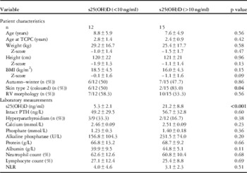
To explore potential associations between the presence of severe vitamin D deficiency and patient characteristics, patients were assigned to two groups according to whether they had a serum 25-hydroxyvitamin D level of <10 ng/ml or >10 ng/ml. The results of this analysis are presented in Table 3. Patients with skin type 2 were more likely to have severe vitamin D deficiency than patients without increased skin pigmentation (50.0 versus 13.3%, p=0.04). No further differences were seen between the two groups. Interestingly, secondary hyperparathyroidism was not more prevalent among Fontan patients in whom a severe vitamin D deficiency was diagnosed (33.3 versus 16.7%; p=0.38).
Sub-group analysis: serum 25-hydroxyvitamin D levels.

BMI=body mass index; NLR=neutrophil-to-lymphocyte ratio; PTH=parathyroid hormone; RV=right ventricle; TCPC=total cavopulmonary connection; 25(OH)D=25-hydroxyvitamin D
Values are expressed as frequencies (percentage) or mean±SD
Significant p-values are indicated in bold
Vitamin D status in protein-losing enteropathy
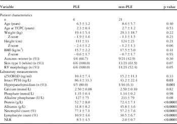
Similar serum 25-hydroxyvitamin D levels were found in Fontan patients with and without protein-losing enteropathy (p=0.33, Table 4). In contrast, the number of patients with hyperparathyroidism (80.0 versus 6.3%, p=0.001) and with increased parathyroid hormone levels (86.4±33.3 versus 43.2±22.4 ng/L, p=0.03) was significantly higher in patients with protein-losing enteropathy (Table 4). A relative lymphocyte count <20.0% was observed in all patients with protein-losing enteropathy. Furthermore, patients with protein-losing enteropathy had a significantly greater neutrophil-to-lymphocyte ratio (8.5±4.5 versus 2.0±0.7, p<0.0001), and were shown to have significantly higher relative neutrophil counts (77.1±7.1 versus 57.2±7.6%, p<0.001). There was a non-significant trend towards patients with protein-losing enteropathy having a morphologically right single ventricle (p=0.058). No further differences were seen between both groups of patients.
Sub-group analysis: protein-losing enteropathy.

BMI=body mass index; NLR=neutrophil-to-lymphocyte ratio; PLE=protein-losing enteropathy; PTH=parathyroid hormone; RV=right ventricle; TCPC=total cavopulmonary connection; 25(OH)D=25-hydroxyvitamin D
Values are expressed as frequencies (percentage) or mean±SD
Significant p-values are indicated in bold
Among all, two Fontan patients with protein-losing enteropathy experienced a pathological femur fracture – 4.5 and 3.2 years after Fontan operation; 2.4 and 2.1 years after protein-losing enteropathy was diagnosed, respectively. They had been chronically treated with systemic corticosteroids. Vitamin D insufficiency (23.7 ng/ml) was present in one patient, and the other patient had severe vitamin D deficiency (7.0 ng/ml). Parathyroid hormone levels were elevated in both patients – 79 and 93 ng/L, respectively.
Correlations between patient characteristics and laboratory measurements
No significant correlation was found between serum 25-hydroxyvitamin D level and parathyroid hormone concentration (r=−0.107, p=0.645; Fig 1a). In contrast, a significant relationship was seen between serum 25-hydroxyvitamin D and calcium levels (r=0.434, p=0.044; Fig 1b). No further associations between serum 25-hydroxyvitamin D and other parameters such as clinical data, anthropometric measures, and laboratory parameters were observed.
Correlation between serum 25(OH)D levels and PTH levels ( a ). Correlation between 25(OH)D levels and serum calcium concentration ( b ). PTH=parathyroid hormone; 25(OH)D=25-hydroxyvitamin D.

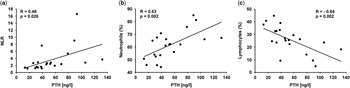
Importantly, significant correlations were observed between parathyroid hormone levels and neutrophil-to-lymphocyte ratio (r=0.484, p=0.026; Fig 2a) and between parathyroid hormone levels and relative neutrophil count (r=0.631, p=0.002; Fig 2b). In addition, a moderately strong inverse relationship was found between parathyroid hormone levels and relative lymphocyte count (r=−0.635, p=0.002; Fig 2c). After exclusion of patients with protein-losing enteropathy from these analyses, all correlations remained significant – relative neutrophil count: r=0.509, p=0.044; neutrophil-to-lymphocyte ratio: r=0.560, p=0.024; relative lymphocyte count: r=−0.566, p=0.022 – suggesting a potential link between the parathyroid hormone, a key calcium-regulatory component, and systemic inflammation in Fontan patients.
Correlations between PTH levels and relative neutrophil count ( a ), relative lymphocyte count ( b ), and NLR ( c ). PTH=parathyroid hormone; NLR=neutrophil-to-lymphocyte ratio.

Vitamin D supplementation
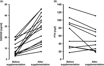
Oral vitamin D supplementation was initiated in all patients with a sub-optimal vitamin D status – 500–2000 IU/day vitamin D3=12.5–50 μg cholecalciferol. Fontan patients with protein-losing enteropathy were started on 1000–2000 IU/day vitamin D3 (25–50 μg cholecalciferol), depending on their vitamin D status and whether they received corticosteroids. Children exposed to repeated courses of corticosteroids and/or following a pathological bone fracture were started on a combined therapy of vitamin D3 and calcium supplementation (500 mg/day), as was recently recommended for children with renal disease.Reference Gruppen, Davin and Oosterveld 28 Follow-up data on vitamin D status were available for 15 patients (Fig 3). Serum 25-hydroxyvitamin D levels increased significantly – pre-treatment: 9.5±6.6 ng/ml, follow-up: 27.7±12.9 ng/ml, p<0.0001 – within 5.8±3.7 months – with a range from 1.0 to 13.0 months – of vitamin D3 supplementation. Vitamin D deficiency resolved in 7/15 patients – 46.7%, serum 25-hydroxyvitamin D level >30 ng/ml – after supplementation. Serum 25-hydroxyvitamin D levels normalised in those patients receiving a daily dose of >1000 IE cholecalciferol and in protein-losing enteropathy patients taking at least 2000 IE/day vitamin D3. Parathyroid hormone levels were available for nine patients and decreased in most patients – n=5/9 (55.6%); 67.9±40.8 versus 49.0±32.0 ng/L, p<0.001. No difference in relative lymphocyte count – n=7; 16.0±9.6 versus 15.5±11.4%, p=0.856 – or neutrophil-to-lymphocyte ratio – n=7; 6.9±5.1 versus 7.7±4.5, p=0.704 – was found after vitamin D3 repletion. Vitamin D supplementation was well tolerated.
Effect of vitamin D3 supplementation on 25(OH)D levels ( a ) and circulating PTH concentrations ( b ), before and after treatment. PTH=parathyroid hormone; 25(OH)D=25-hydroxyvitamin D.

Discussion
This is the largest study to date reporting on the prevalence of vitamin D deficiency and the effect of vitamin D3 supplementation on calcium homoeostasis in a cohort of Fontan patients. A high prevalence of vitamin D deficiency (70.3%) was observed. Although skin type was associated with worse vitamin D status, no other potential risk factor was identified. Hyperparathyroidism was observed in ~24% of patients. Fontan patients with protein-losing enteropathy were more likely to have elevated parathyroid hormone levels. Significant associations were demonstrated between circulating parathyroid hormone levels and white blood cell parameters such as relative lymphocyte count and neutrophil-to-lymphocyte ratio, suggesting a complex pathophysiological interplay between parathyroid hormone levels and low-grade systemic inflammation in Fontan patients. Vitamin D supplementation was associated with higher 25-hydroxyvitamin D levels and a subsequent decrease in parathyroid hormone levels. No effect of vitamin D supplementation was seen on relative lymphocyte count and on the neutrophil-to-lymphocyte ratio.
The discovery of vitamin D receptors in cells of the cardiovascular and immune systems has increased the interest in the role of vitamin D deficiency in cardiovascular disease progression and outcome.Reference Avitabile, Leonard and Zemel 7 , Reference Goldberg, Dodds and Avitabile 12 , Reference Holick, Binkley and Bischoff-Ferrari 14 – Reference Norman and Powell 16 , Reference Lee, O’Keefe, Bell, Hensrud and Holick 20 , Reference Liu, Voors and van Veldhuisen 29 Experimental and clinical studies have established important effects of vitamin D and its metabolites on regulatory pathways involved in maintaining cardiovascular health.Reference Norman and Powell 16 , Reference Liu, Voors and van Veldhuisen 29 A low serum 25-hydroxyvitamin D concentration is common in patients with heart failure and appears to be related to poor clinical outcome.Reference Liu, Voors and van Veldhuisen 29 – Reference Gandhi and Myers 31 The exact mechanisms through which vitamin D deficiency may contribute to a worse prognosis have not been fully understood; however, vitamin D suppresses not only the release of renin, a regulator of the renin–angiotensin–aldosterone system, but also inhibits the production of pro-inflammatory cytokines.Reference Norman and Powell 16 , Reference Liu, Voors and van Veldhuisen 29 – Reference Gandhi and Myers 31 Thus, a sub-optimal vitamin D status may contribute to imbalance in fluid homoeostasis, inappropriate renin–angiotensin–aldosterone system activation, and systemic inflammation, common findings in chronic heart failure patients and in patients with a Fontan circulation.Reference Norman and Powell 16 , Reference Liu, Voors and van Veldhuisen 29 , Reference Inai, Nakanishi and Nakazawa 32 – Reference Tomaschitz, Ritz and Pieske 34
Previous studies have shown vitamin D deficiency in ~25% of Fontan patients with protein-losing enteropathy, but limited data exist on the vitamin D status of Fontan patients without protein-losing enteropathy.Reference Avitabile, Leonard and Zemel 7 , Reference Goldberg, Dodds and Avitabile 12 For the first time, we showed that vitamin D deficiency is highly prevalent among Fontan patients, irrespective of age, time after Fontan procedure, ventricular morphology, or the presence of protein-losing enteropathy. Only skin type was found to be associated with significant lower serum 25-hydroxyvitamin D levels; however, development of hypovitaminosis D in Fontan patients may be multi-factorial. Reduced sunlight exposure, physical activity restriction, dietary habits, and the use of medications such as corticosteroids and coumarin derivates may all contribute to the development of vitamin D deficiency.Reference Avitabile, Leonard and Zemel 7 , Reference Goldberg, Dodds and Avitabile 12 , Reference Holick, Binkley and Bischoff-Ferrari 14 – Reference Norman and Powell 16 , Reference Lee, O’Keefe, Bell, Hensrud and Holick 20 , Reference Avgeri, Papadopoulou and Platokouki 35 – Reference Pappa, Thayu, Sylvester, Leonard, Zemel and Gordon 37 The Fontan circulation per se may augment the risk of a sub-optimal vitamin D status. Malabsorption of cholecalciferol may result from gut inflammation, restricted intestinal blood flow, and vascular congestion, and the synthesis of serum 25-hydroxyvitamin D might be decreased in the presence of liver dysfunction.
Hyperparathyroidism was observed in ~24% of the study group. Recent studies in patients with cardiovascular disease showed that vitamin D deficiency and increase in parathyroid hormone levels have been associated with systemic inflammation, neurohormonal activation, increased severity of heart failure, and subsequent worse clinical outcome.Reference Liu, Voors and van Veldhuisen 29 – Reference Gandhi and Myers 31 , Reference Saiki, Kuwata and Kurishima 33 Parathyroid hormone regulation is complex and incompletely understood.Reference Norman and Powell 16 , Reference Gandhi and Myers 31 , Reference Tomaschitz, Ritz and Pieske 34 Accumulating evidence suggests important bidirectional interactions between the parathyroid hormone and the renin–angiotensin–aldosterone system.Reference Liu, Voors and van Veldhuisen 29 – Reference Gandhi and Myers 31 , Reference Tomaschitz, Ritz and Pieske 34 , Reference Brown and Vaidya 38 Hyperaldosteronism is a common finding in chronic heart failure patients and has been demonstrated in Fontan patients as well.Reference Saiki, Kuwata and Kurishima 33 Excess of aldosterone promotes inappropriate renal and faecal calcium loss, resulting in secondary hyperparathyroidism.Reference Gandhi and Myers 31 , Reference Tomaschitz, Ritz and Pieske 34 , Reference Brown and Vaidya 38 In addition, aldosterone has been shown to increase parathyroid hormone secretion directly by binding to mineralocorticoid receptors located in the parathyroid gland.Reference Norman and Powell 16 , Reference Gandhi and Myers 31 , Reference Tomaschitz, Ritz and Pieske 34 , Reference Brown and Vaidya 38 Hyperparathyroidism has been associated with ventricular hypertrophy, hypertension, arterial stiffness, coronary artery disease, and subsequent cardiovascular morbidity and mortality.Reference Anderson, Vanwoerkom and Horne 30 , Reference Gandhi and Myers 31 , Reference Tomaschitz, Ritz and Pieske 34 , Reference Brown and Vaidya 38 Increased serum parathyroid hormone concentration promotes oxidative stress at the tissue level by inducing intra-cellular calcium overload, the so-called “calcium paradox”, thereby stimulating the release of pro-inflammatory cytokines such as interleukin-6 and tumour necrosis factor-α.Reference Anderson, Vanwoerkom and Horne 30 , Reference Gandhi and Myers 31 , Reference Tomaschitz, Ritz and Pieske 34 These cytokines fuel systemic inflammation in chronic heart failure patients, and possibly in susceptible Fontan patients as well.Reference Sharma, Bolger and Li 39 Systemic inflammation plays a central role in the pathogenesis of protein-losing enteropathy.Reference Ostrow, Freeze and Rychik 40 Addition of tumour necrosis factor-α significantly increased intestinal protein-loss in an experimental model of protein-losing enteropathy.Reference Bode, Salvestrini and Park 41 Taken together, the strong correlation between parathyroid hormone levels and neutrophil-to-lymphocyte ratio seen in our study, a surrogate marker of systemic inflammation,Reference Uthamalingam, Patvardhan and Subramanian 27 and the clinical observation that hyperparathyroidism was present almost exclusively in Fontan patients with protein-losing enteropathy, a “hyperinflammatory” disease state, both point to an important and possibly underestimated pathophysiological effect of the parathyroid hormone in Fontan circulation.
Although a reduced lymphocyte count is commonly encountered in Fontan patients with protein-losing enteropathy,Reference Chueng, Tsang and Kwok 42 we demonstrated a close inverse relationship between parathyroid hormone levels and relative lymphocyte count in Fontan patients. This association was also present after excluding patients with protein-losing enteropathy from the analysis. Although parathyroid hormone levels decreased significantly after vitamin D3 was supplemented, no effect on relative lymphocyte count was observed in the present study. Whether elevated parathyroid hormone levels and reduced relative lymphocyte count are pathophysiologically interrelated or are both surrogate markers of systemic inflammation in Fontan patients remains to be determined.
Further studies are needed to elucidate the clinical role and mechanisms underlying vitamin D deficiency in Fontan patients, the optimal dosing of vitamin D3 supplementation, and whether maximal suppression of the parathyroid hormone should be targeted.
Study limitations
The sample size of our study was relatively small. Moreover, there is a possibility that the patients included in this study created a biased sample, favouring those with more symptoms and advanced disease. Complete data were not available for all the patients – for instance, dual energy X-ray absorptiometry to assess bone mineral density was not performed. Serum aldosterone and renin levels were not available for the analyses. We did not record the dietary habits of our patients, including use of vitamin D-containing products such as fatty fish, dairy products, and meat. No data were available regarding physical activity level and sun exposure.
Conclusions
A high prevalence of vitamin D deficiency was found among Fontan patients, independent of age, time after Fontan procedure, ventricular morphology, and presence of protein-losing enteropathy. Only skin type was found to be associated with significant lower serum 25-hydroxyvitamin D levels. Patients with protein-losing enteropathy were more likely to have elevated parathyroid hormone levels than patients without protein-losing enteropathy.
Acknowledgement
TH is supported by the Koeln Fortune programme (288/2013).
Financial Support
There was no grant or other funding obtained for this study.
Conflicts of Interest
None.
Ethical Standards
This retrospective study was performed according to the local code of research conduct and data protection rules. Approval by the local ethics committee was not required. The study was conducted according to the ethical guidelines and principles of the International Declaration of Helsinki.