INTRODUCTION
Alveolar echinococcosis (AE) is a zoonosis with great public health concerns, caused by a tapeworm Echinococcus multilocularis (Deplazes and Eckert, Reference Deplazes and Eckert2001). AE is mainly distributed in the northern hemisphere, in particular regions of China and the Russian Federation and countries in continental Europe and North America (Craig et al. Reference Craig, McManus, Lightowlers, Chabalgoity, Garcia, Gavidia, Gilman, Gonzalez, Lorca, Naquira, Nieto and Schantz2007). AE is characterized by a tumour-like growth of E. multilocularis metacestodes in the liver of rodents or human beings (Czermak et al. Reference Czermak, Akhan, Hiemetzberger, Zelger, Vogel, Jaschke, Rieger, Kim and Lim2008; Zhang et al. Reference Zhang, Wang, Lu, Li, Lu, Mantion, Vuitton, Wen and Lin2012). It is estimated that approximately 0·3–0·5 million people are suffering from AE worldwide (Zhang et al. Reference Zhang, Zhang, Wu, Shi, Li, Zhou, Wen and McManus2015). The estimated worldwide human burden of AE were 650 000 disability-adjusted life years annually, with most of the disease burden concentrated in western China (Torgerson et al. Reference Torgerson, Keller, Magnotta and Ragland2010). The limited availability of diagnostics is considered to be one of contributing factors for prevalence of echinococcosis (Wang et al. Reference Wang, Gao, Steverding, Wang, Shi and Yang2013). Parasite-specific antibodies are not be detected during the early stages of infection (Yamano et al. Reference Yamano, Miyoshi, Goto and Kawase2014). Therefore, ideal markers with high specificity and high sensitivity for early and specific diagnosis of E. multilocularis are urgently needed.
microRNAs (miRNAs), a class of short endogenous non-coding RNAs, regulate the gene expression. Accumulating studies have shown that miRNAs are key regulators in physiological and pathological processes, such as immune response, inflammatory response and tumour occurrence (Baltimore et al. Reference Baltimore, Boldin, O'Connell, Rao and Taganov2008; Sonkoly and Pivarcsi, Reference Sonkoly and Pivarcsi2009). Using deep sequencing and miRNA microarray, host miRNAs in the relevant cells or tissues have been shown to be dysregulated during parasite infection, demonstrating their important roles in host responses to pathogen challenge (Xu et al. Reference Xu, Zhou, Nisbet, Huang, Fan and Zhu2013; Zheng et al. Reference Zheng, Cai and Bradley2013). Functional prediction revealed that some of them were involved in the immune and inflammatory response to parasitic infection (Judice et al. Reference Judice, Bourgard, Kayano, Albrecht and Costa2016). Recent studies showed that circulating miRNAs can be stably detectable in blood or fluids of humans and animals with helminth infection (Cheng et al. Reference Cheng, Luo, Hu, Cao and Jin2013; Xu et al. Reference Xu, Zhou, Nisbet, Huang, Fan and Zhu2013; Hoy et al. Reference Hoy, Lundie, Ivens, Quintana, Nausch, Forster, Jones, Kabatereine, Dunne and Mutapi2014) and have been extensively studied because of the potential use as diagnostic biomarkers for the early detection of parasite infection (Hoy et al. Reference Hoy, Lundie, Ivens, Quintana, Nausch, Forster, Jones, Kabatereine, Dunne and Mutapi2014; Jia et al. Reference Jia, Chang, Wei, Lu, Yin, Jiang and Chen2014). For example, the host-circulating sja-miR-223 has a potential biomarker for Shistosoma japonicum infection (Wang et al. Reference Wang, Yu, Yu, Bian, Deng, Wan and Zhu2010; He et al. Reference He, Sai, Chen, Zhang, Xu, Zhang and Pan2013). Recent study showed that ovi-miR-192 has the potential to be used as a diagnostic biomarker for Opisthorchis viverrin-induced cholangiocarcinoma (Silakit et al. Reference Silakit, Loilome, Yongvanit, Chusorn, Techasen, Boonmars, Khuntikeo, Chamadol, Pairojkul and Namwat2014). Moreover, accumulating studies have showed that parasite-derived circulating miRNAs can be detected in the serum/plasma of the hosts and served as specific biomarkers for early detection of helminthic infections (Cai et al. Reference Cai, Gobert and McManus2016).
Echinococcus species miRNAs have recently been described (Cucher et al. Reference Cucher, Prada, Mourglia-Ettlin, Dematteis, Camicia, Asurmendi and Rosenzvit2011, Reference Cucher, Macchiaroli, Kamenetzky, Maldonado, Brehm and Rosenzvit2015), providing a possibility of understanding their roles in development and growth, host–parasite interaction, and their future potential use as diagnostic targets. However, circulating miRNA expression associated with E. multilocularis infections have not been profiled. In the study, we demonstrate that several hostcirculating miRNAs are dysregulated in the mouse sera during E. multilocularis infection. We also identify seven parasite-derived miRNAs in the sera of mice infected with E. multilocularis. The results will help us to further understand the molecular basis for host–parasite interactions and to potentiate parasite-derived miRNAs in host serum as a diagnostic target of parasite infection.
MATERIALS AND METHODS
Ethics statement
All animal experiments in the study were handled in strict accordance with good animal practice according to the Animal Ethics Procedures and Guidelines of the People's Republic of China (Regulations for Administration of Affairs Concerning Experimental Animals, China, 1988). All animal experiments in the study were approved by Animal Ethics Committee of Lanzhou Veterinary Research Institute, Chinese Academy of Agricultural Sciences, and were conducted strictly according to the guidelines.
Parasite infections and serum collection
Echinococcus multilocularis Qinghai isolate (Chinese mainland strain, Qinghai population) was maintained in DBA/2 mice in our laboratory. Thirty female DBA/2 mice (6-week-old) were randomly divided into two groups. For blood collection from mice infected with E. multilocularis, one groups of DBA/2 were peritoneally infected with approximately 1000 protoscolecesper mouse. One groups inoculated with phosphate buffer saline (PBS) were used as control. The mice were exsanguinated at different time points after E. multilocularis infection (4, 8 and 12-week post-infection). Serum was prepared and immediately stored at −80 °C prior to RNA extraction.
High-throughput sequencing
Total RNA was extracted from the sera of one healthy mouse and one mouse infected with E. multilocularis at 4-week post-infection using miRNaesy Serum/Plasma Kit (Qiagen, Germany), respectively. Small RNA sequencing was carried out in BGI-Shenzhen (Shenzhen, China). For construction of sequencing libraries, total RNA was subjected to polyacrylamide gel electrophoresis (PAGE) and small RNAs with a size of 18–30 nt were recovered (Jin et al. Reference Jin, Guo, Zhu, Ayaz and Zheng2017). Recovered small RNAs were added with 5′ and 3′ adaptors, respectively, followed by polymerase chain reaction (PCR) enrichment. PCR products were purified by using the PAGE gels and the fraction ranging from 62 to 75 nt was excised, eluted and analysed by Bioanalyser (Agilent). After RNA sequencing, we performed the following bioinformatical analysis according to previously described methods (Cheng et al. Reference Cheng, Luo, Hu, Cao and Jin2013). Briefly, after removing low-quality reads and adaptor sequences, only sequences perfectly matched the mouse genome (http://www.ncbi.nlm.nih.gov/genome/genomes/52) were used for the following analysis. Mouse known miRNAs were identified by BLAST searching against the MirGeneDB database (http://www.mirgenedb.org/index.shtml, release 21) (Cheng et al. Reference Cheng, Luo, Hu, Cao and Jin2013) and the relative miRNA expression levels were analysed using DEGseq method.
To identify E. multilocularis-specific miRNA in the serum of E. multilocularis-infected mouse, we performed the following analyses: small RNAs in the two libraries that were mapped perfectly to the mouse genome were filtered using the miRDeep program (Cucher et al. Reference Cucher, Macchiaroli, Kamenetzky, Maldonado, Brehm and Rosenzvit2015) and the rest were used for further analysis. Only small RNAs were uniquely mapped to the E. multilocularis genome (ftp://ftp.sanger.ac.uk/pub/pathogens/Echinococcus) were retained and taken for further analysed against miRbase for determining the known miRNA.
Targets of differentially expressed miRNAs were predicted using the online software TargetScan (http://www.targetscan.org/vert_71) (Agarwal et al. Reference Agarwal, Bell, Nam and Bartel2015), miRanda (http://www.microrna.org/microrna) (Betel et al. Reference Betel, Wilson, Gabow, Marks and Sander2008) and RNAhybrid (Lai and Meyer, Reference Lai and Meyer2016). The target genes were analysed in terms of Gene Ontology (GO) categories and KEGG pathway enrichment analyses using the Database for Annotation, Visualization and Integrated Discovery (DAVID) gene annotation tool (Dennis et al. Reference Dennis, Sherman, Hosack, Yang, Gao, Lane and Lempicki2003).
Quantitative real-time PCR (qRT–PCR) and a stem-loop-based PCR
A miScript II RT Kit (Qiagen) was used to reversely transcribe RNA to cDNA. The 20 µL reverse transcription reaction contained 2 µL total RNA (approximate 50 ng), 2·2 µL Plasma Spike-In Control (108copies µL−1), 5 µL 5 × miScript HiFlex buffer, 2 µL 10 × miScript Nucleics Mix, 2 µL miScript Reverse Transcriptase Mix and 7·8 µL RNase-free water. The temperature program was 60 min at 37 °C, 5 min at 95 °C and then held at 4 °C. RT–PCR was performed using miScript SYBR Green PCR Kit (Qiagen). The 20 µL PCR reaction included 2 µL of cDNA (1:5 dilution), 10 µL 2 × QuantiTect SYBR Green PCR Master Mix, 2 µL 10 × miScript Universal Primer, 2 µL 10 × miScript Primer Assay and 4 µL H2O. The reactions were conducted in a 96-well plate at 95 °C for 15 min, followed by 40 cycles of 94 °C for 15 s, 55 °C for 30 s and 70 °C for 30 s. The miScript Primers were listed in online Supplementary Table S1. Ct values were median-normalized to synthetic spike – in Caenorhabditis elegans-miR-39 (cel-miR-39) as described previously (Mitchell et al. Reference Mitchell, Parkin, Kroh, Fritz, Wyman, Pogosova-Agadjanyan, Peterson, Noteboom, O'Briant and Allen2008). Relative fold change was calculated as 2−ΔCtn , where Ctn stands for normalized Ct values (Mitchell et al. Reference Mitchell, Parkin, Kroh, Fritz, Wyman, Pogosova-Agadjanyan, Peterson, Noteboom, O'Briant and Allen2008; Hoy et al. Reference Hoy, Lundie, Ivens, Quintana, Nausch, Forster, Jones, Kabatereine, Dunne and Mutapi2014).
For analysis of E. multilocularis-specific miRNA in the sera, the miRNA LNA™ PCR primers were ordered from Exiqon (online Supplementary Table S1). Reverse transcription reactions were performed using miRCURY LNA™ Universal cDNA Synthesis Kit II (Exiqon) according to the manufacturer's protocol. PCR was carried out with miRCURY LNA™ SYBR Green master mix (Exiqon). The fold changes were calculated as the ratio of normalized values in infected serum compared with the background in the uninfected serum.
To examine E. multilocularis-specific miRNA in the infected sera, seven miRNAs were chosen for assay by a stem-loop-based PCR using total RNA from E. multilocularis-infected mouse sera (Salone and Rederstorff, Reference Salone and Rederstorff2015). The stem-loop-based PCR primers were listed in online Supplementary Table S2. First, 50 ng of serum RNA was input for first-strand cDNA synthesis (ThermoFisher Scientific). PCR was conducted with the following steps: 95 °C for 10 min, 40 cycles of 96 °C for 5 s, 54 °C for 5 s and 68 °C for 3 s, and then 72 °C for 1 min. PCR products were resolved on 2% agarose gel, purified and sequenced (Takara, Japan).
Statistical analysis
Data analyses were performed using GraphPad Prism 5 software (Version 5.00 for Windows, GraphPad Software, San Diego, California USA) and a one-tailed unpaired t-test was used for comparison of two groups, with a P value <0·05 being considered to be significantly different.
RESULTS
Sequencing of circulating miRNAs in the serum of E. multilocularis-infected mouse
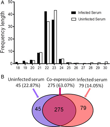
To identify host-circulating miRNAs of which the expression is altered upon E. multilocularis infection, the small RNAs in the sera from the infected and healthy mice were sequenced. In this study, 23 584 745 and 23 900 492 total reads were obtained from the uninfected and E. multilocularis-infected libraries, respectively (online Supplementary Table S3). After removing low-quality reads and adapter sequences, a total of 23, 244, 523 and 23, 497, 669 clean reads were ultimately obtained (online Supplementary Table S3). The size distribution of small RNA reads was similar between the two libraries, and the majority of these miRNA were 21–23 nt in length (Fig. 1A). These results indicated the small RNA libraries were highly enriched in miRNA sequences. In addition to miRNAs, a series of other small RNAs (sRNAs) including rRNAs, tRNAs, snRNAs and snoRNAs, were also detected in two samples (online Supplementary Fig. S1). Both rRNAs and tRNAs accounted for very high proportions in the sequenced reads (online Supplementary Fig. S1).
Size distribution of small RNA sequences and unique miRNAs in two libraries. (A) The size distribution of the small RNA sequences in the sera from the Echinococcus multilocularis-infected and uninfected mice; (B) The distribution of 399 unique miRNAs identified in both uninfected (left, blue circle) and infected mouse sera (right, red circle). The overlapping region indicates co-expressed unique miRNAs.

Identification of host-circulating miRNAs differentially expressed in the serum of E. multilocularis-infected mouse
A total of 355 and 321 known miRNAs was identified in the uninfected and E. multilocularis-infected libraries, respectively (Table 1), and a total of 399 unique miRNAs were obtained from both libraries. As shown in Fig. 1B, 275 of 399 unique miRNAs (63·07%) were co-expressed in both libraries, while 79 (14·05%) and 45 (22·87%) were exclusively expressed in the infected and uninfected mouse, respectively. Moreover, 58 of 399 miRNAs were significantly differentially expressed (fold change ⩾2 and P-value <0·05), of which 21 were upregulated and 37 were significantly downregulated. The detail of miRNA expression data was listed in online Supplementary Table S4. Among these 58 miRNAs, 8 and 18 were specifically expressed in the sera of E. multilocularis-infected and uninfected mouse, and 32 were shared in both libraries.
Summary of known miRNA in each library

Validation of differentially expressed miRNAs by qPCR analysis
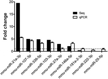
To further validate sequencing data, nine differentially expressed miRNAs were randomly selected and their expression was analysed by qPCR. Consistent with the sequencing results, there was an increase in expression of mmu-miR-103-3p, mmu-miR-107-3p, mmu-miR-339-5p, mmu-miR-146a-5p and mmu-miR-21a-3p, and a reduction in expression of mmu-miR-222-3p in the E. Multilocularis-infected mouse sera compared with the control (Fig. 2). However, mmu-miR-27a-3p, mmu-miR-25-5p and mmu-miR-181a-2-3p did not show different expressions and were not analysed further (online Supplementary Table S5).
Validation of miRNA expression by qPCR in the sera of mice infected with Echinococcus multilocularis 4-week post-infection. Data for the final analysis were from three independent experiments. The dashed horizontal line indicates the relative miRNA expression in each uninfected sera, which was set as 1·0.

Dynamic relative expression of six miRNAs in the mouse sera during E. multilocularis infection by qPCR
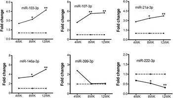
For the 6 miRNAs validated to be differently expressed at 4 weeks post-infection, we next examined their dynamic expression 4–12 weeks post-infection. With the extension of E. multilocularis infection time, the levels of mmu-miR-103-3p, mmu-miR-107-3p, mmu-miR-21a-3p and mmu-miR-146a-5p were significantly upregulated compared with the early stage of infection (4-week post-infection) (Fig. 3). The expression of mmu-miR-339-5p was significantly upregulated at the early stage of infection, but its expression showed no difference after 4-week infection (Fig. 3). Conversely, mmu-miR-222-3p was significantly downregulated in the course of E. multilocularis infection (Fig. 3 and online Supplementary Table S6).
Dynamic expression of host-origin miRNAs in the sera of mice infected with Echinococcus multilocularis 4–12 weeks post-infection. miRNA expression was quantified by qRT–PCR and normalized to cel-miR-39, and fold changes were calculated as the ratio of values from infected vs uninfected mice. The relative miRNA expression in each uninfected sera was set as 1·0. Data were expressed as means ± s.d. (*P < 0·05, **P < 0·01). Data for the final analysis were from three independent experiments.

GO enrichment of differentially expressed miRNAs targets
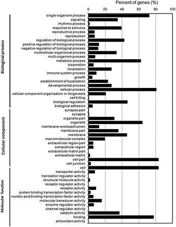
Using the softwares TargetScan (Agarwal et al. Reference Agarwal, Bell, Nam and Bartel2015), miRanda (Betel et al. Reference Betel, Wilson, Gabow, Marks and Sander2008) and RNAhybrid (Lai and Meyer, Reference Lai and Meyer2016) to predict transcript targets from the mouse genome for the identified miRNAs, 16 115 predicted targets were obtained for 58 differentially expressed miRNAs. According to P value of the enrichment analysis of target genes, top 51 enriched GO terms of biological process, molecular function and cellular component were shown in Fig. 4, respectively. The notable GO terms were the immune response, signal transduction, gene expression regulation and metabolism.
GO molecular function annotations of the target genes of differentially expressed miRNAs. According to P value, top GO terms of biological process, molecular function and cellular component were shown, respectively.

Seven E. multilocularis-derived circulating miRNAs were identified in the serum
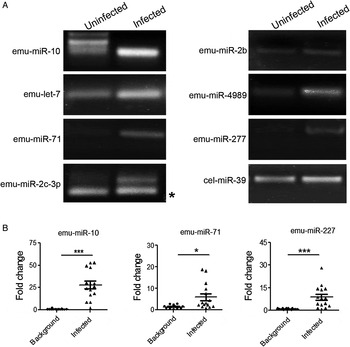
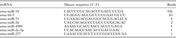
To determine whether E. multilocularis-derived circulating miRNAs are present in the infected serum, we carried out a comparative analysis between the infected library and the control library. After removal of sequences mapping to the mouse genomes and the overlapped sequences in both libraries, only 0·43% sequences were mapped to the E. multilocularis genome. By analysis of reads that mapped to the E. multilocularis genome using the miRDeep program, seven known E. multilocularis miRNAs, including emu-miR-10, emu-let-7, emu-miR-71, emu-miR-2b, emu-miR-4989, emu-miR-2c-3p and emu-miR-277, were identified from the E. multilocularis-infected serum (Table 2). To further confirm the presence of these miRNAs in the infected sera, seven miRNAs were assessed by a stem-loop-based semi-qPCR. Both RT–PCR and sequencing results showed that all seven miRNAs were detected in the sera of E. multilocularis-infected mice (Fig. 5A). Both emu-miR-10 and emu-miR-227 were specifically amplified in all the sera from mice infected with E. multilocularis, but the rest were likely to be nonspecifically amplified in the control probably due to the high sequence identity with mouse miRNAs (Fig. 5A and online Supplementary Table S1). The incorporation of LNA™ in primers can increase the specificity and sensitivity of PCR. Hence, we examined the expression of three parasite-derived miRNAs (emu-miR-10, emu-miR-227 and emu-miR-71) by qPCR using LNA-modified primers (online Supplementary Table S1). Three parasite miRNAs displayed a significant signal level in the serum of E. multilocularis-infected mice compared with the control group (Fig. 5B and online Supplementary Table S7).
Detection of parasite-derived miRNAs in mouse sera during Echinococcus multilocularis infection. (A) Semi-quantitative PCR analysis of E. multilocularis-specific miRNAs identified in the infected and uninfected sera; (B) Detection of parasite-derived miRNAs in mouse sera during E. multilocularis infections. miRNAs were quantified by qRT–PCR and normalized to cel-miR-39, and fold changes were calculated as the ratio of normalized values in infected sera compared with the background in uninfected sera. A total of 15 sera samples in each group were tested and each symbol represents data from one individual mouse (*P < 0·05, *** P < 0·001).

Seven parasite-derived circulating miRNAs in the Echinococcus multilocularis-infected mouse sera

DISCUSSION
Increasing evidence shows that tissue-specific and circulating miRNA in mammalian hosts are dysregulated during an active helminthic infection, hinting their regulatory roles in parasite–host interplay. It was reported that the expression of miRNAs in the sheep intestinal tissue was altered by Echinococcus granulosus infection (Jiang et al. Reference Jiang, Li, Wang, Ban, Hui and Jia2016). Recently, our study also found that E. multilocularis infection disturbed the small RNA-induced silencing pathway via dysregulation of miRNAs (Jin et al. Reference Jin, Guo, Zhu, Ayaz and Zheng2017). To date, no studies have examined the profile of circulating miRNAs in hosts during E. multilocularis infection. In this study, we found that 58 host-circulating miRNAs were dysregulated in the serum of E. multilocularis-infected mouse. The expression levels of six selected differentially expressed miRNAs were consistent with the sequencing data. We further assessed the dynamic expression of six validated miRNAs in the sera from mice with E. multilocularis infection. Besides mmu-miR-339-5p, the expression levels of four upregulated miRNAs were elevated gradually and the mmu-miR-222-3p (one of downregulated miRNAs) was decreased gradually with the extension of E. multilocularis infection time. To understand the regulatory roles of differentially expressed miRNAs, the target genes were predicted. Among these target genes, some are involved in host immune responses. For example, it is worth mentioning that miRNA-146a as a negative regulator is involved in innate immunity and inflammatory response by targeting IRAK1 (interleukin-1 receptor-associated kinase 1) or TRAF6 [tumour necrosis factor (TNF) receptor-associated factor 6) (Taganov et al. Reference Taganov, Boldin, Chang and Baltimore2006; Saba et al. Reference Saba, Sorensen and Booth2014). Its expression was shown to be induced after Plasmodium, Cryptosporidium and Toxoplasma infections (Cannella et al. Reference Cannella, Brenier-Pinchart, Braun, van Rooyen, Bougdour, Bastien, Behnke, Curt, Curt, Saeij, Sibley, Pelloux and Hakimi2014; Judice et al. Reference Judice, Bourgard, Kayano, Albrecht and Costa2016). We also found that the mmu-miR-146a was predominantly upregulated in the mouse sera after E. multilocularis infection. The mmu-miR-103/107, whose expression was also upregulated in the E. multilocularis-infected mouse sera, were predicted to target a negative regulator of the toll-like receptor signalling, TRAF3 (TNF receptor-associated factor 3).
Circulating parasite-derived miRNAs are existed stably in blood plasma or sera of humans or animals during parasite infection. The significance of serum miRNAs as biochemical markers for helminth infections has been gradually recognized (Mitchell et al. Reference Mitchell, Parkin, Kroh, Fritz, Wyman, Pogosova-Agadjanyan, Peterson, Noteboom, O'Briant and Allen2008; Hoy et al. Reference Hoy, Lundie, Ivens, Quintana, Nausch, Forster, Jones, Kabatereine, Dunne and Mutapi2014). To the best of our knowledge, this is the first report of circulating parasite-derived miRNAs in the serum from E. multilocularis-infected mouse. Seven E. multilocularis-derived miRNAs were identified in the infected mouse serum, indicating the secretion of miRNAs by parasites during infection. emu-miR-10 was one of the most highly abundant in E. multilocularis-infected serum. Similarly, sja-miR-10 was also detected in the plasma of rabbits infected with S. japonicum (Cheng et al. Reference Cheng, Luo, Hu, Cao and Jin2013), providing an independent validation for the presence of parasite-derived miRNAs in the serum. Parasite-origin miR-71 was detected in host serum, plasma, body fluids and microenvironments that surround the parasites (Buck et al. Reference Buck, Coakley, Simbari, McSorley, Quintana, Le Bihan, Kumar, Abreu-Goodger, Lear, Harcus, Ceroni, Babayan, Blaxter, Ivens and Maizels2014; Hoy et al. Reference Hoy, Lundie, Ivens, Quintana, Nausch, Forster, Jones, Kabatereine, Dunne and Mutapi2014; Quintana et al. Reference Quintana, Makepeace, Babayan, Ivens, Pfarr, Blaxter, Debrah, Wanji, Ngangyung, Bah, Tanya, Taylor, Hoerauf and Buck2015). miR-71 was already found to be packed into exosomes or exosome-like extracellular vesicles from a trematode (Bernal et al. Reference Bernal, Trelis, Montaner, Cantalapiedra, Galiano, Hackenberg and Marcilla2014, Reference Fromm, Trelis, Hackenberg, Cantalapiedra, Bernal and Marcilla2015; Fromm et al. Reference Fromm, Ovchinnikov, Hoye, Bernal, Hackenberg and Marcilla2016; Zhu et al. Reference Zhu, Liu, Dao, Lu, Li, Gu, Liu, Feng and Cheng2016) and nematodes (Buck et al. Reference Buck, Coakley, Simbari, McSorley, Quintana, Le Bihan, Kumar, Abreu-Goodger, Lear, Harcus, Ceroni, Babayan, Blaxter, Ivens and Maizels2014; Zamanian et al. Reference Zamanian, Fraser, Agbedanu, Harischandra, Moorhead, Day, Bartholomay and Kimber2015). Our recent result showed that emu-miR-71 had a capacity of modulation of NO production by macrophages, suggesting a role in parasite–host interactions (Zheng et al. Reference Zheng, Guo, He, Shao, Zhang, Yang, Shen, Luo and Cao2016). Similarly, sja-miR-227 was identified in the sera of Schistoma mansoni-infected mice and patient (Hoy et al. Reference Hoy, Lundie, Ivens, Quintana, Nausch, Forster, Jones, Kabatereine, Dunne and Mutapi2014; Cai et al. Reference Cai, Gobert, You, Duke and McManus2015). The extensive presence of E. multilocularis-specific miRNAs in host serum may suggest that E. multilocularis releases miRNAs into host serum via exosome-like EVs (Zheng et al. Reference Zheng, Guo, Su, Guo, Ding, Yang, Xiang, Cao, Zhang, Ayaz and Luo2017). The discovery of parasite-specific miRNA profiles in the host serum has raised great interest in their potential as diagnostic and prognostic biomarkers and in the biological functions during the infection of Echinococcus species.
SUPPLEMENTARY MATERIAL
The supplementary material for this article can be found at https://doi.org/10.1017/S0031182017000300.
ACKNOWLEDGEMENTS
The author would like to thank Miss Jing yang and Meng Su for help in serum collection and also thank the reviewer and editor for their constructive suggestions.
FINANCIAL SUPPORT
The study was financially supported by grants from the National Key Basic Research Program (973 program) of China (No. 2015CB150300), the National Natural Science Foundation of China (Nos 31201900 and 31472185) and State Public-interest Institution Basal Research Fund (Nos 1610312016017 and 1610312017017). The funders had no role in study design, data collection and analysis, decision to publish and preparation of the manuscript.