Introduction
Congenital ductal arteriosus aneurysm is characterised by saccular or tubular dilation of the ductus arteriosus. The majority of patients are asymptomatic and it usually presents as an incidental finding. Symptomatic presentation is usually associated with complications, including thromboembolism, infection, spontaneous rupture, or compression of the adjacent structures mainly the airways. In the current report, we review the clinical presentation and surgical management of a neonate with an extensive thrombus originated from a ductal aneurysm with further extension and complete occlusion of the left pulmonary artery.
Case report
Clinical presentation
A full-term male baby was referred to our institution for further management after abnormal systematic saturation screening at 24 hours of life. His pre-ductal and post-ductal saturations were 83 and 95%, respectively. Otherwise, he had no symptoms. The pregnancy and the delivery were uncomplicated. The last fetal ultrasound was performed 6 weeks before birth and was normal (patent ductus arteriosus and normal heart chambers). His birth weight was 4.3 kg.
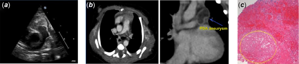
The transthoracic echocardiogram showed a mass, with heterogeneous echogenicity, suspicious of a thrombus (Fig 1a). The mass protruded from the ductal area into the main and left pulmonary arteries. There was a flow acceleration into the left pulmonary artery with a peak gradient of 17 mmHg and with no ductal shunt. There was a right-to-left shunting at the unrestrictive foramen ovale. The right ventricle was severely hypertrophied with qualitatively good biventricular systolic function (Supplementary Video S1). The right ventricular systolic pressure was more than half of the systemic pressure assessed on the moderate tricuspid valve regurgitation. The chest CT angiogram revealed a non-enhancing elongated mass in the aortopulmonary window in keeping with a thrombosed aneurysmal duct. The mass measured 2.1 × 1.2 × 1.6 cm. The mass extended into the main and left pulmonary arteries with near-total occlusion of the left pulmonary artery. The distal portions of the left and right pulmonary arteries were unobstructed (Fig 1b).
Diagnostic of intra-ductus arteriosus aneurysm thrombus: (a) pre-operative echocardiographic 2D image showing thrombus protruding from the ductal area extending into the main and left pulmonary arteries; (b) pre-operative CT angiogram; on the left side, sagittal view showing complete occlusion of the left pulmonary artery with thrombus. On the right side, coronal view showing a large thrombus in the aortopulmonary window measuring 2.1 × 1.4 cm; (c) pathological specimen showing a sub-acute thrombus with focal areas of early organisation.

Medical management
The patient was immediately started on anticoagulation with heparin infusion and observed in the cardiac critical care unit. He remained haemodynamically stable with mild-to-moderate respiratory distress requiring low-flow oxygen to maintain a normal oxygen saturation. However, on the next morning, repeated echocardiogram showed further progression of the thrombus and the decision was undertaken for surgical excision of the mass.
Surgical intervention
The heart was exposed via median sternotomy and a cardiopulmonary bypass was initiated. Inspection of the duct revealed a hugely dilated and purple aneurysmal duct, indicating a thrombus within it. The main pulmonary artery was opened and an organised thrombus was evacuated totally from the left pulmonary artery as one piece. Then, inspection of the duct revealed a fresh gelatinous thrombus that was evacuated with suction. The ductus was closed. Then, the patient was successfully weaned from cardiopulmonary bypass with no complications and remained in normal sinus rhythm (Supplementary Video S2).
Post-operative evolution
Pathological analysis of the specimen showed a pure thrombus with areas of early organisation but also a structure suggesting a subchronic evolution (>1 week delay). There was no evidence of tumour or connective tissue disease disorder (Fig 1c). Post-operative course was uneventful. The patient was discharged home few days later.
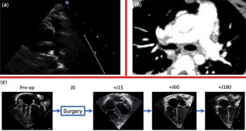
The patient was seen in our cardiology clinic for follow-up at 0.5, 2, 6, and 18 months post-operation. He continued to be completely asymptomatic. Follow-up echocardiograms showed right ventricle remodelling adaptation, unobstructed pulmonary arteries, and normalisation of pulmonary pressures (Fig 2a and c; Supplementary Video S3). The CT scan, done 3 months after the surgery, showed unobstructed pulmonary arteries with no residual ductal lumen, stumps, or thrombi (Fig 2b).
Post-operative assessment: (a) post-operative echocardiographic 2D image showing unobstructed pulmonary arteries; (b) post-operative CT image showing unobstructed pulmonary arteries with no residual stumps of thrombi; (c) remodelling and normalisation of the right ventricle after surgery.

Discussion
Aneurysmal ductus arteriosus has been reported in fetuses, neonates, children, and adults but is commonly seen before 2 months of age. The incidence of ductal aneurysms is highly variable but the diagnosis is clearly underestimated due to the silent presentation and benign course for the majority of these cases. The underlying pathogenesis for ductus arteriosus aneurysm is unclear but multiple theories have been proposed including delayed closure of the aortic end of the duct with subsequent exposure to the high systemic blood pressure, congenital weakness of the ductal wall with defective or diminished elastin fibres, and abnormal intimal cushion formation. Moreover, congenital ductus arteriosus aneurysm might be associated with connective tissue disorder such as Marfan, Loeyz–Dietz syndromes secondary to the abnormal extracellular matrix disposition. Reference Hornberger1–Reference Dyamenahalli, Smallhorn, Geva, Fouron, Cairns, Jutras, Hughes, Rabinovitch, Mason and Hornberger3 However, ductal aneurysms may present with complications including spontaneous rupture with fatal outcome, compression of the surrounding structures mainly the airways and thrombus formation. Reference Masood, Bokowski, Kazmouz and Amin4–Reference Koneti, Kanchi, Kandraju and Jaishankar7 Extensive thrombosis of aneurysmal ductus arteriosus is estimated to be very rare, with only few case reports in the literature, and usually associated with catastrophic outcome. Reference Sheridan and Michelfelder8 Due to diagnostic difficulties and isolated cases, we believe that ductus arteriosus aneurysms with a simple course or about more specifically thrombosed ductus arteriosus aneurysms are largely under-diagnosed and under-reported in the literature.
In our case report, we have two arguments to assert that the occlusion was antenatal (without being able to date it precisely): 1) the major hypertrophy of the right ventricle at birth, proof of pathophysiologic adaptation to an in utero obstacle; 2) the histological exploration is in favour of a chronic thrombus (>1 week). When faced with an antenatal occlusion of the ductus arteriosus, the differential diagnosis to be evoked is ductal aneurysm thrombosis or bronchogenic cyst. Reference McArdle, Paterson and Morris5 Surgical removal and histological exploration allow us to have the final diagnosis.
Echocardiogram is considered as the standard imaging tool for diagnosis and follow-up of ductus arteriosus aneurysm. The successful removal of the thrombus was associated with complete right ventricle remodelling and the right ventricle pressure normalisation. Cross-sectional imaging might help in making or confirming the diagnosis especially in echocardiographic equivocal cases.
There is no clear consensus on the management of ductus arteriosus aneurysms. Surgical resection is recommended in the presence of haemodynamically significantly left-to-right shunt, large aneurysms (>10 mm), evidence of connective tissue disorders, and external compression of the surrounding structures. Reference Hornberger1–Reference Dyamenahalli, Smallhorn, Geva, Fouron, Cairns, Jutras, Hughes, Rabinovitch, Mason and Hornberger3,Reference McArdle, Paterson and Morris5,Reference Koneti, Kanchi, Kandraju and Jaishankar7 For thrombosed ductal aneurysms as in our case, conservative management with antiplatelet and anticoagulation therapy might be effective in mild cases. While, thrombectomy is considered the definitive management in cases complicated with further propagation of the thrombus into the pulmonary arteries or the aorta and/or pressure effect on the airways. Potential serious complications of surgical management include injury of any of surrounding structures or thromboembolism. Reference Hornberger1,Reference Xu, Delpey, Finel and Pennaneach9,Reference Fripp, Whitman, Waldhausen and Boal10 In our case, successful surgical excision of the thrombus was uneventful.
Conclusion
Neonatal ductus arteriosus aneurysms developing antenatally and further progressing postnatally with complete occlusion of the left pulmonary artery represent a surgical emergency. In our case, urgent surgical thrombectomy was successful and the post-operative course was uneventful.
Supplementary Material
To view supplementary material for this article, please visit https://doi.org/10.1017/S1047951120002711
Acknowledgement
None.
Financial Support
None.
Conflict of Interest
None.