Blood cell development is one of the best-understood developmental pathways. This holds true not only for normal development but also for situations where normal differentiation goes astray during tumourigenesis. All haematopoietic development originates from haematopoietic stem cells (HSCs), which have self-renewal activity but also give rise to all mature blood cell types. This is achieved through a series of well-understood differentiation steps, whereby HSCs gradually lose their self-renewal ability and form increasingly restricted precursor cell types (Fig. 1). To be able to generate sufficient blood cells, these precursor cells have a very high proliferative capacity. This is kept in check by sophisticated homeostatic mechanisms rapidly directing precursor cells into differentiation to ensure that the different cell types are produced at the correct frequency. Since rapidly dividing cells are highly sensitive to genotoxic insults, any mutational event that either enhances the growth of undifferentiated precursor cells or blocks differentiation will therefore increase the risk that such cells are subverted into leukaemic cells, where normal homeostatic control is obliterated. Indeed, leukaemic cells come in a variety of phenotypes, all resembling the approximate state where normal differentiation has been derailed. In this review we touch upon the molecular mechanisms by which this happens and outline recent ideas of novel therapeutic strategies that are aimed at reversing malignant phenotypes.
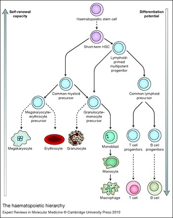
The haematopoietic hierarchy. Haematopoietic stem cells (HSCs) develop into different precursor cells whose developmental potential becomes more and more restricted.

Basic principles of gene regulation at the epigenetic level
Establishing cellular identities
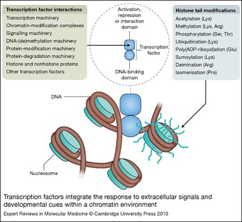
The identities of cells in multicellular organisms are defined by the individual gene expression programmes of different cell types. Since all cell types contain the same DNA, specific mechanisms must therefore be developed to differentially use this genetic information. Epigenetics describes the molecular mechanisms by which gene expression patterns are established and, most importantly, how they are maintained during cell division (Refs Reference Berger1, Reference Ptashne2). This implies the presence of molecules in the nucleus of the cell that can recognise individual genes, and switch them on or off in response to developmental signals. These molecules are the sequence-specific transcription factors (Fig. 2), which are capable of performing multiple functions: recognising a specific DNA sequence, responding to external signals, interacting with other transcription factors binding to neighbouring sequences, and, importantly, recruiting other, non-DNA-binding factor complexes that cooperate to either maintain the active state or initiate the establishment of an inactive state that is maintained by other means.
Transcription factors integrate the response to extracellular signals and developmental cues within a chromatin environment. The figure lists the different molecules and complexes interacting with transcription factors in the nucleus. The ‘transcription machinery’ includes RNA polymerase and associated factors, and ‘chromatin-modification complexes’ can be activating or repressing. Transcription factors bind to DNA in the context of chromatin, which consists of DNA wrapped around nucleosomes. The nucleosome on the right is shown with its protruding histone tails, which can be modified in different ways, as indicated.

Gene activation occurs in chromatin
The activation and silencing of tissue-specific genes in eukaryotic cells requires an alteration of their chromatin structure. The basic structural unit of chromatin is the nucleosome, in which ~147 bp of DNA are wrapped 1.65 times around the histone octamer, which is made up of two copies of histones H2A (H2AF), H2B (H2BF), H3 (H3F) and H4 (H4F) (Ref. Reference Kornberg and Lorch3). The N-terminal tails of histones protrude from the nucleosome core and are susceptible to covalent modifications and proteolytic cleavage (Ref. Reference Luger4). Further folding of nucleosomes leads to compact higher-order structures (Ref. Reference Robinson and Rhodes5). Condensed chromatin fibres repress transcription by preventing most transcription factors and the basal transcription machinery interacting with promoter/enhancer regions. Therefore, for active transcription to take place, chromatin needs to be actively unwound and rendered accessible to the transcription machinery. To achieve this, transcription factors need to recruit enzymatic activities that facilitate transcription by covalently modifying histones, and ATP-dependent chromatin remodellers, such as SWI/SNF-type complexes, that disrupt nucleosome–DNA interactions.
Histone modifications include lysine acetylation, lysine and arginine methylation, serine and threonine phosphorylation, lysine ubiquitination, glutamate poly(ADP-ribosyl)ation, lysine sumoylation, arginine deimination and proline isomerisation (Ref. Reference Kouzarides6). Each of these modifications, which are mostly found at different amino acids within the long N-terminal tails of histones (Fig. 2), is installed by different families of enzymatic activities, including histone acetyltransferases (HATs), histone methyltransferases, kinases and ubiquitin ligases. Every histone modification has its matching enzymatic activity that removes it. For example, acetyl groups are removed by histone deacetylases (HDACs) and methyl groups are removed by histone demethylases, all of which belong to extended families of enzymes with different substrate specificity. Each histone modification serves a distinctive purpose to support either the maintenance of the active (e.g. histone H3 Lys9 acetylation) or the inactive (e.g. histone H3 Lys9 methylation) transcriptional state. Moreover, the active transcriptional state also involves the replacement of canonical histones with variant histones, such as H2AZ (H2AFZ) or H3.3 (H3F3) (Ref. Reference Henikoff7). However, it is important to note that the placement of active or inactive histone marks and the concomitant recruitment of the respective histone modification activities is a highly dynamic process. In each transcription cycle, positive and negative regulators such as HATs and HDACs cooperate to reset the transcriptional state of a gene to the ground state after RNA polymerase has finished transcribing (Ref. Reference Wang8), thus ensuring that continuous signalling is required to maintain a certain transcription frequency (Ref. Reference Metivier9) and the cell can rapidly respond to changing environmental conditions.
Mediators of heritable gene silencing
In the absence of transcriptional activators, the chromatin of genes adopts a heritable silent or heterochromatic state with distinct structural features. DNA within heterochromatin is often methylated at CpG dinucleotides. This modification is introduced by DNA methyltransferases, which either methylate cytosines de novo (such as DNMT3A/B) or require a methylated cytosine at one strand of DNA (DNMT1) to maintain previously installed methylation states during replication (Ref. Reference Chen and Li10). In addition, heterochromatic genes are tightly packed and carry inactive histone marks such as methylated histone H3 Lys9 or Lys27. These modifications, in turn, serve as binding sites for protein complexes recognising them. The best-characterised example for such interactions is the binding of heterochromatin protein 1 (HP1/CBX) family members (Refs Reference Lachner11, Reference Bannister12) to trimethylated Lys9 of histone H3. Another example is the interaction of polycomb complexes with methylated Lys27 of histone H3 (Ref. Reference Wang13), which is described in more detail in the next section. CpG methylation is recognised by different methyl-binding proteins (Ref. Reference Klose and Bird14). All of these proteins associate with macromolecular complexes that include histone-modifying enzymes, which either re-install the inactive mark or remove active marks and thus sustain an inactive chromatin structure. For example, DNA methyltransferase DNMT1 associates with HDACs, and HP1 interacts with both HDACs and DNA methyltransferases (Ref. Reference Smallwood15).
However, in addition to silencing occurring by default in the absence of activators, silencing of chromatin can also be induced. These two mechanisms are exemplified by the silencing of the interleukin 4 (IL4) genes in T cell maturation, which involves the recruitment of DNMT1 (Ref. Reference Makar16), and the active silencing of the CD4 gene in CD8+ T cells by members of the RUNX family of transcription factors, which associate with corepressor complexes. In the latter case, the silenced state is stable even after the removal of the binding site for the repressive factor (Refs Reference Taniuchi17, Reference Taniuchi18), illustrating that, once established, patterns of heterochromatin are faithfully maintained. Heritability of the silent state is most likely mediated by the ability of certain heterochromatin-forming complexes such as histone methylases to interact with the replication machinery (Ref. Reference Esteve19).
Epigenetic regulatory proteins involved in haematopoiesis
Many chromatin-modifying and -remodelling complexes are involved in the regulation of cell differentiation processes in the mammalian haematopoietic system. Since these proteins are pleiotropic regulators of gene expression, it is not surprising that they regulate haematopoietic differentiation as well. Examples of this are an absolute requirement for the HAT known as MYST histone acetyltransferase (MOZ or MYST3/4) for HSC development (Ref. Reference Katsumoto20), an impediment of HSC self-renewal in mice lacking the histone H3 methyltransferase MLL1 (MLL; ‘myeloid/lymphoid or mixed-lineage leukemia’) (Refs Reference Jude21, Reference McMahon22), multiple haematopoietic defects in mice lacking MLL5 (Ref. Reference Heuser23), a disturbance of T cell differentiation in mice lacking the methyl-CpG-binding protein MBD3 (Ref. Reference Hutchins24), and a requirement for the SWI/SNF-type chromatin remodeller BRG1 (SMARCA4) for erythropoiesis and the activation of globin genes (Refs Reference Bultman, Gebuhr and Magnuson25, Reference Griffin, Brennan and Magnuson26). Recently, it was shown that normal levels of DNA methylation are necessary for normal haematopoietic differentiation (Ref. Reference Broske27). However, in the context of this review, the most interesting molecules are polycomb complexes.
HSCs have the ability to self-renew and thus maintain a heritable pattern of gene expression. In molecular terms, this means that the same transcription factor complexes reassociate with the same active genes and programme the same chromatin structure after DNA replication, and inactive genes stay inactive. As in so many cases, research in fruit flies provided the clue for identifying some of the molecular players involved in this process. In Drosophila melanogaster the pattern of expression of homeobox (Hox) genes specifies segment identity. This ‘Hox-code’ is established early in development and is maintained during ontogenesis. Polycomb proteins form large macromolecular assemblies that are essential to maintain this expression pattern and have therefore been identified as key regulators of epigenetic memory (Ref. Reference Schwartz and Pirrotta28). It turned out that this process is highly conserved in evolution and is also a hallmark of haematopoiesis: HSCs express a specific pattern of HOX genes, and polycomb proteins are crucial in controlling self-renewal of HSCs (Refs Reference Park29, Reference Lessard and Sauvageau30). Expression of HOXB4 enhances the self-renewing ability of HSCs and transient amplifying precursor cells (Ref. Reference Kyba, Perlingeiro and Daley31). Overexpression of the polycomb proteins BMI1 and EZH2 increases HSC self-renewal as well, whereas loss of function of BMI1 causes rapid cell differentiation (Ref. Reference Iwama32). As indicated above, polycomb proteins bind to HOX family genes and other genes and repress chromatin via their association with complexes installing negative histone-modification marks. Polycomb complexes come in two types: PRC1 and PRC2. Complexes from the PRC1 type bind to histone H3 Lys27, but also interact with PRC2 complexes containing the methyltransferase EZH2 that modifies histone H3 Lys27, thus reinforcing the silent state (Refs Reference Wang13, Reference Wang33, Reference Cao, Tsukada and Zhang34, Reference Cao35).
However, polycomb complexes are recruited by transcription factors (Ref. Reference Mahmoudi36) and repress promoters even in the presence of co-occupying transcriptional activators and bound polymerase (Refs Reference Breiling37, Reference Dellino38). The molecular mechanism by which this occurs involves polycomb complexes from the PRC1 type containing BMI1, which repress gene expression by ubiquinating histone H2A and thus interfere with transcriptional elongation (Ref. Reference Stock39). Thus, gene silencing by polycomb proteins at specific genes is a dynamic process that is distinct from heterochromatisation.
Transcriptional regulation of haematopoiesis
HSCs are primed to express multiple genes
During development, proliferating precursor cells change their identities and differentiate along one specific pathway, while losing the ability to give rise to alternative pathways. This principle also operates in blood cell development, but for many years the underlying molecular basis of this process was unclear. This changed after techniques were developed to examine the gene expression profile of rare HSCs. It was found that HSCs already express multiple specific lineage-associated genes at low level and this was proposed to reflect the pluripotent character of these cells (Refs Reference Hu40, Reference Enver and Greaves41, Reference Miyamoto42, Reference Gorczynski43, Reference Mansson44). It also was shown that the chromatin of multiple lineage-specific genes in early precursor cells was already reorganised and bound by transcription factors (Ref. Reference Tagoh45). Furthermore, it was demonstrated that lineage specification and the restriction of developmental potential involve the selective upregulation of lineage-appropriate genes while lineage-inappropriate genes are successively silenced (Ref. Reference Mansson44). This finding implies that HSCs express a unique combination of transcription factors that are capable of priming the majority of blood-cell-specific genes, and subsequent experiments demonstrated that this was indeed the case.
Transcription factors crucial for haematopoiesis
Transcription factor expression in development is regulated in a hierarchical fashion, whereby factors such as RUNX1 (runt-related transcription factor 1), GATA2 (GATA-binding protein 2) or TAL1 (T cell acute lymphocytic leukaemia 1; also known as SCL) important for the initial formation of the haematopoietic system are the first to be expressed in development, followed by factors important for the formation of specific lineages, such as PU.1 (SPI1; spleen focus-forming virus proviral integration oncogene spi1), C/EBPα [CEBPA; CCAAT/enhancer-binding protein (C/EBP), α] or GATA1. If the precise timing of expression is altered, blood cell development is disturbed (Ref. Reference Iwasaki46).
One of the most important transcription factors for haematopoiesis is TAL1, which is a member of the basic helix–loop–helix family of transcription factors. Without it, no haematopoietic cells are formed because it is required to form the haemogenic endothelium from which haematopoietic cells emerge (Refs Reference Lancrin47, Reference Robb48, Reference Shivdasani, Mayer and Orkin49), and it is also required for erythroid and megakaryocytic differentiation. Another transcription factor that is absolutely essential for haematopoiesis is LMO2 [(LIM domain only 2 (rhombotin-like 1)] (Refs Reference Warren50, Reference Yamada51, Reference Osada52). LMO2 does not bind to DNA, but is required to form a bridging complex that stabilises TAL1-containing transcription factor complexes (Ref. Reference Osada52). In the absence of RUNX1, the haemogenic endothelium is formed, but it is unable to give rise to definitive haematopoietic cells and mice die in utero at E12.5 (Refs Reference Okuda53, Reference Chen54). RUNX1 can act as a transcriptional repressor, but its main function appears to be as a transcriptional activator. RUNX1 does not act alone: for high-affinity DNA binding it requires a partner, CBFβ (CBFB; core-binding factor, beta subunit), which is also absolutely required for haematopoiesis (Ref. Reference North55). By contrast to TAL1 and LMO2, RUNX1 is expressed in all haematopoietic lineages and is only downregulated in maturing erythrocytes (Ref. Reference North56). An important feature of many transcription factors contributing to stem cell development is the fact that they form recursive regulatory loops, meaning that transcription factors important for stem cell formation tend to bind to each other's cis-regulatory elements and thus maintain the self-renewing state (Refs Reference Pimanda57, Reference Swiers, Patient and Loose58).
The balance of lineage-specific transcription factors determines cell fate
RUNX1 is absolutely required for the activation of important haematopoietic regulator genes, including those encoding the transcription factors PU.1, C/EBPα and GATA1 (Refs Reference Lacaud59, Reference Lacaud60, Reference Hoogenkamp61, Reference Okada62). However, RUNX1 is largely dispensable at later stages of haematopoietic development (Refs Reference Chen54, Reference Hoogenkamp61, Reference Ichikawa63, Reference Growney64). The molecular explanation for this is that RUNX1-induced transcription factors can still form stable complexes on the cis-regulatory elements of their target genes in the absence of RUNX1 (Ref. Reference Hoogenkamp61). Moreover, some of these factors, such as C/EBPα, also fulfil roles in stem cell homeostasis (Ref. Reference Zhang65), thus stabilising the stem cell transcriptional network as a whole.
The differentiation of multipotent precursor cells into defined mature blood cell lineages is regulated by the balance of transcription factor expression. Several transcription factors function in a mutually antagonistic fashion and their concentration in the cell determines the lineage fate. A good example for this notion is the cross-antagonism between PU.1 and GATA1, which determines the differentiation outcome from the short-term HSC via the common myeloid precursor into the granulocyte–monocyte precursor or megakaryocyte–erythrocyte progenitor (Fig. 1). PU.1 is essential for the formation of myeloid cells and B cells, whereas GATA1 is required to form erythroid and megakaryocytic cells (Refs Reference Scott66, Reference McKercher67, Reference Pevny68, Reference Pevny69). GATA1 and PU.1 functionally antagonise each other through direct physical interaction via the DNA-binding domains of both proteins, using distinct gene-specific mechanisms (Refs Reference Zhang70, Reference Rekhtman71, Reference Nerlov72). Depending on who ‘wins’, either the myeloid or the erythroid differentiation programme is activated, whereas the reciprocal programme is extinguished.
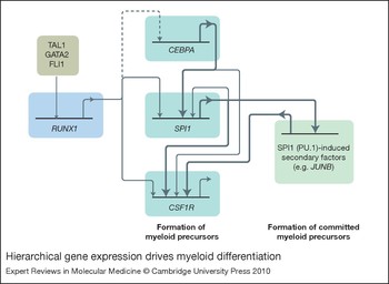
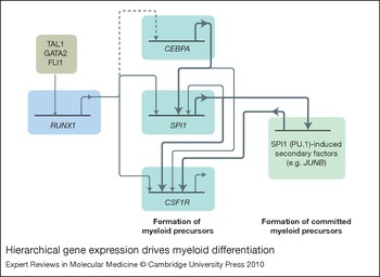
The common theme in cell fate decision processes in the haematopoietic system is that they are highly dynamic. This is exemplified in Figure 3 for myelopoiesis. Blood cell development starts out with a gene expression programme that favours self-renewal and is established by early-acting transcription factors that regulate their own expression but also prime multiple lineage-specific genes. However, simultaneously these factors drive differentiation by activating the expression of downstream transcription factors necessary for the development of specific lineages. These then form stable transcription factor complexes on lineage-specific genes that drive high-level expression. A good example for this principle is the upregulation of the gene encoding the colony-stimulating factor 1 receptor (CSF1R), which is the major growth factor receptor for the macrophage lineage. During haematopoiesis, this gene is upregulated by a two-step mechanism. Its promoter is already fully occupied in HSCs and this activation requires PU.1, but in these cells expression levels are very low. A high level of CSF1R mRNA and protein are only expressed once an enhancer element is activated in committed macrophage precursors (Refs Reference Krysinska73, Reference Tagoh74). This delayed enhancer activation is regulated by the PU.1-dependent expression of transcription factors such as EGR2 (early growth response 2) in macrophage precursors (Ref. Reference Laslo75), thus ensuring that a high level of CSF1R is expressed only in macrophages and nowhere else.
Hierarchical gene expression drives myeloid differentiation. The picture shows different myeloid transcription factor genes whose expression must be controlled in a strictly hierarchical fashion. The general principles are that transcription factors important for the formation of the haemogenic endothelium such as TAL1 (SCL) are expressed first, and then switch on the expression of factors such as RUNX1 that are required to form haematopoietic stem cells. RUNX1, in turn, switches on genes encoding for factors important for the formation of specific haematopoietic lineages, such as SPI1 (encoding PU.1). Dashed line: although expression of CEBPA [encoding CCAAT/enhancer-binding protein (C/EBP) α] is dependent on RUNX1, it is not clear whether it is directly bound by RUNX1. PU.1 starts another feed-forward loop by upregulating factors such as EGR2 that are required for the expression of differentiation-specific genes, such as the gene encoding the colony-stimulating factor 1 receptor (CSF1R) (Refs Reference Hoogenkamp61, Reference Krysinska73, Reference Laslo155). It is important to note that at each step, factors occupying one level on the hierarchy regulate each other as well, thus promoting self-renewal until terminal differentiation is reached.
Epigenetic mechanisms involved in leukaemogenesis
Translocations leading to the expression of fusion proteins comprising gene-regulatory proteins
Experiments using conditional knockout mice have demonstrated that haematopoietic development is extraordinarily stable. Once the system is established, many transcription factors that are essential at early stages of development become less important and in their absence differentiation may proceed less efficiently but with surprisingly few defects, as demonstrated for the transcription factors RUNX1 and TAL1 (Refs Reference Chen54, Reference Hoogenkamp61, Reference Ichikawa63, Reference Growney64, Reference Mikkola76). However, there are several ways normal cell differentiation can grind to a halt. The most important mechanism in the context of this review is the formation of aberrant transcription factors or epigenetic regulatory proteins by chromosomal translocations generating fusion proteins between different partners. As explained in Figure 2, normal transcriptional regulators interact with other factors in a regulated and balanced way, thus forming the highly dynamic transcription factor complexes that regulate differential gene expression. These interactions are disrupted at the target genes for leukaemic transcription factors. This can happen in two ways. First, the DNA-binding domain of a transcription factor might be fused with a novel interaction domain, as exemplified by translocations fusing the DNA-binding domain of RUNX1 to various partners, many of which recruit repressive chromatin-modification activities to normally activated genes and/or act as dominant negative inhibitors of RUNX1 function (Refs Reference Alcalay77, Reference Shigesada, van de Sluis and Liu78). Second, chromatin-modifying complexes can be recruited to ectopic sites by fusion of a chromatin modifier to a DNA-binding moiety or an interaction surface recognising other transcription factors. This is seen with MLL fusions, which interfere with the proper regulation of histone H3 Lys4 trimethylation at promoters, as discussed further below (Refs Reference Guenther79, Reference Daser and Rabbitts80).
Upregulation of gene expression by aberrant gene regulation
Another way of deregulating transcription in leukaemia is by deregulating or overexpressing epigenetic regulatory proteins. This can happen in several ways, including translocating the coding regions next to a powerful enhancer, viral insertion, gene amplification or the generation of aberrant transcriptional activators. Classical examples in humans for the first mechanism is the t(8;14) translocation in Burkitt lymphoma where expression of the MYC locus, which encodes a transcription factor involved in global chromatin organisation (Ref. Reference Knoepfler81), is controlled by IgH locus cis-regulatory elements (Refs Reference Battey82, Reference Madisen and Groudine83). The second mechanism is found in leukaemias caused by the insertion of retroviruses into the EVI1 (MECOM) locus, which encodes an important haematopoietic regulator and is aberrantly activated by the insertion (Refs Reference Goyama and Kurokawa84, Reference Goyama85). The third mechanism, gene amplification, is exemplified with childhood acute lymphoblastic leukaemia, where the RUNX1 locus is often amplified, although it is not clear whether this is causative for leukaemia development (Refs Reference Harewood86, Reference Perez-Vera87).
In the fourth mechanism, translocation products are expressed that comprise epigenetic regulators unable to respond to developmental signals, as exemplified by MLL-fusion proteins. The normal MLL protein is a homologue of the yeast methyltransferase Set1, which is part of the basal transcription machinery and is recruited to active promoters containing the elongating form of RNA polymerase II (Ref. Reference Ng88). Here it installs the histone H3 Lys4 trimethylation mark at nucleosomes downstream of the polymerase, which reinforces the active transcriptional state by cooperating with elongation complexes and other histone-modification activities (Ref. Reference Dou89). In all MLL-fusion proteins known so far, the histone methyltransferase domain is usually deleted and the rest of the protein is fused to different partners (Ref. Reference Slany90). However, the most predominant of MLL-fusion proteins, such as MLL–ENL (MLLT1), are still capable of stimulating transcription elongation and recruit other histone-modification activities, but their activities appear to be deregulated. For example, in a mouse model of acute lymphoblastic leukaemia, MLL–AF4 (AFF1) cooperates with the histone methylase DOT1L to impose an aberrant histone H3 Lys79 methylation pattern (Refs Reference Bitoun, Oliver and Davies91, Reference Krivtsov92). This leads to deregulation of gene expression and a block in differentiation (Ref. Reference Guenther79). Last, but not least, the expression of mutant transcription factors contributes to the development of leukaemia. This has been clearly shown for mutations of RUNX1 (Ref. Reference Osato93) and mutations of C/EBPα, which frequently occur in acute myeloid leukaemia (Ref. Reference Pabst94). In a mouse model, expression of different types of mutant C/EBPα interfered with the role of C/EBPα in stem cell homeostasis and initiated stem cell expansion (Ref. Reference Bereshchenko95).
Another hallmark of leukaemic cells whose molecular cause is not known is lineage-inappropriate gene expression, which involves the activation of genes not normally expressed in a specific cell type, including a number of different aberrantly expressed transcription factors and signalling molecules. An interesting example for this type of aberrant gene regulation is found in classical Hodgkin lymphoma. The malignant Hodgkin-/Reed–Sternberg (HRS) cells of Hodgkin lymphoma originate from mature B cells but have lost the expression of many B-cell-specific genes. In turn, various genes normally suppressed in B cells are expressed in HRS cells, resulting in a unique phenotype among human lymphomas (Refs Reference Schwering96, Reference Mathas97, Reference Kuppers98).
Aberrant expression of microRNAs
MicroRNAs (miRNAs) bind to mRNAs and form duplex molecules that are either degraded or are unable to be translated (Ref. Reference Chekulaeva and Filipowicz99), thus functioning as a fine-tuning mechanism to ensure that the correct quantity of protein is produced. miRNAs are transcribed as long precursors and are subsequently processed into mature miRNAs of 19–24 nucleotides. miRNA targets include mRNA for transcription factors and signalling proteins that are required for proper haematopoiesis. Similar to transcription factor genes, miRNA expression is often developmentally regulated and its deregulation can lead to severe disturbances in blood cell development (Refs Reference Chen100, Reference Zhou101, Reference Xiao and Rajewsky102). Moreover, miRNA expression can be regulated by the very transcription factors they control (Ref. Reference Fazi103), thus forming feedback loops controlling each other's expression. It is therefore not surprising that genes encoding miRNAs are also deregulated or mutated in leukaemia (Ref. Reference Fabbri, Croce and Calin104). Global expression studies demonstrated that the pattern of miRNA expression not only varies between haematopoietic lineages but is differentially deregulated in different types of leukaemia (Ref. Reference Mi105). The deletion of a miRNA cluster in the 13q14.3 region is associated with chronic lymphocytic leukaemia (Ref. Reference Calin106). One member of this cluster is miR-15a/16-1, which targets the antiapoptotic protein BCL2; loss of this miRNA therefore leads to overexpression of this protein, thus contributing to the pathogenesis of chronic lymphocytic leukaemia (Ref. Reference Calin107). Another example of the contribution of miRNAs to leukaemogenesis is provided by miR223, which is important for the development of the myeloid lineage. It was recently shown that the gene encoding this miRNA is a target for the leukaemic oncoprotein RUNX1–ETO (RUNX1T1), which downregulates its expression and contributes to the differentiation block in myeloid precursors (Refs Reference Fazi103, Reference Fazi108).
Gene silencing by aberrant DNA methylation
DNA methylation patterns of tissue-specific genes are established during embryonic development and are controlled by at least three different DNA methyltransferases: DNMT1, DNMT3A and DNMT3B. Of these, only DNMT3A and DNMT3B are capable of installing the methyl mark de novo; DNMT1 maintains methylation patterns on hemimethylated DNA, as indicated above (Ref. Reference Chen and Li10). DNMT1 knockout is lethal in mice (Ref. Reference Li, Bestor and Jaenisch109) and reduced expression is tumourigenic (Ref. Reference Gaudet110). It was previously thought that methylation maintenance by DNMT1 occurs only at replication foci; however, it has recently been shown that DNMT1 can be loaded onto DNA outside of S phase (Ref. Reference Easwaran111). Currently it is not known whether methylation patterns are established by the specific recruitment of de novo DNA methylases to specific genes by sequence-specific DNA–protein complexes or whether they reflect the progressive methylation of inactive genes in the absence of transcription factor binding in specific cell lineages. It is likely that both mechanisms play a role.
In tumour cells the transcriptional and signalling network that regulates growth, survival and differentiation has been subverted by oncogenic mutations. DNA methylation patterns are altered in leukaemic cells (Ref. Reference Boultwood and Wainscoat112) and malignant cells express a transcriptional network that is geared towards tumour cell survival (Ref. Reference Baylin and Ohm113). This very often involves the aberrant methylation of genes that normally balance cell differentiation with cell growth and would normally hold growth in check. An example for this is the cell cycle regulator gene CDKN2B, encoding p15, which is repeatedly methylated in many different cancer types (Refs Reference Uchida114, Reference Galm115). However, methylation patterns are not uniform even within one tumour type and the combination of differentially silenced regulator genes influences disease progression (Ref. Reference Hess116). Currently, the cause of aberrant methylation at the molecular level is unclear, but it is likely that the same mechanisms that differentially programme DNA methylation during development are also responsible for the establishment of aberrant methylation patterns in tumour cells. As with normal cells, constant expression of DNA methyltransferases is required to maintain aberrant epigenetic silencing in leukaemic cells and maintain the tumour-specific epigenetic state. This was shown by global reduction of DNA methylation levels by miRNA-mediated downregulation of all three DNA methyltransferases, which leads to the re-expression of previously silenced tumour suppressor genes (Ref. Reference Garzon117).
From studies of a large number of genes, and increasingly also from high-throughput studies, it is clear that there is a close link between the pattern of transcription factor expression in a given tumour cell type and the recruitment of specific chromatin-modification activities that keep the silencing machinery at bay. In a few cases it has been shown that oncogenic transcription factors can directly recruit the de novo methylases DNMT3A/B to specific gene loci and an example for this is PML–RAR (PRAM1; PML–RARA-regulated adaptor molecule 1) (Ref. Reference Di Croce118). Another example for this notion is the finding that MLL family members that regulate transcriptional elongation at active promoters are also involved in maintaining correct DNA methylation patterns in haematopoietic and nonhaematopoietic cells (Refs Reference Heuser23, Reference Terranova119, Reference Glaser120). In addition, the recruitment of polycomb complexes to tumour suppressor genes can be an initiating factor for DNA methylation by recruiting DNA methyltransferases (Refs Reference Vire121, Reference Mohammad122, Reference Schlesinger123). In this case it is interesting to note that CpG island promoters that are frequently silenced by tumour-specific DNA methylation have polycomb complexes bound in normal tissues, but only associate with DNA methyltransferases in tumour cells, indicating that in normal tissues other factors abolish this interaction (Refs Reference Schlesinger123, Reference Straussman124). It is likely that these will be activators of transcription such as sequence-specific DNA-binding proteins and components of the transcription machinery itself (Ref. Reference Straussman124). This idea is supported by the fact that the MLL-induced active histone mark H3 Lys4 trimethylation inhibits binding of DNMT3L (Ref. Reference Ooi125) and that transcription factors can prevent the methylation of their recognition sites (Refs Reference Macleod126, Reference Lin and Hsieh127).
In summary, establishing correct DNA methylation patterns is intricately linked to differential gene expression as the basis of all cell differentiation processes. If this process is disturbed, cells become malignant. Currently a model emerges by which a genetic defect such as the expression of an aberrant oncoprotein deregulates specific genes by altering their chromatin structure, thus setting a cascade of events in motion that selects for cells in which the normal expression pattern of silenced and active genes has been reprogrammed. The main challenges in the next years will be to identify the precise molecular mechanisms of reprogramming in different types of cancer and devise ways to correct it.
Reversion of the leukaemic phenotype by targeting leukaemic fusion proteins
Extensive work with mouse and cell line models has discovered several specific chromosomal alterations that cause leukaemia. The findings summarised so far demonstrate that as a result of mutational events in leukaemic cells the normal transcriptional network is altered, leading to a block in cell differentiation and aberrant cell growth. However, as a result of increasing genomic instability, leukaemic cells acquire significant numbers of chromosomal aberrations that enhance the leukaemic phenotype and promote clonal expansion. In many cases, the primary mutation is also not enough to cause cancer and a second mutation is necessary. For example, this is true for acute myeloid leukaemia with the t(8;21) translocation expressing the RUNX1–ETO fusion protein (Refs Reference Rhoades128, Reference Higuchi129).
With respect to therapy, the question now arises as to what extent the tumour phenotype is reversible and whether eliminating the primary cause driving aberrant cell differentiation – that is, expression or activity of a leukaemic fusion protein – is sufficient for this reversal. It has turned out that in many cases it is. Two examples for this notion are the selective pharmacological inhibition of the BCR–ABL kinase causing chronic myeloid leukaemia (Ref. Reference Druker130) and the treatment of acute promyelocytic leukaemia caused by the PML–RARα fusion protein by all-trans retinoic acid (ATRA). Both treatments lead to loss of the leukaemic phenotype in circulating blast cells, and cell differentiation. The molecular mechanism of the reversal of PML–RARα action is well understood. In the absence of ligand, the retinoic acid receptor (RAR) binds to target genes and represses gene expression by interacting with corepressor molecules, whereas it is converted into a transcriptional activator in the presence of ligand. Fusion with PML turns RAR into a potent repressor that interacts with polycomb repressive complexes, DNA methyltransferases and transcriptional corepressors and does not respond to physiological concentrations of retinoic acids (Refs Reference Villa131, Reference Villa132). Treatment of acute promyelocytic leukaemia cells with high concentrations of ATRA leads to a dissociation of these repressive complexes and the concomitant activation of RAR target genes required for cell differentiation to proceed. These examples demonstrate that the epigenetic alterations caused by leukaemic fusion proteins can be reversed at least temporarily. However, it still remains unclear if these treatments can alone effect permanent cures. Although some acute promyelocytic leukaemia patients are reported with long-term survival following ATRA alone, the majority require additional therapies to achieve a high cure rate for this disease. Similarly, chronic myeloid leukaemia can be suppressed to an undetectable level in a minority of patients by imatinib mesylate therapy (targeting the ABL kinase), and clinicians are now beginning to investigate the consequences of stopping therapy in such good responders (Ref. Reference Hughes133).
The development of compounds for transcription therapy
An exciting new development in recent years has been the design of compounds that directly target leukaemic transcription factors without affecting their normal counterparts. For example, inhibitory RNAs (siRNAs) designed to specifically block the translation of RUNX1–ETO resulted in the differentiation of leukaemic cell lines and cells from patients (Refs Reference Dunne134, Reference Heidenreich135). Another strategy was the development of peptides that target the ETO or PML–RARα oligomerisation domains that are necessary for leukaemogenesis (Refs Reference Wichmann136, Reference Racanicchi137). The block in cell differentiation of leukaemic blasts was also reversed here, again indicating that targeting the primary cause of leukaemia is a promising therapeutic strategy. A problem with both strategies is the difficult delivery of these molecules into leukaemic cells. A very promising approach is therefore identification of small-molecule compounds, which penetrate cells readily, to disrupt protein–protein interactions. For example, such compounds have been designed to disrupt the interaction between RUNX1 and its interaction partner CBPβ (Ref. Reference Gorczynski43). While in this case the activity of the normal RUNX1 protein is affected as well, the examples above demonstrate the very real possibility of the future development of similar molecules that specifically target aberrant oncoproteins.
Therapeutic targeting of aberrant epigenotypes in myeloid malignancy
The increasing recognition that epigenetic aberration contributes to carcinogenesis has prompted renewed interest in therapies targeting the epigenome. Epigenetic therapeutic agents in clinical trials for haematological malignancy include DNA methyltransferase inhibitors targeting aberrant methylation and HDAC inhibitors targeting aberrant chromatin silencing.
The pyrimidine analogue 5-aza-2′-deoxycitidine (DAC) and its ribose congener 5-azacitidine (AZA) were synthesised in the 1960s. The antileukaemic activity of DAC was first identified in animal models in 1968, leading to therapeutic trials of both agents in the 1980s; however, the mechanism of action of both agents remains to be comprehensively elucidated. Both are clearly incorporated into cellular DNA during de novo DNA synthesis, resulting in trapping of DNA methyltransferases (Ref. Reference Schermelleh138). Subsequent cell division is then deficient in de novo genome methylation due to a reduced availability of these enzymes. The consequences of this may be cytotoxicity, or the re-expression of hitherto silenced genes at loci critical for cellular differentiation. Global hypomethylation has been demonstrated in vivo in both clonal (leukaemic) and nonclonal (normal) cells following treatment of patients with hypomethylating agents, demonstrating the proof of principle for on-target effects (Ref. Reference Mund139), although the clear association between response to therapy and alterations in genome methylation (such as reversal of hypermethylation at the INK4B (p15) tumour suppressor gene locus) (Refs Reference Soriano140, Reference Daskalakis141, Reference Fandy142) has yet to be established. In addition, the ribonucleoside analogue AZA is prominently incorporated into RNA (Ref. Reference Li143), potentially influencing gene expression independent of cell division. As a deoxyribonucleoside analogue, DAC is predominantly incorporated into DNA without a requirement for conversion to the 2′-deoxy variant (necessary for AZA), which may explain the greater toxicity of DAC in terms of bone marrow suppression. In vitro study of the Kasumi-1 acute myeloid leukaemia cell line exposed to AZA and DAC also indicates significant differences in downstream pathway changes (Ref. Reference Flotho144).
Following in vitro studies demonstrating hypomethylating and differentiating activity at low concentration, the clinical efficacy of AZA at low doses was confirmed in early clinical trials (Ref. Reference Silverman145). Simultaneously, large Phase II studies of DAC were conducted in Europe demonstrating activity in high-risk myelodysplastic syndrome patients who have a short expected survival (less than 1 year) and a high rate of progression/transformation to acute myeloid leukaemia (Ref. Reference Wijermans146).
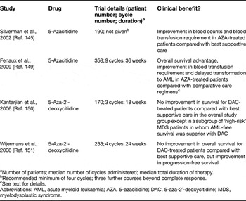
The subsequent drug development programme led to the licensing of both AZA and DAC for the treatment of myelodysplastic syndromes and acute myeloid leukaemia in the USA, although activity of these agents in other myeloid malignancies is limited (Refs Reference Mesa147, Reference Quintas-Cardama148). The Cancer and Leukaemia Group B (CALGB) 9221 study was a prospective randomised controlled study of AZA versus best supportive care (BSC) for patients with myelodysplastic syndromes defined by the French–American–British (FAB) classification (Ref. Reference Silverman145). The study design allowed crossover from the BSC to the AZA arm, impairing the potential to demonstrate a survival advantage for AZA. Responses occurred in 60% of patients on the AZA arm, comprising 7% complete response (with restoration of normal blood counts and bone marrow appearance), 16% partial response (some improvement in blood counts) and 37% ‘improved’ (smaller improvement but still potentially useful for patients). In patients previously dependent on red-cell transfusion, 45% became independent of transfusion. An improvement in the time to leukaemic (acute myeloid leukaemia) transformation or death was noted (21 months for AZA versus 13 months for BSC), indicating that AZA had clearly modified the natural history of myelodysplastic syndromes in these patients (Table 1).
Phase III randomised controlled trials of hypomethylating therapy for myelodysplastic syndromes

aNumber of patients; median number of cycles administered; median total duration of therapy.
bRecommended minimum of four cycles; three further courses beyond complete response.
cSee text for details.
Abbreviations: AML, acute myeloid leukaemia; AZA, 5-azacitidine; DAC, 5-aza-2′-deoxycitidine; MDS, myelodysplastic syndrome.
The larger AZA 001 study was recently concluded, leading to licensing of AZA in Europe in late 2008 (Ref. Reference Fenaux149). Patients with high-risk myelodysplastic syndromes (N = 358) were randomised to receive AZA or conventional care regimens (CCRs) comprising one of BSC, low-dose cytosine arabinoside or induction chemotherapy. The median overall survival was superior in the AZA arm compared with CCR (24 months versus 15 months). Patients receiving AZA had prolonged time to acute myeloid leukaemia transformation or death, and lower transfusion dependence, infection and hospitalisation compared with CCR patients. The median number of courses of AZA was nine; responding or stable patients continued AZA until progression, loss of response or death. Toxicity was mild – predominantly myelosuppression, toxic skin reactions to subcutaneous injections and gastrointestinal toxicity (Table 1).
A large Phase II study of DAC (N = 66) enrolled predominantly high-risk myelodysplastic syndrome patients as well as some lower-risk patients with heavy transfusion dependence (Ref. Reference Wijermans146). Of these, 49% of patients responded, but for a relatively short median duration of 31 weeks. A subsequent randomised Phase III trial of DAC versus supportive care (N = 170) demonstrated a modest advantage for DAC therapy, restricted to high-risk myelodysplastic syndrome patients (Ref. Reference Kantarjian150). Finally, a recent Phase III study in elderly patients with high-risk myelodysplastic syndromes has failed to show a survival advantage for DAC versus BSC but a median of only four cycles was administered (Ref. Reference Wijermans151) (Table 1). The AZA 001 data would indicate that prolonged therapy with hypomethylating agents is required to maximise response. However, complete normalisation of cytogenetic abnormalities in the bone marrow has been clearly documented following treatment with DAC, perhaps indicating greater cytotoxicity than with AZA (Ref. Reference Lubbert152).
In summary, single-agent therapy with hypomethylating agents has shown meaningful clinical activity in the treatment of patients with myelodysplastic syndromes and acute myeloid leukaemia, and clearly alters the natural history of these diseases. They have become the standard of care for patients with ‘high-risk’ myelodysplastic syndromes.
Ongoing epigenetic therapeutics and future outlook
Attention is now shifting to combination therapy with hypomethylating and other novel therapeutic agents, some of which also target the epigenome. As discussed above, histone acetylation is associated with gene silencing and is controlled by the balance between the activity of HATs and HDAC enzymes. Inhibition of HDACs derepresses gene expression and may act in synergy with hypomethylating agents to further promote the re-expression of genes associated with cellular differentiation. Single-agent therapy with the HDAC inhibitor sodium valproate produced responses in patients with myelodysplastic syndromes, albeit predominantly short-lived (Ref. Reference Kuendgen153). Preliminary data indicate that sequential or simultaneous therapy with HDAC inhibitors and hypomethylating agents has promising clinical activity in myelodysplastic syndromes and acute myeloid leukaemia (Refs Reference Soriano140, Reference Gore154). The near future will see the emergence of more class-specific and potent HDAC inhibitors into clinical trials. Ongoing Phase II and III studies are now addressing clinically challenging areas such as the attempt to reduce relapse risk in myelodysplastic syndromes and acute myeloid leukaemia, with hypomethylating therapy in the peritransplant period or as maintenance therapy after induction chemotherapy. Other ongoing or planned studies are combining hypomethylation with other effective agents in myelodysplastic syndromes such as the immunomodulatory agent lenalidomide or the nucleoside analogue clofarabine. Successful therapeutic strategies are likely to evolve through these combination treatments. Further challenges will include more precise prediction of responders, perhaps based on individual epigenetic profiling and an understanding of the factors that underlie resistance to current therapeutic agents.
Acknowledgements and funding
Funding for haematopoiesis research in C. Bonifer's laboratory comes from Leukaemia & Lymphoma Research (UK), Cancer Research UK, the Biotechnology and Biological Sciences Research Council (BBSRC), the European Union (Integrated Project EuTRACC, grant. no. LSHG-CT-2007-037445) and Candlelighters. Research funding for D. Bowen is from Cancer Research UK, Yorkshire Cancer Research and Celgene Corporation. The authors thank the referees for their helpful comments.