INTRODUCTION
Interactions between a parasite and its host can be studied in many ways. Most classically, the populations of the parasites within experimentally infected hosts may be followed by serial necropsy (e.g. Eriksen et al. Reference Eriksen, Nansen, Roepstorff, Lind and Nilsson1992; Roepstorff et al. Reference Roepstorff, Eriksen, Slotved and Nansen1997; Kringel and Roepstorff, Reference Kringel and Roepstorff2006) while simultaneously investigating the immune responses mounted against the parasites. Basic information on dynamics of the parasite populations as well as the host responses is crucial in order to understand epidemiological and diagnostic data and to optimize parasite control programmes. In addition, such information is crucial for optimal planning of new experimental studies, i.e. to sample the right organ at the right time-point (Miquel et al. Reference Miquel, Roepstorff, Bailey and Eriksen2005; Steenhard et al. Reference Steenhard, Kringel, Roepstorff, Thamsborg and Jungersen2007).
Trichuris suis is found sporadically under intensive indoor production systems (Roepstorff et al. Reference Roepstorff, Nilsson, Oksanen, Gjerde, Richter, Ortenberg, Christensson, Martinsson, Bartlett, Nansen, Eriksen, Helle, Nikander and Larsen1998; Joachim et al. Reference Joachim, Dulmer, Daugschies and Roepstorff2001; Weng et al. Reference Weng, Hu, Li, Li, Lin, Xie, Gasser and Zhu2005). In contrast, free range or organic systems seem to favour higher prevalences (Roepstorff et al. Reference Roepstorff, Jørgensen, Nansen, Henriksen, Skovgaard-Petersen and Andreasen1992) and more recently 9 out of 10 and 37·5% organic (outdoor) farms were shown to be positive for T. suis in Denmark and The Netherlands, respectively (Carstensen et al. Reference Carstensen, Vaarst and Roepstorff2002; Eijck and Borgsteede, Reference Eijck and Borgsteede2005). When pigs ingest infective T. suis eggs, the L1 larvae are released in the intestine. The larvae penetrate the caecal and colonic mucosa and enter the cells lining the crypts. The histotrophic phase lasts about 2 weeks whereafter the posterior part of the larvae starts to protrude into the gut lumen and mating takes place after 4 moults when the adult stage is reached. Eggs can be found in the faeces 6–7 weeks p.i. (Beer, Reference Beer1973; Kringel and Roepstorff, Reference Kringel and Roepstorff2006). In pigs single-infected with T. suis a worm expulsion phase has been shown to take place around week 9 p.i. (Kringel and Roepstorff, Reference Kringel and Roepstorff2006), but no studies have investigated the population dynamics of experimental trickle infections in pigs.
The life cycle of Trichuris trichiura, which infects around 1 billion people worldwide (Chan et al. Reference Chan, Medley, Jamison and Bundy1994), is expected to be similar to that described for T. suis in pigs (Bundy and Cooper, Reference Bundy and Cooper1989). For this reason and because of the morphological and physiological similarities of the gastro-intestinal tracts of pigs and humans (Miller and Ullrey, Reference Miller and Ullrey1987), as well as the capability of T. trichiura to infect pigs and T. suis to infect man (Beer, Reference Beer1976), T. suis in pigs has been suggested as a good model for T. trichiura infections in humans (Boes and Helwigh, Reference Boes and Helwigh2000). The recent use of repeated T. suis dosing in Inflammatory Bowel Disease (IBD) patients (Summers et al. Reference Summers, Elliott, Urban, Thompson and Weinstock2005a, Reference Summers, Elliott and Weinstockb; Elliott et al. Reference Elliott, Summers and Weinstock2007), in which the parasite seems to provoke a beneficial immunomodulation, makes such a study even more important.
In the present paper we describe the population dynamics of T. suis in trickle-infected pigs. The long-term repeated exposure is expected to simulate the ‘natural’ situation for both pigs and humans, living in contaminated environments. We have applied the recently developed EDTA method which allows us to isolate the small and immature T. suis worms (Kringel et al. Reference Kringel, Roepstorff and Murrell2002). The pigs were concurrently infected with Ascaris suum (Nejsum et al. 2009 b).
MATERIALS AND METHODS
Inoculation material
Our strain of T. suis was originally isolated from an organic pig farm (Roepstorff and Murrell, Reference Roepstorff and Murrell1997) and has since been passed 3 times in pigs. The eggs were isolated from faeces of experimentally infected pigs and subsequently embryonated in moist vermiculite at room temperature for around 6 months. The batch (DCEP batch 2002) had an infectivity of approximately 100%, as described by Kringel and Roepstorff (Reference Kringel and Roepstorff2006).
Experimental design
Twenty-three crossbreed Danish Landrace/Yorkshire/Duroc pigs of both sexes (females and castrated males) 8 weeks of age were obtained from a specific-pathogen-free farm. After stratification according to sex and litter, the pigs were randomly allocated to 4 helminth-free paddocks of equal size. The pigs were infected with T. suis (5 egg/kg/day) in the feed twice weekly until necropsy, and faecal samples were collected from all pigs at weeks 0, 6, 7, 8, 9, 10, 12, and 14 p.i. and at necropsy. A concentration McMaster technique with a lower detection limit of 20 eggs per gram of faeces (epg) was used to estimate the faecal egg count (FEC) (Roepstorff and Nansen, Reference Roepstorff and Nansen1998). Seven, 8, and 8 pigs were necropsied at weeks 4, 8, and 14 p.i., respectively, for recovery of worms from the caecum and colon. Serum samples were collected from all pigs at day 0 p.i. and from pigs necropsied at weeks 4, 8 and 14 p.i.
Necropsy
The pigs did not receive feed on the day of necropsy. They were killed by a captive bolt pistol and exsanguination. The intestine was immediately removed. The colon and caecum were cut open and the mucosa was gently washed free of luminal content with 0·9% NaCl solution at 37°C. The worms were recovered from the mucosa of the large intestine by a slight modification of the EDTA method described by Kringel et al. (Reference Kringel, Roepstorff and Murrell2002). Briefly, the washed intestine was cut into smaller pieces and incubated in 10 mm EDTA in 0·9% NaCl (pH 7) at 37°C for 6 h. The liberated worms were collected on a 212 μm sieve. In order to pick up non-liberated worms the intestine was subsequently washed twice in 0·9% NaCl water by pulling the intestine through 2 fingers, and the washing water was also poured onto a 212 μm sieve. Worms were stored in 70% alcohol at 5°C. Worms from the intestinal contents were recovered by washing a 5% subsample on a 212 μm sieve. Subsamples were fixed in iodine (80 g iodine and 400 g potassium iodine in 800 ml distilled water) for later recovery of worms under a dissecting microscope after decolourization with 30% sodium thiosulphate solution. The lengths of worms were measured (longer or shorter than 1 cm) and the sex determined. Worms were stored in 70% alcohol at 5°C.
ELISA
The presence of IgG1, IgG2 and IgA serum antibodies specific for adult T. suis excretory/secretory (ES) antigen was determined by an indirect enzyme-linked immunosorbent assay (ELISA) as described by Kringel and Roepstorff (Reference Kringel and Roepstorff2007). T. suis ES antigen was obtained from in vitro culture fluid of adult T. suis worms. Maxisorp 96-well plates (NUNC, Denmark) were coated overnight at 4°C with a pre-optimized concentration of adult T. suis ES antigen (5 μg/ml, 100 μl per well) in carbonate buffer, pH 9·6. The plates were washed 3 times in PBS+0·05% Tween 20 (PBST). The washing step was repeated between all steps until colour development. Pre-optimized 2-fold dilution series of serum samples and of a reference serum were incubated on the plates (100 μl per well) for 1.5 h at 37°C. The reference serum was selected as the serum giving the highest optical density (OD) value of several sera from pigs infected with T. suis. Bound antibody was detected by adding monoclonal antibodies specific for porcine IgA (K61-1B4, Serotec), IgG1 (K139-3C8, Serotec), or IgG2 (K68-Ig2, Serotec) in pre-optimized dilutions (1:400) in PBST (100 μl per well) and incubating for 1.5 h at 37°C. Bound monoclonal antibodies were visualized by adding goat anti-mouse IgG (Fc specific) conjugated to horseradish peroxidase (Serotec) at 1:10 000 in PBST (100 μl per well) and incubating for 1.5 h at 37°C. The TMB microwell peroxidase substrate system (K&P Lab) was used to develop the reaction and the OD was read at 450 nm with 650 nm reference filter. A standard curve was constructed from the reference serum, which was assigned an arbitrary ELISA unit (EU) of 106. The quantities of antibody in the serum samples were determined by interpolating the OD values of the serial dilutions that fell within the linear portion of the standard curve. EU of serum samples are expressed relative to the standard.
Statistical analysis
To describe how worm aggregation changes over time, the parameter k of the negative binomial distribution was estimated on the number of worms recovered from the large intestine and on the FEC as follows: k=mean2/(variance-mean).
Differences in antibody responses between weeks 0 and 4, 0 and 8 and 0 and 14 p.i. for each isotype were analysed by paired t-test and for the other week combinations by analysis of variance (ANOVA) using pairwise comparisons of means at the different time-points using SAS® (version 9.1). Correlations between antibody responses and worm counts and FEC at week 8 p.i. were performed by linear regression. In order to obtain variance homogeneity, the natural logarithm (ln) was used to transform the ELISA responses, worm counts, and FEC.
RESULTS
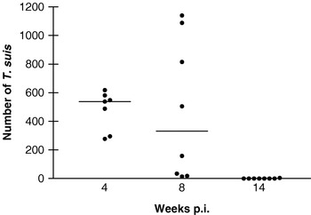
By weeks 4, 8, and 14 p.i. each pig had received approximately 4200, 11 200, and 29 800 T. suis eggs, respectively. The median worm numbers (min–max) recovered from the pigs were 538 (277–618), 332 (14–1140), and 0 (0–4) by weeks 4, 8, and 14 p.i., respectively (Fig. 1). The parameter k of the negative binominal distribution was 14·6, 1·1, and 0·2 at 4, 8, and 14 weeks p.i., respectively, suggesting an increase in the worm aggregation with time.
Individual worm counts (•) and median (—) numbers of Trichuris suis in trickle-infected pigs as a function of time post-first infection (p.i.).

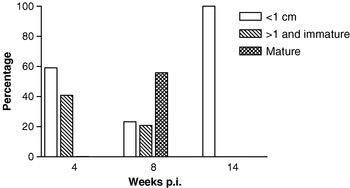
At week 4 p.i. the main population of worms (59%) was <1 cm long while the remaning worms (41%) were >1 cm but still immature (Fig. 2). By week 8 p.i., 23% were <1 cm, 21% >1 cm but immature while 56% of the worm population consisted of matured adults. At week 14 p.i., 1 pig harboured 4 small worms (<1 cm) and all others were negative.
The relative composition of worm populations in relation to weeks post-first inoculation (p.i.) in pigs trickle-infected with Trichuris suis. At week 14 p.i. only 4 worms were recovered from 1 pig.

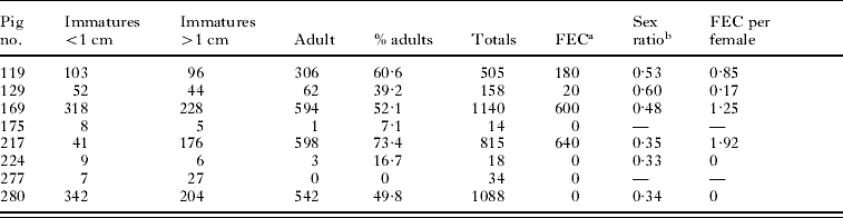
Worm populations of the individual pigs by week 8 p.i. are shown in Table 1, together with FECs obtained at necropsy, sex ratio of adult worms, and FEC per female. Even though all pigs were worm-positive at this time-point only 4 pigs (50%) were FEC positive. Pig 280 had a high burden of adult worms but had an FEC of 0, which was also the case for a faecal sample obtained 2 days earlier from the same pig. In addition, this pig had an FEC of 0 and 20 at weeks 6 and 7 p.i., respectively.
Number of Trichuris suis belonging to different size catagories recovered from pigs trickle infected for 8 weeks. In addition, FEC at 8 week p.i., sex ratio and FEC pr. female are shown
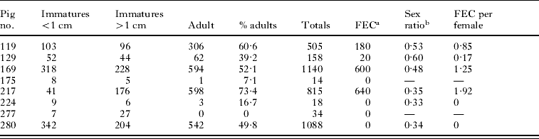
a FEC, fecal egg count (epg) obtained the day of necropsy.
b Proportion of males.
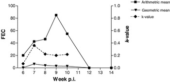
All pigs were FEC-negative at weeks 0 and 4 p.i. Fig. 3 shows the arithmetic and geometric mean FEC as a function of time p.i., together with the parameter k of the negative binominal distribution. In contrast to the k-value of the worm counts, the FEC k-values indicate a constant high degree of aggregation over time (weeks 7–10), which is in agreement with some pigs being worm positive without excreting eggs (Table 1).
Arithmetric and geometric mean faecal egg counts, FECs, as a function of time in Trichuris suis trickle-infected pigs together with the parameter k of the negative binominal distribution. Weeks 6–8 p.i.: n=16; Weeks 9–14 p.i.: n=8.

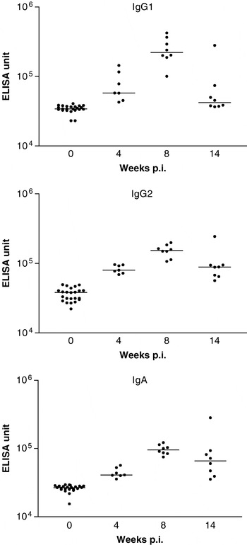
The serum levels of specific T. suis ES IgG1, IgG2, and IgA all followed the same pattern with a maximum reached at week 8 p.i. followed by a decline at week 14 p.i. (Fig. 4). All levels of IgG1, IgG2, and IgA at weeks 4, 8 and 14 p.i., respectively, were significantly (P<0·01) increased in comparison to week 0 except for IgG1 at week 14 p.i. (P=0·06). All other week combinations were significantly different from each other at P<0·05 except between weeks 4 and 14 p.i. for IgG1 and IgG2. At week 8 p.i., there was a significant positive correlation between worm counts and IgA level (r2=0·61; P<0·05) and borderline significance for IgG2 (r2=0·48; P=0·06). Interestingly, the highest isotype levels at week 14 p.i. were from the only worm-positive pig at that time-point, although it only had 4 worms.
Individual (•) and median (—) serum levels of adult Trichuris suis excretory/secretory antigen specific IgG1, IgG2, and IgA antibodies in trickle-infected pigs. The antibody levels are expressed in ELISA units according to an arbitrary standard which has been given the value 106. All levels of IgG1, IgG2, and IgA at weeks 4, 8, and 14 p.i., respectively, were significantly (P<0·01) increased in comparison to week 0 except for IgG1 at week 14 p.i. All other week combinations were significantly different from each other (P<0·05) except between weeks 4 and 14 for IgG1 and IgG2.

DISCUSSION
We here present the first T. suis trickle-infection study in pigs using serial necropsy. Despite a continuous re-infection regime with all pigs initially (week 4 p.i.) T. suis-positive with a non-aggregated worm load, this pattern changed by week 8 p.i. with a noticeable decrease in the k-value and a lower median worm burden due to a higher accumulation of worms in some pigs while others had expelled the majority of their worms. By week 14 p.i. only 1 single pig was infected with 4 small (<1 cm) worms. The pig host therefore seems to acquire nearly sterile immunity against T. suis. Interestingly, the population dynamics in these trickle-infected pigs follow a pattern similar to that described for single infections (Kringel and Roepstorff, Reference Kringel and Roepstorff2006). However, the latter authors found an almost 100% establishment of a 5000 egg single dose at weeks 3–7, i.e. before the main expulsion started. The present week-4 pigs received almost the same total egg dose but only ~10% of the inoculated eggs were recovered as larvae. We find several reasons for this discrepancy. First, since T. suis larvae burrow deeply in the mucosa for the first 2 weeks (Beer, Reference Beer1973) and therefore are partly out of reach of the EDTA isolation (Kringel et al. Reference Kringel, Roepstorff and Murrell2002), the worms recovered by week 4 mostly represents worms from the first 2 weeks of inoculation. Secondly, in pigs repeatedly inoculated with A. suum, the numbers of larvae reaching the liver is dramatically reduced with time (Urban et al. Reference Urban, Alizadeh and Romanowski1988; Eriksen et al. Reference Eriksen, Nansen, Roepstorff, Lind and Nilsson1992). For T. suis, a comparable decrease in establishment may take place already during the first weeks of exposure, perhaps combined with a gradual prolonging of the histotrophic phase, and this may explain the relative low worm number at week 4 p.i. Lastly, these pigs were concurrently infected with A. suum and these migrate through the colon on their way to the liver and it is not known whether A. suum interferes negatively with the establishment of T. suis.
In epidemiological studies, T. suis is found especially when the pigs are reared under traditional conditions with access to outdoor runs (Roepstorff and Nansen, Reference Roepstorff and Nansen1994). In infected herds, young growing pigs have the highest prevalences while sows are rarely infected (e.g. Jacobs and Dunn, Reference Jacobs and Dunn1969; Roepstorff et al. Reference Roepstorff, Jørgensen, Nansen, Henriksen, Skovgaard-Petersen and Andreasen1992; Carstensen et al. Reference Carstensen, Vaarst and Roepstorff2002). Furthermore, naturally infected pigs usually have very low faecal egg counts like in the present study and very far from the high egg counts that often are obtained in experimental single infections (Pedersen and Saeed, Reference Pedersen and Saeed2000; Kringel and Roepstorff, Reference Kringel and Roepstorff2006). The transitory and low egg excretion may therefore have considerable consequences for the diagnostic value of faecal samples and the interpretation of sporadic low egg counts and may not reflect the level of environmental contamination. A high exposure level may thus be difficult to reveal by faecal examination, although it may cause severe retarded growth and even death in pigs (Jensen and Svensmark, Reference Jensen and Svensmark1996).
The worm population may be affected in several ways by the host. Thus, the degree of immunity elicited by the host may affect the ability of worms to establish in the host and therefore the total number of worms. At week 8 p.i. the variation in worm load is increasing (higher aggregation) compared to week 4 p.i. Furthermore, the fecundity of the worm population may be influenced by the host immunity. Intriguingly, we observed 1 pig with a high worm load, but no FEC. Roepstorff et al. (Reference Roepstorff, Bjørn, Nansen, Barnes and Christensen1996) infected pigs twice weekly with 3 doses (100, 1000 and 10 000) of Oesophagostomum dentatum larvae and found the highest number of worms but also the lowest geometric mean FEC in the highest dose group, in which severely stunted adults without egg production were observed. Given these regulation mechanisms, care should be taken when using FEC as a measure of worm load, although positive correlations between FEC and worm burden have been found in single infections (Kringel and Roepstorff, Reference Kringel and Roepstorff2006).
T. suis infection in pigs has been suggested as a model for T. trichiura in humans (Boes and Helwigh, Reference Boes and Helwigh2000) due to the great similarities in intestinal morphology and physiology of the gastrointestinal systems of the two hosts (Miller and Ullrey, Reference Miller and Ullrey1987) and the potential of the two species to cross-infect their main hosts (Beer, Reference Beer1976). However, the population dynamics seem to differ. In pigs, T. suis primarily infects young animals (e.g. Carstensen et al. Reference Carstensen, Vaarst and Roepstorff2002) while T. trichiura infections in man are chronic by nature (Stephenson et al. Reference Stephenson, Holland and Cooper2000). This may be due to differences in the underlying immune responses elicited by the two hosts or by the degree of exposure (i.e. pigs are expected to show a higher degree of geophagia) or it may reflect differences in nutritional status. Pedersen et al. (Reference Pedersen, Saeed, Michaelsen, Friis and Murrell2002) showed that experimentally infected pigs fed with a ‘low protein’ diet had higher numbers of infected pigs than a normal diet group. In addition, a lower aggregation (higher k value) was found in the depleted group compared to the controls. However, in undernourished pigs e.g. scavenging pigs in Burkina Faso (Tamboura et al. Reference Tamboura, Banga-Mboko, Maes, Youssao, Traore, Bayala and Dembele2006), Ghana (Permin et al. Reference Permin, Yelifari, Bloch, Steenhard, Hansen and Nansen1999), and Zimbabwe (Marufu et al. Reference Marufu, Chanayiwa, Chimonyo and Bhebhe2008), T. suis is still found at a low prevalence, so poor nutrition may not be the only explanation for the difference between the two host-parasite systems.
A caveat to this study is the potential interaction between T. suis and A. suum. If such an interaction exists we expect it to be limited since we in the present study found similar population dynamics as described for T. suis in single-infected pigs (Kringel and Roepstorff, Reference Kringel and Roepstorff2006) and because the two worm species are located in two different compartments of the intestine. For example, no interaction has been demonstrated between A. suum (small intestine) and O. dentatum (large intestine) based on actual worm load (Helwigh et al. Reference Helwigh, Christensen, Roepstorff and Nansen1999).
The present study may also provide suggestions for the fate of T. suis in IBD patients dosed at 2 or 3 week intervals (Summers et al. Reference Summers, Elliott, Urban, Thompson and Weinstock2005a, Reference Summers, Elliott and Weinstockb) although it should be emphasized that parasite population dynamics may depend markedly on the interactions with the particular host species. By endoscopy, Summers et al. (Reference Summers, Elliott and Weinstock2006) have only rarely observed large worms in T. suis-treated patients. It is evident that T. suis eggs may hatch and the larvae may presumably establish for a short while in humans, even in the late doses of a treatment regime, as the repeated dosing seems to mount a positive immunomodulatory effect. These observations may indicate that the population dynamics of T. suis in pigs and man are different, with expulsion taking place a few weeks within the patent period of the first cohorts in pigs while man responds more quickly expelling the worms before patency. In the late phase of a repeated exposure neither pigs nor man have any large worms and hardly any detectable worms at all except for a small minority of low-responding individual pigs (Nejsum et al. Reference Nejsum, Roepstorff, Jørgensen, Fredholm, Göring, Anderson and Thamsborg2009a) and patients (Kradin et al. Reference Kradin, Badizadegan, Auluck, Korzenik and Lauwers2006).
Despite the continuous exposure regime in the present study, we have found T. suis-specific IgG1, IgG2, and IgA response patterns similar to those described for T. suis single-infected pigs, in which the antibody response peaked around weeks 7 and 9 p.i. (Kringel and Roepstorff, Reference Kringel and Roepstorff2007). Since adult worm ES was used as antigen in the ELISA the responses in both studies may merely reflect the presence of adult worm. The antibody kinetics may therefore have been different if ES products had been obtained from immature worms.
IgG1 and IgG2 have been suggested to reflect a Type 2 and Type 1 immune response, respectively (Crawley et al. Reference Crawley, Raymond and Wilkie2003) and T. suis has been shown to induce a Type 2 skewed immune response in the swine host (Kringel et al. Reference Kringel, Iburg, Dawson, Aasted and Roepstorff2006). However, we only found a weak positive correlation between antibody levels (IgA) and worm burdens which also was the case in pigs single-infected with T. suis (Kringel and Roepstorff, Reference Kringel and Roepstorff2007). Trichuris species in general elicit strong antibody responses in their natural hosts (Lillywhite et al. Reference Lillywhite, Bundy, Didier, Cooper and Bianco1991; Hill et al. Reference Hill, Romanowski and Urban1997; Blackwell and Else, Reference Blackwell and Else2002; Kringel and Roepstorff, Reference Kringel and Roepstorff2007), but the regulatory function of the antibodies remains uncertain. Blackwell and Else (Reference Blackwell and Else2002) used lipopolysaccharides for in vitro stimulation of mesenteric lymph node cells and found that the production of T. muris-specific antibodies was reflected in the expulsion kinetics, suggesting an effect at the local level in contrast to the systemic level.
In conclusion, pigs continuously exposed to T. suis eggs seem to obtain nearly sterile immunity despite an initial establishment in all pigs. The main worm expulsion takes place around week 8 p.i. and the antibody level against adult ES declines accordingly.
The skilful technical assistance of Niels Peter K. Hansen, Frederik Andersen, Trine Andersen, Margrethe Pearman, Anders Rasmussen, Henrik B. Lassen, Maria Langkjær, Peter Kristiansen, Niels Midtgaard, and Pernille B. Larsen is greatly appreciated. The Danish National Research Foundation is acknowledged for the financial support of this project under the auspices of the Danish Centre for Experimental Parasitology. All animals were treated according to Danish legislation (13 October 2000, Allan Roepstorff: 2000/561-321).