Introduction
Perforation of the tympanic membrane, with intermittent or persistent discharge from the ear, is termed chronic suppurative otitis media (CSOM). Chronic suppurative otitis media is a neglected disease, with a global prevalence estimated to be as high as 360 million, leaving up to 200 million with significant hearing impairment and a disease burden of over 2 million disability-adjusted life years.Reference Acuin1, Reference Monasta, Ronfani, Marchetti, Montico, Brumatti and Bavcar2 The disease is associated with socioeconomic deprivation, and several countries, particularly those in the Pacific region and South-East Asia, report a disease prevalence at greater than 4 per cent, reaching the World Health Organization (WHO) classification of a massive public health problem.Reference Acuin1, Reference Monasta, Ronfani, Marchetti, Montico, Brumatti and Bavcar2
The WHO has stipulated that programmes to tackle CSOM must incorporate primary prevention, including improvements to socioeconomic factors and medical therapies.3 However, many CSOM cases will persist or recur following medical treatment, and they can then only be cured by surgical intervention. Surgical treatment of CSOM ranges in complexity depending on the disease manifestation, but for isolated mucosal disease tympanoplasty provides barrier protection against middle-ear infection, and often improves hearing, with associated gains to quality of life.Reference Acuin1, Reference Jung, Cho, Hong, Chung, Lee and Hong4 Chronic suppurative otitis media with cholesteatoma requires more aggressive surgical treatment to remove the cholesteatoma, and prevent potential intratemporal and extratemporal complications.Reference Bhutta, Williamson and Sudhoff5 Such surgery aims to provide a safe ear by removing disease, with the secondary objectives of ensuring the ear is dry and functional through ossicular and tympanic reconstruction where possible.
Unfortunately, few low- and middle-income countries are currently equipped with the human and material resources required for a locally staffed otological surgery service. Many of the regions with the highest prevalence of CSOM paradoxically have a shortage of trained ENT surgeons, and, of these, very few are able to perform tympanoplasty or mastoidectomy.6 Tympanoplasty and mastoidectomy are technically difficult procedures that require a period of sustained training and the use of specialised equipment.Reference Palva and Ramsay7
To fill the need for tympanoplasty and mastoidectomy, short term visits by ENT surgeons from high-income countries have been undertaken, with acceptable reported surgical outcomes.Reference Barrs, Muller, Worrndell and Weidmann8–Reference Lehnerdt, van Delden and Lautermann11 However, these missions are limited in their duration, and do not provide the sustainability of a locally delivered service. As the surgical disease burden in low- and middle-income countries has gained more attention, there has been a move towards building local capacity through training and task shifting to local surgeons.Reference Riviello, Ozgediz, Hsia, Azzie, Newton and Tarpley12, Reference Aliu, Pannucci and Chung13 This has included the development of long-term partnerships between hospitals from high- and low-resourced countries in which sustained general otolaryngology training and education has been established.Reference Waller, Larsen-Reindorf, Duah, Opoku-Buabeng, Edwards and Brown14
The value of ‘vertical’ surgical missions that focus on a single pathology and its management has been well established in other surgical subspecialties. This has led to infrastructure for the management of conditions such as cleft palate and cataracts.Reference Farmer and Kim15 Whilst there has been recognition of this impetus on local training during otological surgical missions, there is a dearth of formal reporting on such training programmes or the outcomes produced.
Cambodia is a lower- to middle-income nation in a region of high CSOM prevalence.Reference Acuin1 National healthcare challenges include a predominantly rural population, a gross national income per capita of US$ 1070 and a health expenditure being borne primarily by the patient as an out-of-pocket expense.16, 17 The Cambodian healthcare system is still recovering from the loss of medical expertise suffered during the Khmer Rouge regime, and, prior to our intervention, it included (to our knowledge) only one local surgeon trained in tympanoplasty.
In October 2014, we instigated a sustained in-country training programme in Cambodia to develop local proficiency in type I tympanoplasty. This study aimed to describe this novel training programme, and its qualitative educational outcomes (as determined by interviews with the two local trainees) and quantitative surgical outcomes (as determined by surgical closure of the tympanic membrane and improvement to hearing). A subsequent four-month long training programme in mastoidectomy and ossiculoplasty was implemented, and we report the preliminary surgical and educational outcomes from this.
Materials and methods
Study design and setting
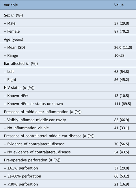
The Institutional Review Board of the Children's Surgical Centre reviewed and approved this study. The tympanoplasty training programme was instigated in October 2014 at the Children's Surgical Centre, a multidisciplinary surgical charity hospital in Phnom Penh.18 The Children's Surgical Centre provides free rehabilitative surgery to the impoverished population of Cambodia, attracting many patients from rural communities beyond Phnom Penh. The patient population suffering from CSOM have a wide age range and advanced disease profile (Table 1).
Patients’ demographics and clinical parameters used to assess tympanoplasty outcome

SD = standard deviation; HIV = human immunodeficiency virus
Prior to our intervention, the Children's Surgical Centre had an ENT department staffed by locally trained nurses and two Cambodian ENT surgeons. The two surgeons had completed medical degrees and post-graduate otolaryngology residency programmes within Cambodia, but this included very limited exposure to otological surgery given the inaccessibility of necessary material resources and local expertise. They had experience of observing and assisting in tympanoplasty and mastoidectomy, but neither individual had previously performed the procedures as the primary surgeon.
Tympanoplasty training programme
An initial scouting visit by a UK-trained ENT consultant determined equipment and training needs. This was followed by procurement, from UK and US suppliers, of a surgical microscope (with a teaching side arm) and microsurgical instruments, and transportation of these to Cambodia. Equipment and transportation costs were subsidised by donations from medical supply companies and UK hospital foundations where possible, with the remaining costs remunerated by the Children's Surgical Centre directly.
A training fellow (CTH) delivered six-month continuous in-country training in type I tympanoplasty to the two local ENT surgeons. The training fellow (CTH) is a UK-qualified ENT surgeon who at that time had just completed his training. A UK operating theatre nurse accompanied the training fellow for the first two weeks of his residency, providing parallel training to the local operating theatre nurses.
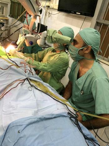
Training for the surgeons included themes of applied surgical anatomy, clinical judgment in peri-operative disease recognition and management, microsurgical technique, and intra-operative judgment and problem solving. Training was provided via traditional formats such as didactic lectures and paper-based resources, as well as online resources and model trainers for practical skill exercises.Reference Clark, Westerberg and Mitchell19 Trainees were allowed to perform relevant stages of tympanoplasty as soon as deemed appropriate (Fig. 1).
One of the Cambodian surgeons being trained by author MFB.

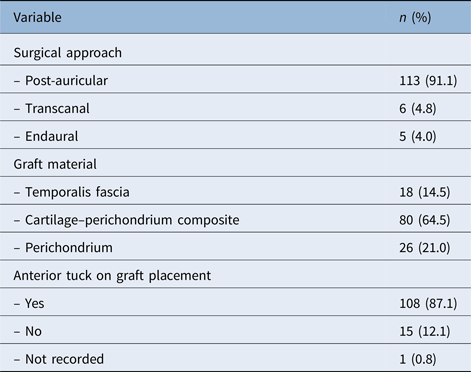
In order to facilitate learning, we utilised a standardised technique with general anaesthetic, using a post-auricular approach, an underlay composite tragal cartilage and perichondrium graft, an anterior tuck where required (for anteriorly based perforations), underlay and overlay gelatine packing to support the graft, and a head bandage. The post-auricular approach and composite graft were deemed appropriate methods for the typically large perforations and disease severity found in our population, although other approaches and graft materials were also discussed and taught (Table 2). All tympanoplasties performed during and subsequent to the training programme were performed with consent obtained by the local Khmer speaking surgeons.
Surgical parameters used to assess tympanoplasty outcome

By the end of the six-month training period, the Cambodian surgeons were performing tympanoplasty as the primary surgeon. They had the option of contacting their trainer in the UK via internet or phone if any concerns or queries arose.
Mastoidectomy training programme
After completion of the tympanoplasty training programme, another otology fellow (MFB) provided four months’ continuous in-country training in mastoidectomy and homograft ossiculoplasty for the treatment of cholesteatoma (Fig. 1). This built upon the themes and surgical skills focused on during the tympanoplasty training programme, with more detail on temporal bone anatomy and surgical drilling skills.
Pettigrew plastic temporal bones were used as an aid to teaching.Reference Bhutta20 Again, a standardised operative technique was utilised, with a post-auricular incision, a ‘front to back’ canal wall down approach to minimise risk of residual or recurrent disease (follow up of our patient population was not assured), lowering of the facial ridge, and meatoplasty where required. Surgery could comprise atticotomy, atticoantrostomy or mastoidectomy, depending upon extent of disease.
We did not routinely utilise pre-operative computed tomography scanning. Facial nerve monitoring was not available.
If ossiculoplasty was deemed appropriate, it was performed simultaneously (again in light of the uncertainty of follow up, with potentially no opportunity for staged surgery). Where a stapes was present and the reconstructed middle-ear cavity was low in depth, a cartilage cap was placed on the stapes capitulum; otherwise, the incus was refashioned for use as a replacement ossicle, or a strut of bone carved from the calvarium in cases where the incus was eroded.
Educational outcomes evaluation
Learner outcomes were assessed one year after the tympanoplasty training programme and again one year after the mastoidectomy training programme. Using a five-point Likert scale, the trainees were asked to rate their surgical confidence and ability in various aspects of performing tympanoplasty, mastoidectomy and homograft ossiculoplasty; they were asked to rate these parameters prior to the onset of the training programme and at the present time.
A researcher who had no role in training (author RFA) conducted private semi-structured interviews with the two trainee surgeons, to gather their opinions on the structure of the training programme and on personal learning outcomes. Interviews were conducted in English (a second language for our trainee surgeons). These interviews were audio recorded and transcribed, and the results were evaluated using thematic analysis.
Clinical outcomes evaluation
We retrospectively evaluated the clinical outcomes of all type I tympanoplasties, performed by the trainee surgeons, for a period of 12 months after the resident trainer (CTH) departed (March 2015 to March 2016). These operations were performed independently of the trainer. We included patients with a chronically discharging perforation and excluded those with cholesteatoma.
We collected data on patient demographics and surgical pathology. Surgical success was measured by evidence of an intact tympanic membrane on otoendoscopy at six weeks’ follow up (we lost a large proportion of patients to follow up after six weeks, making analysis at later time points unreliable).
We evaluated audiological outcomes using pure tone audiometry performed by a partner organisation (All Ears Cambodia), comparing audiometric thresholds pre-operatively and at three months post-operatively. Our primary audiological outcome was change in the air–bone gap.
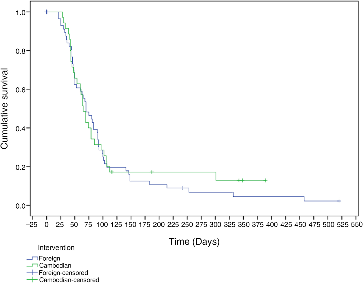
We also evaluated all mastoidectomies performed for cholesteatoma, with or without ossiculoplasty, from January 2016 to July 2017, where the primary surgeon was one of the Cambodian trainees (the mastoidectomy training programme ran from January to June 2016). We assessed tympanic membrane closure, the proportion of cases with a dry ear, time to dry ear, and audiological results (assessed as described previously). Survival analysis was carried out for the time until dry ear, with patients lost to follow up censored at day 0, and those who never achieved a dry ear censored at the last day of recorded follow up. In order to create a comparison group, we performed the same analysis of outcomes from mastoidectomies conducted from November 2014 to July 2017 where the primary surgeon was non-Cambodian (foreign).
Results
Educational outcomes
At completion of the tympanoplasty training period, the more senior of the Cambodian trainees felt able (and was deemed able) to perform type I tympanoplasty without supervision. The junior trainee, with the supervision of her senior local colleague, reached the same stage only two months later.
In the interviews conducted one year after completion of the tympanoplasty training programme, the trainees confirmed confidence in their ability to perform the steps of type I tympanoplasty, stating: ‘Confidence in performing tympanoplasty is 100 per cent’ and ‘100 per cent all cases no problem’.
Both trainees were deemed safe to perform mastoidectomy after completion of the mastoidectomy training programme.
Interviews revealed that the trainees preferred prolonged training with a single resident teaching fellow over the previous experience of multiple short-term visits by visiting surgeons, with the trainees stating: ‘when we start the first time we cannot want more teacher, only the one’, ‘I know the person in the short term, but sometimes I cannot show myself and what I can do. In the long term I can do or if I have a problem I can ask’ and ‘One week is a very short time, I had the experience already I didn't get anything’.
The programme also succeeded in delivering analytical skills. The trainees stated: ‘I learnt from the international surgeon, we need to think not just about us, we need to think about the patient, about what is good for the patient’, ‘they make me to think, they teach me to think’ and ‘when we have problem during the operation we find by our self’.
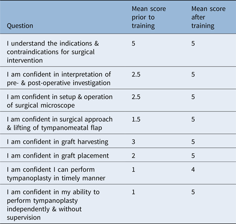
The Likert scale evaluation of confidence and ability in tympanoplasty revealed that both Cambodian surgeons had an increase in surgical confidence across all aspects questioned (Table 3). The mean improvement across all aspects was 2.35 points, and their overall confidence to perform the operation independently improved by 4 points (out of a possible 4).
Self-rated confidence in performing tympanoplasty*

The scale scores ranged from 1 (not confident) to 5 (very confident).
* As rated by the two trained surgeons
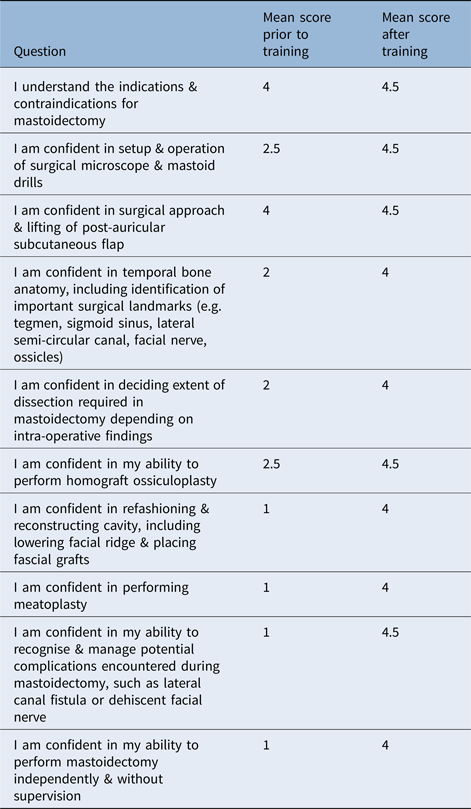
A similar increase in surgical confidence was witnessed after the mastoidectomy training programme (Table 4). The mean improvement across all aspects assessed was 2.28 points, and the overall confidence to perform mastoidectomy independently improved by 3 points (out of a possible 4).
Self-rated confidence in performing mastoidectomy*

The scale scores ranged from 1 (not confident) to 5 (very confident).
* As rated by the two trained surgeons
Tympanoplasty clinical outcomes
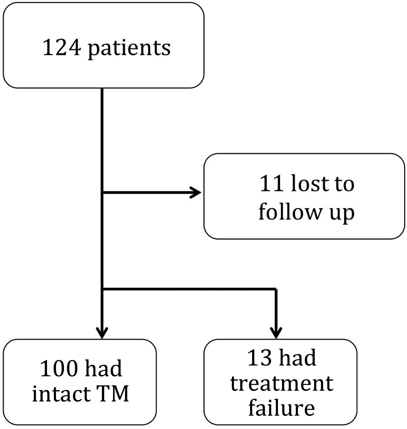
There were 124 patients in the evaluation period (demographic characteristics are summarised in Table 1). Of these, 11 patients (8.9 per cent) were lost to follow up at the six-week review. Of the remaining 113 patients, 100 (88.5 per cent) were found to have an intact tympanic membrane, 11 (9.7 per cent) had a residual perforation estimated at less than 5 per cent and 2 (1.8 per cent) had a residual perforation of undocumented size (Fig. 2).
Surgical outcomes in patients undergoing tympanoplasty in the year after completion of the training programme. TM = tympanic membrane

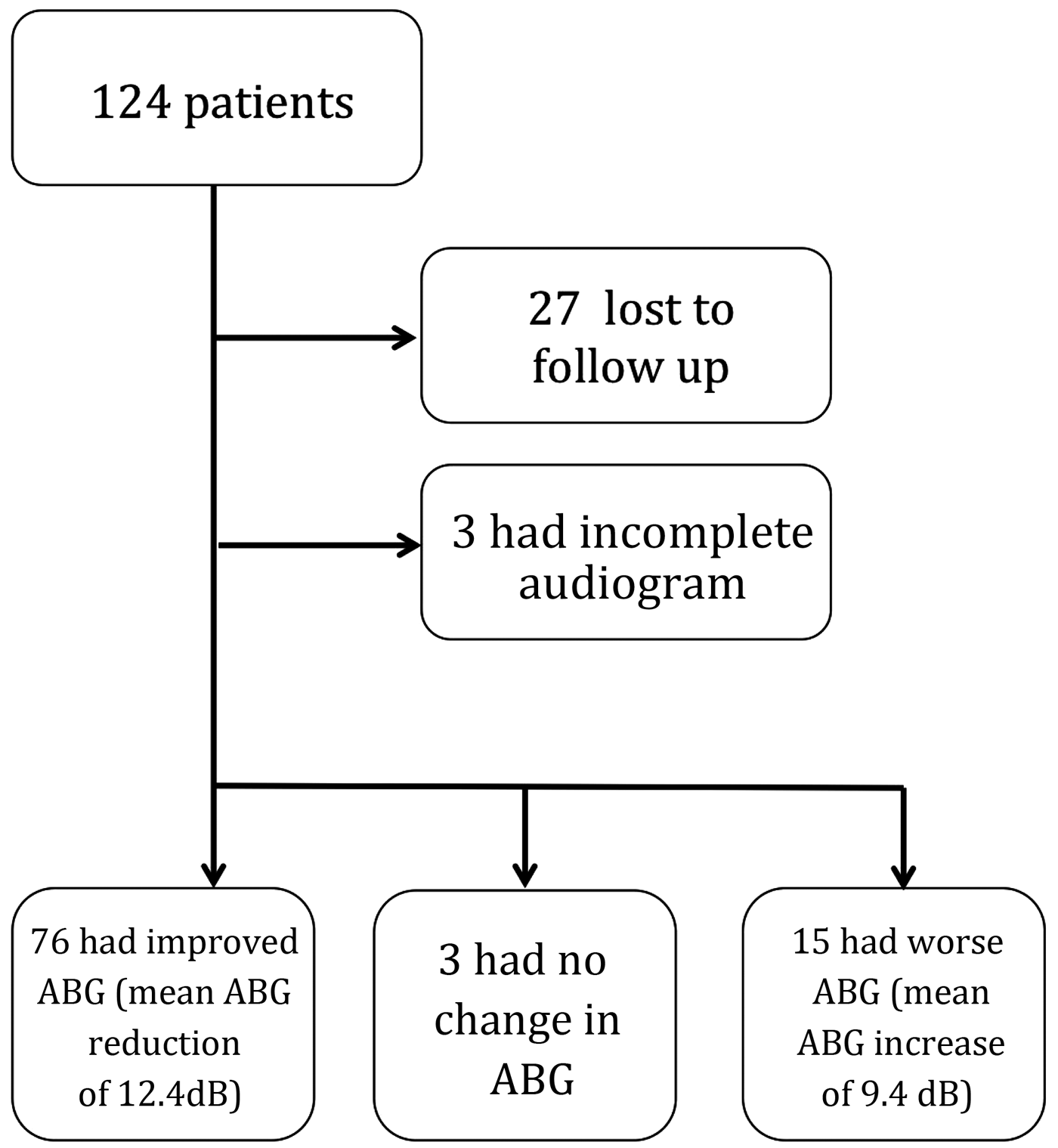
Post-operative audiometry data were available in 94 patients, with 27 (21.8 per cent) lost to follow up and a further 3 patients excluded because of incomplete audiograms. Across the 94 patients, there was a mean reduction in air–bone gap of 12.4 dB (range, −32.5 to 43.8 dB). There were 76 patients (80.9 per cent) who had audiometric improvement (mean reduction in air–bone gap of 17.1 dB), 3 who had no change in air–bone gap and 15 with an increased air–bone gap (mean increase in air–bone gap of 9.4 dB) (Fig. 3). Those patients with a residual perforation had a mean reduction in air–bone gap of 15.1 dB. The average post-operative air–bone gap was 19.5 dB, with 84 patients (89.4 per cent) having a post-operative air–bone gap of 30 dB or lower.
Audiological outcomes in patients undergoing tympanoplasty in the year after completion of the training programme. ABG = air–bone gap

There were no major complications such as complete sensorineural hearing loss, facial nerve palsy, vertigo or death.
Mastoidectomy clinical outcomes
A total of 112 mastoidectomies were performed at the Children's Surgical Centre in the evaluation period; 66 (58.9 per cent) were primarily performed by overseas-trained surgeons and 46 (41.1 per cent) were carried out by Cambodian surgeons. Ninety-one patients (81 per cent) attended for clinical follow up at three months; 63 patients (56 per cent) underwent a hearing test, with the remainder lost to follow up.
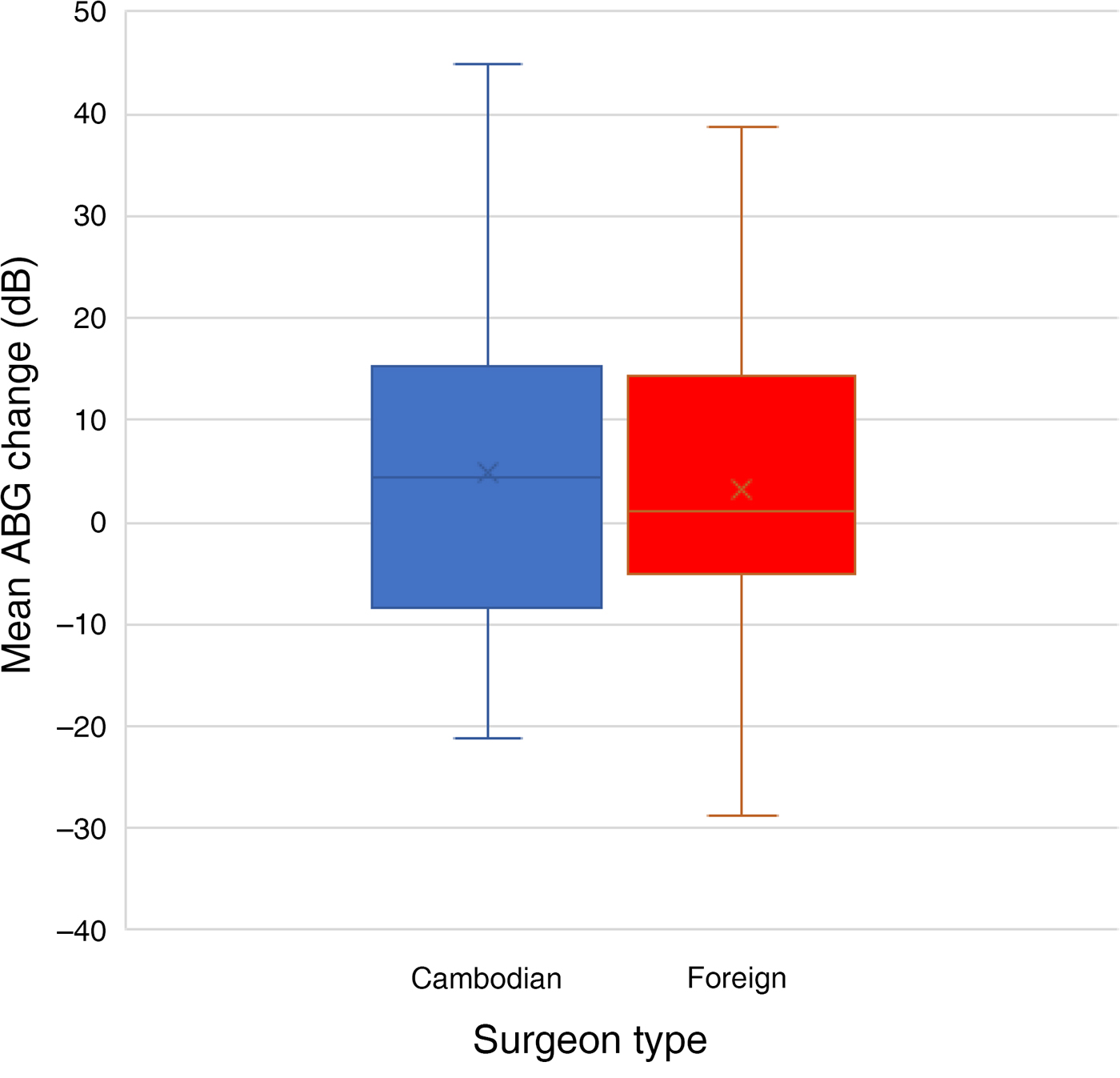
There were no significant differences between operation outcomes for Cambodian versus foreign surgeons in terms of rates of tympanic membrane closure, the proportion of cases with a dry ear or the time taken to achieve a dry ear (Table 5, and Figs. 4 and 5). There was no significant mean change in audiological thresholds in either group (Table 5); however, using the classification of 15 dB change in air–bone gap as a clinically significant change in hearing, five patients operated on by Cambodian surgeons had improved hearing and two patients had worse hearing. The equivalent figures for patients operated on by foreign surgeons were nine with improved hearing and three with worse hearing.
Time to dry ear in patients undergoing mastoidectomy by Cambodian and foreign surgeons.

Mean change in air–bone gap (ABG) in patients operated on primarily by Cambodian and foreign surgeons. The whiskers indicate the minimum and maximum mean air–bone gap change; the boxes represent the first and third quartiles of the mean air–bone gap change; the horizontal line within each box indicates the median air–bone gap change; and the cross within each box represents the mean air–bone gap change across the study population.

Mastoidectomy outcomes at three months*

* For patients operated on by Cambodian and foreign surgeons. ABG = air–bone gap
There were no major complications in either group, such as complete sensorineural hearing loss, cerebrospinal fluid leak, persistent vertigo or facial paralysis.
Discussion
This is the first report from a resource-limited setting demonstrating successful outcomes from a training programme in tympanoplasty and mastoidectomy. We attribute our success to the provision of training fellows who were resident in-country for four to six months, and so were able to provide a sustained period of training. Other authors have recognised the importance of training local surgeons in tympanoplasty in resource-limited settings, and have conducted such training during short-term surgical missions, but have not reported or demonstrated educational benefits from their efforts.Reference Isaacson21–Reference Isaacson, Drum and Cohen23
Our training programme resulted in excellent educational and clinical outcomes. In interviews, the two Cambodian surgeons reported a strong preference for having a resident fellow (over and above short-term visits) to meet their training needs. They also reported marked improvements in all aspects of their assessment, surgical technique and decision-making in the care of patients with CSOM, as evidenced through their comments in the semi-structured interview and in their self-assessment surveys. We acknowledge that these qualitative outcomes were assessed retrospectively, with no baseline data obtained at initiation of either training programme, increasing the risk of recall bias. We also acknowledge that the trainees self-reported ‘100 per cent’ confidence may reflect a lack of experience of the difficulties in some cases of tympanoplasty, although, to date, the trainee surgeons have not felt the need to abandon a case.
The clinical outcomes are also commendable. The results from the first year in which our surgeons were performing tympanoplasty independently show an 88.5 per cent success rate in tympanic membrane closure. The WHO regards a successful tympanoplasty programme as one where success rates are over 80 per cent.3 Studies from high-income countries report rates of 80–85 per cent, with similar results reported by surgeons performing otological missions in resource-limited environments.Reference Horlbeck, Boston, Balough, Sierra, Saenz and Heinichen9–Reference Lehnerdt, van Delden and Lautermann11, Reference Hardman, Muzaffar, Nankivell and Coulson24 One limitation of our study is that evaluation of the tympanic membrane was conducted at six weeks after the operation, which may over-estimate success. Late failure is known to occur, but could not be assessed here as few of our patients attended for long-term follow up. This logistical difficulty is well recognised, with our study displaying a relatively low loss to follow up in comparison to other studies in similar settings.Reference Barrs, Muller, Worrndell and Weidmann8–Reference Lehnerdt, van Delden and Lautermann11, Reference Isaacson21–Reference Isaacson, Drum and Cohen23
The audiological outcomes demonstrated a modest improvement in the majority of tympanoplasty cases, with a mean reduction in air–bone gap across all cases of 12.4 dB. This too is comparable to reports from local and visiting surgeons in other low- and middle-income countries.Reference Horlbeck, Boston, Balough, Sierra, Saenz and Heinichen9–Reference Lehnerdt, van Delden and Lautermann11, Reference Kamath, Sreedharan, Rao, Raj and Raju25, Reference Mishra, Sonkhya and Mathur26 Again, a limitation of our study is that we evaluated audiological outcomes at three months post-operatively because obtaining long-term audiological data was logistically problematic. Even at three months, there was significant loss to follow up (21.8 per cent), which, whilst somewhat less than that seen in other similar studies, could limit the validity of the audiological outcomes reported.
Early outcomes from the mastoidectomy and ossiculoplasty training programme are also encouraging, suggesting non-inferiority of the Cambodian trainees in comparison to the overseas-trained surgeons. The foreign and Cambodian surgeons had similar success in achieving a dry ear, and no difference in audiological outcomes. There are some limitations to this analysis. Operations by foreign surgeons occurred predominantly early in the evaluation period, allowing a longer period of follow up, which potentially increases rates of dry ear because of the late resolution of otorrhoea. Conversely, cases predicted to be more complex had surgery delayed until foreign expertise was available, which may bias foreign surgeon results to worse outcomes. We also recognise that this dataset is small, making it underpowered to detect small differences between the two groups of surgeons.
A notable feature of this training programme is its demonstrable sustainability. The surgeons who were trained have now been performing tympanoplasty independently for over two years, and have been undertaking mastoidectomy and ossiculoplasty for over one year. The volume of cases being referred to them has increased as their reputation has become known. A new Cambodian surgeon joined the ENT department a year ago, and is now performing tympanoplasty solo, having been trained by the local team. We expect that in the future many more ENT surgeons will be trained in tympanoplasty and mastoidectomy by the team at the Children's Surgical Centre hospital, helping to expand the ENT skill base and provision of surgical therapy in Cambodia. The important measures in the evaluation of any surgical initiative in a low- or middle-income country are intermediate and long-term success and sustainability, and here we have strong indicators that our programme has been successful.Reference Ibrahim, Cadotte and Bernstein27
There are some potential limitations to the generalisability of our programme. Tympanoplasty requires the use of a microscope and specialised micro-instruments, which may be difficult to source, and may require periodic repair or replacement. Repair can be difficult in low- and middle-income countries, where biomedical engineering departments are scarce. Equipment can be sourced through donations direct from manufacturers, as used equipment from healthcare providers in high-income settings or through e-commerce retailers. However, consumable costs in performing tympanoplasty are minimal. The specialised nature of a type I tympanoplasty training programme also necessitates sound background otolaryngology knowledge and surgical skills in the prospective trainees, which in the programme reported here was met through the training of surgeons with previous formal ENT training. For this training to be emulated successfully in other low- and middle-income countries, trainees may need similar background knowledge.
• Chronic suppurative otitis media affects up to 360 million people globally, and is associated with socioeconomic deprivation
• Tympanoplasty or mastoidectomy are often required for cure, but are not readily available in many low- and middle-income countries
• This paper describes a tympanoplasty and mastoidectomy training programme in Cambodia
• Success was demonstrated through learning outcomes of locally trained surgeons, with surgical cure rates comparable to that of foreign surgeons
• The programme is sustainable, with the trained surgeons now training others; this model can be emulated in other settings
In summary, our training programme has produced demonstrably self-confident and technically able surgeons in Cambodia, who are capable of successfully performing tympanoplasty and mastoidectomy. We believe that this model can be emulated to develop the infrastructure and capacity for the surgical therapy of CSOM in other resource-limited settings.
Acknowledgements
We would like to acknowledge All Ears Cambodia for their provision of pure tone audiometry for many of the patients in this study. We thank Ana Santos for her provision of operating theatre nurse training to the ENT department. We also thank the staff of the Children's Surgical Centre for welcoming us to work with them. The training programme was funded by general charitable donations to the Children's Surgical Centre from several philanthropic sources.
Competing interests
None declared