Tissue complexity in nature
Tissue development and maintenance (i.e., homeostasis) involve complex and dynamic regulation of interactions between (stem) cells, cell growth factors, hormones and cytokines, and extracellular matrix (ECM) that influence cell fate and tissue organization through the set-up of local gradients of soluble or insoluble factors and local physical forces.Reference Discher, Mooney and Zandstra1–Reference Engler, Sen, Sweeney and Discher5 At the cellular level, cell behavior, such as apoptosis, quiescence, proliferation, migration, or differentiation, has been shown to be governed by nano- and microtopography,Reference Davidson, Fromigué, Marie, Hasirci, Reiter and Anselme6 matrix stiffness,Reference Winer, Oake and Janmey7 ligand density (such as peptides containing the amino-acid sequence RGD:arginine, glycine, and aspartic acid),Reference Huebsch, Arany, Mao, Shvartsman, Ali, Bencherif, Rivera-Feliciano and Mooney4 growth factor activity, and cell neighbors.Reference Grellier, Bordenave and Amédée8 At the tissue level, tissue morphogenesis (i.e., the ability of cell populations to organize themselves into anatomically correct structures)Reference Lecuit and Le Goff9 results from specific/orientated cell division and cell rearrangements,Reference Ingber10 as well as cell migration through chemical gradients (haptotaxis and chemiotaxis) or mechanical gradients (durotaxis).
This suggests a dynamic reciprocity between form and functionReference Engler, Humbert, Wehrle-Haller and Weaver11, Reference Sung, Kam and Shuler12 and underlies the importance of engineering adequate geometries of cells, cues, or even drugs to engineer realistic tissue surrogatesReference Singhvi, Kumar, Lopez, Stephanopoulos, Wang, Whitesides and Ingber13, Reference Phillips, Burns, Le Doux, Guldberg and García14 or to promote proper tissue healing. Developing tools to create, manipulate, and analyze the cell-matrix interactions in vitro and in vivo at cellular scale resolution, including the location and shape of cell patterns, is thus essential.Reference Baquey15
Tissue engineering and regenerative medicine
Since the late 1980s and the creation of the first workable definition of hybrid artificial organs,Reference Langer and Vacanti16 an increasing number of research groups throughout the world have begun to develop various novel tissue-engineering approaches. As stated by Langer and Vacanti,Reference Petersen, Calle, Zhao, Lee, Gui, Raredon, Gavrilov, Yi, Zhuang, Breuer, Herzog and Niklason17 these approaches apply the principles of engineering and life sciences toward the development of biological substitutes that restore, maintain, or improve tissue function or a whole organ. In other words, generating biological tissues in vitro involves the use of engineering and material methods, the appropriate combination of cells, and the suitable biochemical and physicochemical factors to mimic both the microenvironment of cells and the microarchitecture of tissues in the body. Tissue engineering aims to overcome the limitations of current treatments based on organ transplantation and biomaterial implantation, as well as to produce tissue-engineered realistic surrogates that should serve in vitro for drug screening, chemical toxicity testing,Reference Singhvi, Kumar, Lopez, Stephanopoulos, Wang, Whitesides and Ingber13 and basic cell biology.
Manufacturing approaches
From the manufacturing perspective, two tissue engineering strategies have been implemented: the top-down and bottom-up approaches. The top-down approach is based on the design and fabrication of a scaffold, either synthetic or natural, or a decellularized organ,Reference Uygun, Soto-Gutierrez, Yagi, Izamis, Guzzardi, Shulman, Milwid, Kobayashi, Tilles, Berthiaume, Hertl, Nahmias, Yarmush and Uygun18, Reference Hutmacher19 onto which cells are subsequently seeded, followed by the maturation of this as-made assembly into a bioreactor, such as an artificial incubator, or in vivo, prior to transplantation in situ. Several requirements have been identified as being crucial for the production of tissue-engineering scaffolds.Reference Sachlos and Czernuszka20 The scaffold should (1) possess interconnecting pores of appropriate scale to favor tissue integration and vascularization, (2) be made from material with controlled biodegradability or bioresorbability so that tissue will replace the scaffold eventually, (3) have appropriate surface chemistry to interact with cells (e.g., decorated by biomolecules to promote cell adhesion and differentiation), (4) possess adequate mechanical properties to match the host site of implantation and/or to orient cell differentiation, (5) not induce any adverse immunological response directly or due to degradation byproducts, and (6) be easily fabricated into a variety of shapes and sizes.Reference Place, Evans and Stevens21 The top-down approach has led to scientific progress and clinical successes over the past two decades, especially for the regeneration of thin or avascular tissues, such as skin, cartilage, connective tissues or the bladder, or tissues with a high power of healing and remodeling, such as bone.Reference Perez-Castillejos22 However, since this approach does not allow precise control of cell location and its microenvironment, it has not yet become a suitable method for generating 3D structures with a physiologically relevant size, a functional organization, and a supporting vascular network.Reference Sakai, Huang, Hanada and Niino23
The bottom-up approach relies on the paradigm of assembling building blocks mimicking in vivo functional units into larger tissue constructs using layer stacking, random packing, and 3D printing.Reference Nichol and Khademhosseini24–Reference Steinberg27 The principle of bottom-up tissue engineering is not to merely seed cells randomly onto a macroscopic, cell-instructive scaffold, but rather to pattern the individual components of one tissue according to a predefined pattern that will guide the maturation of the tissue construct toward a functional tissue architecture thanks to the promotion of cellular self-sorting and self-assembly capabilitiesReference Lecuit and Le Goff9 and morphogenesis mechanisms.Reference Nigam, Wu and Bush28 Bottom-up technologies have been recently reviewed.Reference Guillotin and Guillemot29 They mainly involve different biofabrication methods based on the use of cell-laden hydrogels,Reference Du, Lo, Ali and Khademhosseini30 cell sheets,Reference Yang, Yamato, Shimizu, Sekine, Ohashi, Kanzaki, Ohki, Nishida and Okano31 and bioprinting technologies (see later in text).
Bioprinting technologies
Among biofabrication technologies, advances in the fields of automation, miniaturization, and computer-aided design and computer-aided machining (CAD-CAM) have accompanied the development of bioprinting technologies,Reference Mironov, Boland, Trusk, Forgacs and Markwald32–Reference Guillemot, Mironov and Nakamura34 which have been defined as “the use of computer-aided transfer process for patterning and assembling living and non-living materials with a prescribed 2D or 3D organization, in order to produce bioengineered structures serving in regenerative medicine, pharmacology, and basic cell biology studies.”Reference Guillemot, Mironov and Nakamura34 As demonstrated during the International Conference on Bioprinting and Biofabrication in BordeauxReference Guillemot, Mironov and Nakamura34 and during the last Biofabrication Conference held in Philadelphia in 2010, the international community in bioprinting has been rapidly evolving, now bringing together physicists, biologists, and clinicians. From the technological aspect, the following methods are already available for tissue engineers.
Inkjet printing
Since pioneering work performed by Klebe et al.,Reference Klebe35 commercial inkjet printers (e.g., piezoelectric and thermal) have been modified to print micro-droplets of biological materials, including viable cells, with microscale precision.Reference Boland, Xu, Damon and Cui36, Reference Nakamura, Iwanaga, Henmi, Arai and Nishiyama37 Even if this technology is compatible with cell viability,Reference Saunders, Gough and Derby38, Reference Nair, Gandhi, Khalil, Yan, Marcolongo, Barbee and Sun39 as-made tissue constructs might contain a low cell density,Reference Cui and Boland40 which is in contrast with the cellular volume fraction found in most living tissues. This limitation comes from the need to use low cell concentrations (up to 5 × 10⁶ cells per mL)Reference Xu, Jin, Gregory, Hickman and Boland41 in the bio-ink to limit clogging issues in the nozzle, owing to cell sedimentation and aggregation.Reference Saunders, Gough and Derby38
3D printing
Mechanical extrusion has been implemented to control the geometry of engineered tissues.Reference Wang, Yan and Zhang26 For instance, a pneumatic dispensing system has been utilized to print 3D structures by depositing a continuous stream of cell-laden hydrogel.Reference Fedorovich, Swennen, Girones, Moroni, van Blitterswijk, Schacht, Alblas and Dhert42 This approach takes advantage of CAD/CAM rapid prototyping procedures to build a cellularized scaffold with geometric control of its internal structure and external shape. Moreover, 3D printing can be used to print cell aggregates, used as building blocks, without hydrogel.Reference Mironov, Visconti, Kasyanov, Forgacs, Drake and Markwald25 While these methods offer a great opportunity to handle highly concentrated cell-containing media, their typical features have lateral dimensions of a few hundred micrometers.
Emerging technologies
Interestingly, the capability to encapsulate a single to a few cells with micrometer precision, high viability, and controlled directionality was demonstrated a few years ago using two distinct nozzle-free ejection technologies using either an acoustic fieldReference Demirci and Montesano43 or a laser pulse.Reference Ringeisen, Kim, Barron, Krizman, Chrisey, Jackman, Auyeung and Spargo44 This article focuses on laser-assisted bioprinting (LAB) and how this new manufacturing technology may help in dealing with tissue complexity.
Laser-assisted bioprinting
Besides laser processing methods such as pulsed laser deposition,Reference Koch, Johnson, Kumar, Jelinek, Chrisey, Doraiswamy, Jin, Narayan and Mihailescu45 laser direct writing,Reference Chrisey46 local surface annealing,Reference Guillemot, Prima, Tokarev, Belin, Porté-Durrieu, Gloriant, Baquey and Lazare47, Reference Guillemot, Prima, Tokarev, Belin, Porté-Durrieu, Gloriant, Baquey and Lazare48 and two photon-induced polymerization techniquesReference Claeyssens, Hasan, Gaidukeviciute, Achilleos, Ranella, Reinhardt, Ovsianikov, Shizhou, Fotakis, Vamvakaki, Chichkov, Farsari and Langmuir49 (which may provide medical engineers with advanced functional biomaterials such as titanium),Reference Guillemot50, Reference Guillemot, Prima, Bareille, Gordin, Gloriant, Porté-Durrieu, Ansel and Baquey51 LAB technologyReference Guillemot, Souquet, Catros and Guillotin52, Reference Schiele, Corr, Huang, Raof, Xie and Chrisey53 has emerged since the initial work and patents by researchers at the Naval Research LaboratoryReference Ringeisen, Kim, Barron, Krizman, Chrisey, Jackman, Auyeung and Spargo44, Reference Young, Auyeung, Piqué, Chrisey and Dlott54 as a very promising method for engineering artificial tissues. Based on the laser-induced forward transfer (LIFT) principle described later in the text, LAB is a direct write method that can achieve virtual deposition of droplets containing single cells and biomaterials at a MHz range speed.
Principle
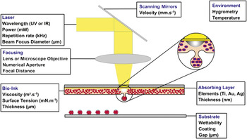
Basically, a typical LAB setup is generally composed of three elements, a pulsed laser source, a target coated with the material to print (the ribbon), and a receiving substrate facing the ribbon at a few hundred μm distance from the ribbon (see Figure 1). The ribbon is a multiplex structure made of a transparent glass slide onto which a thin layer of liquid (bio-ink) with embedded heat sensitive biological material (e.g., biomaterials, cells, biomolecules) is spread. Depending on the optical properties of the bio-ink with respect to the laser properties (wavelength and pulse duration), a solid laser absorbing interlayer may be necessary to transmit the energy from the laser beam (i.e., if the bio-ink is optically transparant to the laser), and this is placed between the support and the bio-ink film. Hence, the terminology of biological laser printing (BioLP)Reference Barron, Wu, Ladouceur and Ringeisen55 or absorbing film assisted-LIFT (AFA-LIFT)Reference Hopp, Smausz, Kresz, Barna, Bor, Kolozsvári, Chrisey, Szabó and Nógrádi56 is used to distinguish this method from matrix-assisted pulse laser evaporation—direct write (MAPLE-DW) (see the Riggs et al. article in this issue),Reference Barron, Ringeisen, Kim, Spargo and Chrisey57 in which the laser triggers direct vaporization of the first molecular layer of the liquid. Such a solid interlayer reduces direct interaction between the beam and the bio-ink, since 99% of the non-reflected incident beam may be transmitted in the case of MAPLE-DW. The interlayer consists of a thin film (tens of nm) of metal (Au, Ti, Ag), metal oxide (TiO2), or photo-decomposing volatile polymer (triazene). Interestingly, it has been recently proposed to substitute this metallic interlayer with a polyimide layer, which deforms when absorbing the laser energy.Reference Brown, Kattamis and Arnold58 This later variant, named blister-actuated LIFT, avoids the printing of undesired debris produced by a metallic interlayer because of potential harm to cell biology in the long term.
Schematic of laser-assisted bioprinting featuring the experimental parameters that govern droplet deposition. Ejection regimes are associated with laser pulse conditions, focusing conditions, as well as optical and mechanical properties of the absorbing layer and the bio-ink. Substrate properties mainly affect printing resolution.

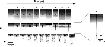
The generation of micro-droplets of bio-ink by LAB proceeds through a multiple-step mechanism:Reference Mezel, Hallo, Souquet, Breil, Hebert and Guillemot59 (1) first the laser energy (in the order of 1 J/cm2) is deposited either into the skin depth of the absorbing layer or into the bio-ink film;Reference Duocastella, Fernández-Pradas, Morenza and Serra60 (2) this induces, as a function of the laser irradiation intensity, the generation of a bubble that (3) expands until the bio-ink film is deformed, thereby (4) yielding a high speed liquid jet (from 20 to 100 m.s‒1) (see Figure 2). Eventually, the jet may touch the substrate, resulting in the printing of a micro-sized droplet of bio-ink.
Time-resolved observation of jet formation as a function of laser energy at different times after laser pulse deposition (in μs). First, second, and third rows correspond to sub-threshold, jetting, and plume regimes. For jetting conditions, at 30 μs, the jet is cut by the receiving substrate facing the ribbon, thus generating a pL volume droplet.

Experimentally, the jet formation requires precise control of laser pulse parameters, which are recapitulated in Figure 1. Within jetting conditions, it has been shown that the volume of deposited material depends linearly on the laser pulse energy, whereas the droplet volume can also be tuned by controlling the spot diameter,Reference Colina, Duocastella, Fernandez-Pradas, Serra and Morenza61 the thickness of the absorbing layer,Reference Hopp, Smausz, Kresz, Barna, Bor, Kolozsvári, Chrisey, Szabó and Nógrádi56 as well as some of the bio-ink properties such as viscosityReference Lin, Huang and Chrisey62 and film thickness.Reference Duocastella, Patrascioiu, Dinca, Fernández-Pradas, Morenza and Serra63, Reference Dinca, Farsari, Kafetzopoulos, Popescu, Dinescu and Fotakis64 Otherwise, sub-threshold (i.e., bio-ink deformation without forming a jet) and plume regimes (i.e., splashing)Reference Young, Auyeung, Piqué, Chrisey and Dlott54 are observed using lower and higher laser pulse energies, respectively (see Figure 2).
Main achievement for tissue-engineering applications
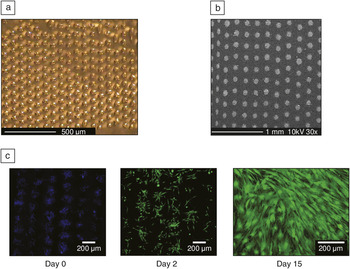
Over the last few years, it has been experimentally demonstrated that tiny amounts (femto- to nanoliters) of biological solutions (including hydrogelsReference Unger, Gruene, Koch, Koch and Chichkov65 and biomoleculesReference Colina, Serra, Fernández-Pradas, Sevilla and Morenza66, Reference Ringeisen, Wu, Kim, Piqué, Auyeung, Young, Chrisey and Krizman67) and suspensions (including nanoparticlesReference Catros, Fricain, Guillotin, Pippenger, Bareille, Remy, Lebraud, Desbat, Amédée and Guillemot68 and living cellsReference Ringeisen, Kim, Barron, Krizman, Chrisey, Jackman, Auyeung and Spargo44, Reference Koch, Kuhn, Sorg, Gruene, Schlie, Gaebel, Polchow, Reimers, Stoelting, Ma, Vogt, Steinhoff and Chichkov69) with a wide range of rheological properties could be deposited by LAB with high accuracyReference Guillotin, Souquet, Catros, Duocastella, Pippenger, Bellance, Bareille, Rémy, Bordenave, Amédée and Guillemot70 and high throughputReference Guillemot, Souquet, Catros, Guillotin, Lopez, Faucon, Pippenger, Bareille, Rémy, Bellance, Chabassier, Fricain and Amédée71 without affecting material integrity (e.g., viability and function of stem cellsReference Koch, Kuhn, Sorg, Gruene, Schlie, Gaebel, Polchow, Reimers, Stoelting, Ma, Vogt, Steinhoff and Chichkov69, Reference Raof, Schiele, Xie, Chrisey and Corr72) in vitro (see Figure 3), and also in vivo.Reference Keriquel, Guillemot, Arnault, Guillotin, Miraux, Amédée, Fricain and Catros73 Moreover, it has been recently demonstrated that 3D structures could be built-up using LAB.Reference Catros, Guillemot, Nandakumar, Ziane, Moroni, Habibovic, van Blitterswijk, Rousseau, Chassande, Amédée and Fricain74 LAB thereby overcomes some of the limitations of other bioprinting technologies such as ink-jet printing and pressure-operated mechanical extrusion that are, namely, the clogging of print heads or capillaries and the difficulties in printing high cell density droplets with a printing resolution that compares favorably with the geometrical complexity and cell density of in vivo tissues.Reference Guillotin and Guillemot29 Accordingly, LAB has been used to print endothelial capillary-like patterns, opening a new approach for vascular engineering,Reference Wu and Ringeisen75 and for fabricating a cardiac patch hosting both mesenchymal stem cells and endothelial cells.Reference Gaebel, Ma, Liu, Guan, Koch, Klopsch, Gruene, Toelk, Wang, Mark, Wang, Chichkov, Li and Steinhoff76
Laser-assisted bioprinting of (a) alginate microdroplets, (b) hydroxyapatite nanoparticles, and (c) human osteoprogenitor cells, which proliferate for 15 days.

Summary: Dealing with tissue complexity using laser-assisted bioprinting
Laser-assisted bioprinting (LAB) is a promising tool for dealing with tissue complexity and creating functional tissue-engineered 3D constructs. On this road, LAB would enable creation of “on-demand” manipulation and study of in vitro cell micro-environments by controlling intensity and shape of cell patterns and morphogen gradients (e.g., growth factors). Generating artificial cell nichesReference Lutolf and Blau77 by the co-deposition of suitable combinations of stem cells with extra-cellular matrix components should also be addressed. To engineer macroscopic 3D structured complex tissues, LAB may be integrated with other laser-assisted processes (e.g., machining, polymerization) and coupled with additional bottom-up methods.
Acknowledgments
The authors would like to thank the Région Aquitaine, GIS Advanced Materials in Aquitaine, and ANR for financial support.